Leonard, E. J., Witztum, J. L., and Steinberg, D. (1991) Proc. Natl. Acad. Sci. ... Nelken, N. A., Coughlin, S., Gordon, D., and Wilcox, J. N. (1991) J. Clin. Invest.
THE JOURNAL OF BIOLOGICAL CHEMISTRY © 1997 by The American Society for Biochemistry and Molecular Biology, Inc.
Vol. 272, No. 26, Issue of June 27, pp. 16404 –16413, 1997 Printed in U.S.A.
Cloning, in Vitro Expression, and Functional Characterization of a Novel Human CC Chemokine of the Monocyte Chemotactic Protein (MCP) Family (MCP-4) That Binds and Signals through the CC Chemokine Receptor 2B* (Received for publication, June 20, 1996, and in revised form, April 2, 1997)
Theo A. Berkhout‡, Henry M. Sarau§, Kitty Moores‡, John R White¶, Nabil Elshourbagy¶, Edward Appelbaum¶, Theresa J.Reape‡, Mary Brawner¶, Jayneeta Makwana‡, James J. Foley§, Dulcie B Schmidt§, Christine Imburgia¶, Dean McNulty¶, Jane Matthews¶, Kevin O’Donnell¶, Daniel O’Shannessy¶, Miller Scott¶, Pieter H. E. Groot‡i, and Colin Macphee‡ From the ‡Department of Vascular Biology, SmithKline Beecham Pharmaceuticals, The Frythe, Welwyn, United Kingdom and the Departments of §Pharmacology and ¶Biopharmaceutical Sciences, SmithKline Beecham Pharmaceuticals, King of Prussia, Pennsylvania 19406
Here we describe the characterization of a novel human CC chemokine, tentatively named monocyte chemotactic protein (MCP-4). This chemokine was detected by random sequencing of expressed sequence tags in cDNA libraries. The full-length cDNA revealed an open reading frame for a 98-amino acid residue protein, and a sequence alignment with known CC chemokines showed high levels of similarity (59 – 62%) with MCP-1, MCP-3, and eotaxin. MCP-4 cDNA was cloned into Drosophila S2 cells, and the mature protein (residues 24 –98) was purified from the conditioned medium. Recombinant MCP-4 induced a potent chemotactic response (EC50 5 2.88 6 0.15 nM) and a transient rise in cytosolic calcium concentration in fresh human peripheral blood monocytes but not in neutrophils. Binding studies in monocytes showed that MCP-4 and MCP-3 were very potent in displacing high affinity binding of 125I-MCP-1 (IC50 for MCP-4, MCP-3, and unlabeled MCP-1 of 2.1 6 1.4, 0.85–1.6, and 0.7 6 0.2 nM respectively), suggesting that all three chemokines interact with the CC chemokine receptor-2 (MCP-1 receptor). This was confirmed in binding studies with Chinese hamster ovary cells, stably transfected with the CC chemokine 2B receptor. Northern blot analysis in extracts of normal human tissues showed expression of mRNA for MCP-4 in small intestine, thymus, and colon, but the level of protein expression was too low to be detected in Western blot analysis. However, expression of MCP-4 protein was demonstrated by immunohistochemistry in human atherosclerotic lesion and found to be associated with endothelial cells and macrophages.
Chemokines are structurally and functionally related 8 –10kDa polypeptides, involved in the recruitment of white blood cells into areas of inflammation and their subsequent activation (1, 2). In addition, some chemokines are able to regulate the proliferative potential of hematopoietic progenitor cells, endothelial cells, and certain types of transformed cells (3, 4). Based on whether the first two cysteine moieties are separated * The costs of publication of this article were defrayed in part by the payment of page charges. This article must therefore be hereby marked “advertisement” in accordance with 18 U.S.C. Section 1734 solely to indicate this fact. i To whom correspondence should be addressed: Dept. of Vascular Biology, SmithKline Beecham, The Frythe, Welwyn (Herts), AL6 9AR, United Kingdom. Tel.: 44-1438-782405; Fax: 44-01438-782580.
by one amino acid moiety or are adjacent, chemokines belong to the a-chemokine or CXC chemokine family (e.g. interleukin-8) or the b-chemokine or CC chemokine family (e.g. RANTES1 and MCP-1). CXC chemokines preferentially attract and affect neutrophils, while CC chemokines chemoattract and affect eosinophils, monocytes, and T cells with relative potencies that differ between the different members of this family. Chemokines express their biological responses through interaction with chemokine receptors (5). Two human CXC chemokine receptors (the interleukin-8A and interleukin-8B (6, 7)) and six human CC chemokine receptors (the MIP-1a/RANTES receptor (CC-CKR-1) (8, 9), the MCP-1A and -B receptors (CCCKR-2A and -B) (10, 11), the eotaxin/RANTES receptor (CCCKR-3) (12–14), the promiscuous receptor on basophils CCCKR-4 (15), and a new MIP-1a/MIP-1b/RANTES receptor (CCCKR-5) (16)) have to date been cloned. Here we describe the identification, cloning, and in vitro expression of a novel human CC chemokine, tentatively named MCP-4. This chemokine shows a high level of amino acid sequence homology (62%) with human MCP-1. Following expression of MCP-4 in Drosophila S2 cells, an N terminus-processed protein (amino acid residues 24 –98) was purified from the conditioned medium, and alignment with the sequences of other mature forms of members of the CC chemokine family suggests that this polypeptide represents mature MCP-4. MCP-4 (residues 24 –98) binds and induces functional responses in freshly isolated human monocytes but not in polymorphonuclear leukocytes. We also show that binding of MCP-4 (residues 24 –98) to monocytes is at least in part due to the CC-CKR-2B receptor, a conclusion supported by data obtained in recombinant CHO cells expressing this receptor. Using immunohistochemical methods, we also show that MCP-4 is expressed in human atherosclerotic carotid and coronary arteries and that the expression is associated with vascular endothelial cells and macrophages. EXPERIMENTAL PROCEDURES
Materials Recombinant human MCP-1, MCP-2, MCP-3, RANTES, and MIP-1a were from R & D Systems, Europe Ltd. and Advanced Protein Products. 1 The abbreviations used are: RANTES, regulated on activation normal T-cell expressed and secreted; MCP, monocyte chemotactic protein; CC-CKR, CC chemokine receptor; MIP, macrophage inflammatory protein; PCR, polymerase chain reaction; PBS, phosphate-buffered saline; CHO, Chinese hamster ovary; [Ca21]i, cytosolic free calcium concentration.
16404
This paper is available on line at http://www.jbc.org
Characterization of MCP-4 125
I-Labeled MCP-1 and RANTES were from Amersham International UK (specific activity, 2000 Ci/mmol). Dextran T500 was obtained from Pharmacia Biotech Inc.; NycoPrepTM1.068 was from Nycomed UK; PBS and RPMI 1640 were from Life Technologies, Inc.; HEPES (ultrapure) and ionomycin were from Calbiochem; Fura-2/AM was from Molecular Probes, Inc.; bovine serum albumin (fatty acid-free) and naphthyl-butyrate esterase kit were purchased from Sigma; Diff-Quik stain was from B. M. Browne Ltd.; multiple tissue blots for Northern blotting and a tissue-specific protein solution of normal human tissues for Western blotting were purchased from CLONTECH Inc. (Palo Alto, CA). Ready made human multiple tissue blots from DNA Technologies Inc. (Gaithersburg, MD) were also used for Western blotting. All other reagents were analytical grade (AnalaR).
Methods Cloning and Expression of Recombinant Human MCP-4 MCP-4 was initially identified at Human Genome Sciences (Rockville, MD) by random sequencing of expressed sequence tags in a cDNA library from an 8-week-old human fetus, and a full-length clone was identified and sequenced subsequently. MCP-4 was expressed in a Drosophila cell culture system (17, 18). The MCP-4 coding region was amplified by polymerase chain reaction (PCR) using the cDNA clone as substrate and the following pair of primers: 59-TCCCCGCGGCCACCATGAAAGTTTCTGCAGTGCTT-39 (SacII site and initiator methionine codon underlined) and 59-GCTCTAGACTATTAAGTCTTCAGGGTGTGAGCT-39 (XbaI site underlined). The PCR product was digested with SacII and XbaI, cloned into SacII/ XbaI-digested vector pMTAL, and sequenced to ensure the absence of PCR-generated mutations. This placed the open reading frame between a copper sulfate-inducible metallothionein promoter and an SV40 late polyadenylation site. This vector was introduced into Drosophila Schneider 2 (S2) cells by co-transfection with a vector carrying a hygromycin B resistance gene. Hygromycin B-resistant cells were selected as a stable polyclonal population, induced with copper sulfate, and examined for expression by Western blot analysis using an antiserum raised in rabbits immunized with a MCP-4 peptide. Cells were adapted to serum-free medium (19) and scaled to 10 liters to generate conditioned medium for purification.
Purification of MCP-4 from Drosophila-conditioned Media Ten ml of 0.5 M EDTA (pH 7.4) was added per liter of conditioned medium, which was then diluted 1:1 (v/v) with deionized/distilled water. The diluted medium was filtered through a 0.45-mm membrane filter to remove particulates and subsequently loaded, via a peristaltic pump, at 14 –15 ml/min onto a 10-ml bed volume Sepharose-S Fast Flow column previously equilibrated with 10 mM HEPES, pH 7.0. After sample loading, the column was washed with 10 mM HEPES, pH 7.0, until the absorbance at 280 nm of the effluent returned to base line. Bound material was then eluted from the column with 1 M NaCl in 10 mM HEPES, pH 7.0. The eluate was concentrated using an Amicon Stir Cell concentrator using a YM3 3,000-Da cut-off membrane. The concentrated eluate was applied to a Superdex 75 size exclusion column equilibrated in 20 mM sodium phosphate, 150 mM NaCl, pH 7.0, and developed using the same buffer. Fractions were analyzed by 17.5% SDS-PAGE, and those containing MCP-4 were pooled, concentrated in an Amicon Stir Cell concentrator using a YM3 membrane, filter-sterilized through a 0.22-mm membrane filter, aliquoted, and stored at 220 °C. Samples of the purified MCP-4 were subjected to N-terminal sequence analysis, quantitative amino acid analysis, and matrix-assisted laser desorption time-of-flight mass spectrometry to confirm the integrity and concentration of the product.
Amino Acid and N-terminal Sequence Analysis of MCP-4 MCP-4 was hydrolyzed in vacuo under 6 N HCl for 20 h at 110 °C. The resulting hydrolysate was analyzed by ion exchange amino acid analysis using postcolumn ninhydrin detection on a Beckman 6300 analyzer equipped with a System Gold data acquisition system (Beckman Instruments, Fullerton, CA). N-terminal sequence analysis was performed on an Applied Biosystems 470A gas phase protein sequencer (Applied Biosystems, Inc., Foster City, CA) equipped with a Beckman 126/166 system (Beckman Instruments, Fullerton, CA) for on-line phenylthiohydantoin analysis. Data were acquired using System Gold chromatography software (Beckman Instruments, Palo Alto, CA). Samples were spotted directly onto Polybrene-coated GF/C filters (Applied Biosystems), and standard ABI sequencing cycles were used.
16405
Mass Spectrometry Matrix-assisted laser desorption mass spectrometry data for MCP-4 were obtained on a Vestec LaserTec Research laser desorption time-offlight mass spectrometer (Vestec Corp., Houston, TX). Samples were prepared for analysis by mixing MCP-4, in PBS, pH 7.0, with sinapinic acid (saturated solution in 33% CH3CN, 0.1% trifluoroacetic acid (v/v)) for a final concentration of 1 mM. Apomyoglobin (Sigma) was included as an internal calibrant (MH21 5 8476.8 Da). Desorption/ionization was accomplished using a 337-nm nitrogen laser. Spectra were averaged over ;50 laser scans. Calibrations were carried out using a customized version of IGOR on a Macintosh personal computer.
Monocyte Isolation Human peripheral blood monocytes were prepared from the blood of normal healthy volunteers, essentially as described by Boyum (20). Blood was collected into anticoagulant (1 part 50 mM EDTA, pH 7.4, to 9 parts blood) and then centrifuged for 5 min at 600 3 g. The upper layer of platelet-rich plasma was removed and centrifuged for 15 min at 900 3 g to pellet the platelets. The upper layer of platelet-poor plasma was removed and added back to the packed red cells; the pelleted platelets were discarded. Dextran T500 was added (10 volumes of EDTA blood to 1 volume of 6% (w/v) dextran in 0.9% (w/v) NaCl), and the erythrocytes were allowed to sediment at unit gravity for 30 min. The resultant leukocyte-rich plasma was removed and centrifuged for 5 min at 400 3 g. The cell pellet was resuspended in 5 ml of the supernatant, and the suspension was underlayered with 3 ml of NycoPrepTM1.068 and then centrifuged for 15 min at 600 3 g. The mononuclear layer at the interface between the plasma and the NycoPrepTM1.068 was removed and washed through PBS by centrifugation for 5 min at 400 3 g. The mononuclear layer typically contained $80% monocytes, determined by staining cytocentrifuge preparations for nonspecific esterase using a-naphthyl butyrate. Cell viability (typically .95%) was assessed as the ability to exclude trypan blue.
Preparation of Chinese Hamster Ovary Cells, Expressing the Human CC-CKR2B Receptor Oligonucleotide primers 59-TGGGGTACCCCACAACATGCTGTCCACATCTCGTTCTCG-39 (sense) and 59-CAAACGGATCCTCGTTTTATAAACCAGCCGAG-39 (antisense) containing KpnI and BamHI restriction sites, respectively (underlined), were synthesized based on the published sequence of the human CC-CKR-2B receptor (10). These primers were used to produce a full-length cDNA for this receptor by PCR with human placenta cDNA as the template. The expected product of 1.1 kilobase pairs was digested with KpnI and BamHI and inserted into the equivalent sites of the mammalian expression vector pCDN. The insert of the resulting construct was completely sequenced to confirm its identity and orientation. To obtain a stable cell line expressing the CC-CKR-2B receptor, the pCDN:CC-CKR-2B expression plasmid was electroporated into Chinese hamster ovary cells. Stable cell lines were isolated by growing cells in the absence of nucleosides. Those cell lines that grew out of selection were then analyzed for CC-CKR-2B expression using a radiolabeled MCP-1 binding assay. The cell line used here, C11, was found to express approximately 200,000 receptors/cell.
Chemokine Binding and Binding Displacement Studies Monocytes were suspended in RPMI 1640 medium without bicarbonate containing 0.2% bovine serum albumin and 0.1% azide. 125I-Labeled chemokines (MCP-1 or RANTES, about 2 3 104 cpm) were incubated with 1–2 3 106 cells in the presence or absence of increasing concentrations of unlabeled chemokines (MCP-1, MCP-3, MCP-4, RANTES, or MIP-1a) for 15 min at 37 °C in a 96-well plate in a final volume of 0.2 ml. After the incubation, 0.5 ml of ice-cold wash buffer (20 mM Tris, 0.5 M NaCl, pH 7.4) was added, and cells were collected onto a polyethyleneimine-treated Whatman GF/C filter using a Brandell cell harvester. Filters were washed with 4 ml of cold wash buffer, and the radioactivity bound to the filters was counted in a g-counter. For competition studies, the IC50 was calculated with a curve fitting program (GraFit, Erithacus Software, London), using a four-parameter logistic, cpmbound 5 cpms max/(1 1 ([L]/IC50) )1 cpmns, where cpmmax represents the binding without competitor, [L] is the competitor concentration, cpmns is the nonspecific binding, and s is the slope factor. The cpmbound was corrected for “no cell” controls. To obtain the Kd and capacity of binding for individual chemokines, specific binding data from homologous displacement experiments were fitted into a single site ligand binding equation using the GraFit best fit program. Chemokine binding to the CHO cells, expressing the human CC-CKR-2B receptor, were performed essentially as described above except that 125I-labeled chemokines were di-
16406
Characterization of MCP-4
luted with unlabeled chemokine to a specific activity of 400 Ci/mmol and used in a final concentration of 0.6 nM. Cells were incubated with 125 I-labeled chemokines plus or minus unlabeled chemokines for 30 min at 37 °C.
Measurement of [Ca21]i For measurements of cytosolic Ca21 concentrations, monocytes were incubated with 0.5 mM Fura-2/AM for 30 min at 37 °C in HEPESbuffered saline (145 mM NaCl, 5 mM KCl, 1 mM MgCl2, 10 mM HEPES, and 10 mM glucose), pH 7.4, at 37 °C, supplemented with 1% albumin (w/v) and 1 mM CaCl2. After loading with Fura-2, the cells were centrifuged for 5 min at 300 3 g and then resuspended in buffer containing no added albumin, to a cell density of 1.5 3 106 cells/ml, and kept at room temperature until use. This protocol resulted in a cytosolic Fura-2 concentration of ;100 mM. Serial dilutions of chemokines in PBS plus 0.1% albumin (w/v) (sterile filtered) were added to aliquots (0.7 ml) of cell suspension. The Fura-2 fluorescence of monocyte suspension was measured at 37 °C in a single excitation, single emission (500 nm) wavelength Perkin-Elmer LS5 fluorometer. [Ca21]i was calculated from changes in fluorescence measured at a single excitation wavelength of 340 nm, as described by Grynkiewicz et al. (21). [Ca21]i measurements in CHO cells, expressing the human CC-CKR-2B receptor were performed essentially as described above for monocytes. After loading with Fura-2/AM, cells (1 3 106/ml) were kept in ice-cold medium (118 mM NaCl, 4.6 mM KCl, 25 mM NaHCO3, 1 mM KH2PO4, 11 mM glucose, 50 mM HEPES, 1 mM MgCl2, 1 mM CaCl2, 0.1% gelatin (pH 7.4). Aliquots (2 ml) of cell suspension were prewarmed at 37 °C for 5 min in 3-ml plastic cuvettes, and fluorescence was measured in a fluorometer (Johnson Foundation Biomedical Group) with magnetic stirring and temperature controlled at 37 °C. Excitation was set at 340 nm, and emission was set at 510 nm. [Ca21]i was calculated as described for monocytes. For studies in monocytes on cross-desensitization of calcium responses, chemokines were added sequentially with a 2-min interval, and [Ca21]i transients were recorded. The concentrations used in these studies varied for each chemokine and were set at levels known to induce the maximal response for [Ca21]i mobilization (see above).
Monocyte Chemotaxis Assay Cell migration was evaluated using a 48-well modified Boyden microchemotaxis chamber (Neuroprobe, Cabin John, MD). 25-ml aliquots of chemokines, diluted in RPMI 1640 containing 0.1% bovine serum albumin (w/v) were placed in the wells of the lower compartment, and 50-ml aliquots of monocyte suspension in RPMI 1640 (1.5 3 106 cells/ml) were placed in the upper wells of the chamber; the two components were separated by a polycarbonate filter with a 5-mm pore size. The chamber was incubated at 37 °C in humidified air containing 5% CO2, for 60 min. After incubation, the filter sheet was removed, and the nonmigrated cells were scraped from its upper surface. The filter was stained with Diff-Quik, and the number of migrated cells was counted (5 fields/well), using a 3 40 objective and 3 10 ocular containing a 10-mm2 counting grid.
MCP-4 mRNA Expression Multiple Tissue Blot Analysis—Multiple tissue blots were commercially purchased and prepared as described in the accompanying literature from the manufacturer. Denatured MCP-4 cDNA (50 ng) was labeled with [32P]CTP using Klenow reagent. Multiple tissue blots were probed using ExpressHyb solution and after washing were exposed on X-Omat (Kodak) x-ray development film for between 48 and 96 h. Expression in Human Pulmonary Smooth Muscle Cells—Total RNA (3 mg/sample) isolated from quiescent human pulmonary smooth muscle cells and cells treated with serum for the indicated times were immobilized on nitrocellulose filters and hybridized to either 32P-labeled MCP-4 cDNA or glyceraldehyde-3-phosphate dehydrogenase cDNA, as described previously (22). Antibodies to MCP-4 —Initial antibodies used for the monitoring of the expression of recombinant MCP-4 were raised in rabbits after injection of a synthetic MCP-4 peptide (cysteine 58 to isoleucine 73). Subsequent polyclonal antibodies against MCP-4 were raised in rabbits using purified E. coli-expressed His-tagged MCP-4. The antiserum CS09, used for immunodetection of MCP-4 in the present study, was highly specific for MCP-4 (E. coli- as well as Drosophila-expressed MCP-4) and showed no cross-reactivity whatsoever with related chemokines (MCP-1, MCP-2, MCP-3, eotaxin, MIP-1a, or RANTES) in Western blot analysis. Western Analysis of Human Tissue Extracts—MCP-4 protein expression in human tissues was studied by Western blot analysis. Tissue-
specific protein solutions of normal human tissues were obtained from CLONTECH, fractionated (75 mg of protein/lane) on a 16% polyacrylamide gel and blot on nitrocellulose membranes. Commercial, ready made human multiple tissue blots (75 mg of protein extracts of normal human tissues) were also used and obtained from DNA Technologies, Inc. (Gaithersburg, MD). Immunoblots were probed with antiserum CS09 (1:2000 dilution) using the ECS (Amersham) detection system. The lower limit of detection was determined using dilutions of pure MCP-4 and found to be ;1 ng of MCP-4. Immunohistochemistry—Tissues used in this study were coronary arteries (n 5 6) from heart transplant recipients and carotid endarterectomy specimens (n 5 2) from patients undergoing vascular reconstructive surgery for arterial occlusive disease. Tissue specimens were snap frozen in liquid nitrogen and stored at 270 °C. Specimens were mounted in Tissue-Tek OCT compound embedding medium (Agar Scientific Ltd, Essex, UK) for cryostat sectioning. Serial transverse tissue sections (10 mm) were thaw mounted onto Superfrost1 slides (BDH, Leicestershire, UK). Sections were fixed in acetone for 10 min and stored at 270 °C until use. Immunohistochemistry was performed using the avidin-biotin-peroxidase complex method and the rabbit polyclonal antiserum for MCP-4 CS09 (see above). All incubations were done at room temperature unless otherwise stated. Briefly, endogenous peroxidase activity was blocked by incubating slides in 0.6% H2O2 in methanol for 15 min. Nonspecific binding was blocked by incubating sections in normal swine serum before applying the primary antibody. Optimal primary antibody concentrations were predetermined by titration, and slides were incubated overnight at 4 °C in a 1:1000 dilution. Slides were then incubated in biotinylated swine anti-rabbit antibody (Dako, Buckinghamshire, UK) for 30 min, washed, and incubated with avidin/biotinylated horseradish peroxidase complex (Vector Laboratories, Peterborough, UK) for 30 min. Slides were then stained with diaminobenzidine tetrahydrochloride substrate (Vector Laboratories). The sections were counterstained with hematoxylin, dehydrated through a graded series of alcohols, and mounted in DPX. Negative controls were omission of primary antibody and substitution of primary antibody with normal rabbit serum. The cellular composition of the plaques was determined using the following mouse monoclonal antibodies anti-CD68, (a specific human monocyte/macrophage marker), anti-a-actin (smooth muscle), and antiVon Willebrand factor (endothelial cells), all from Dako. The method was the same as above except that primary antibody incubations were for 30 min at room temperature. These were followed by incubation with a biotinylated rabbit anti-mouse secondary antibody (Dako). RESULTS
Sequence Comparison with Other Chemokines—The derived amino acid sequence of the full-length open reading frame for the novel chemokine is given in Fig. 1. Full-length MCP-4 consists of 98 amino acid residues, and an amino acid sequence alignment with known human chemokines showed a high homology to MCP-1 (62% similarity), MCP-3 (61%), and eotaxin (59%) with lower homologies to MCP-2, MIP-1a, MIP-1b, and RANTES (52, 37, 39, and 29%, respectively). Based upon these sequence homologies and functional properties (see below), we propose to adopt the name MCP-4 as used throughout this manuscript. Expression and Purification of Recombinant MCP-4 —To allow functional studies with this novel chemokine, MCP-4 was expressed in Drosophila S2 cells, a system previously shown to correctly recognize and remove human cytokine signal sequences (18, 23). Growth medium from a stably transfected Drosophila S2 cell line carrying the expression vector for MCP-4 was analyzed by immunoblot, using an antiserum raised against a synthetic MCP-4 peptide. A 9-kDa protein band was detected that was absent from medium from control cells (Fig. 2A). After purification by ion exchange and size exclusion chromatography, the final preparation contained a 9-kDa species as visualized on a stained polyacrylamide gel (Fig. 2B). Mass spectrometry showed a predominant peak with a mass of 8583 Da, corresponding to the size expected (8580 Da) for a protein extending from glutamine 24 (cyclized to pyroglutamatic acid) to the end of the open reading frame (Fig. 2C). A minor peak at 8782 Da was also observed, which is close to the
Characterization of MCP-4
16407
FIG. 1. Alignment of MCP-4 with other members of the human CC chemokine family. Alignment was done by the clustral method with a PAM 100 residue weight table using the DNAStar Lasergene package. Shaded, boxed letters indicate residues that are identical to the CC chemokine consensus sequence. The arrowheads indicate the start of the mature, functionally active forms of these chemokines.
FIG. 2. Expression of MCP-4 in Drosophila S2 cells and characterization of purified protein. A, immunoblot analysis of conditioned medium from copper sulfate-induced Drosophila S2 cells carrying an MCP-4 expression vector (lane 1) or vector without MCP-4 (lane 2). Positions of molecular weight markers (sizes in kDa) are indicated to the left. B, SDS-PAGE of purified MCP-4 (2 mg of MCP-4, stained with Coomassie Blue (lane 1); molecular weight markers (lane 2). C, matrixassisted laser desorption mass spectrometry analysis of purified MCP-4.
mass expected (8780 Da) for a molecule extending from leucine 22 to the end of the open reading frame. N-terminal sequence analysis indicated that most of the material in the preparation was blocked, as expected for a protein with an N-terminal pyroglutamatic acid. However, minor sequences starting at unblocked glutamine 24 (QPDALNVPST) and at leucine 22 (LAQPDALNVP) were detected. These results show that over
90% of the material in this preparation has glutamine 24 (cyclized or free) at the N terminus. This N-terminal sequence aligns very well with the consensus sequence QPDAV for mature human CC chemokines (compare Fig. 1) and is likely to represent the product of MCP-4 maturation in humans. The nature of the minor sequence in Drosophila cell expression (MCP-4 starting at leucine 22) is presently unknown; however, MCP-4 maturation by signal sequence cleavage at the glycine 21–leucine 22 bond and subsequent removal of the leucine 22-alanine 23 dipeptide seems unlikely, since proline at a position 3 residues upstream of a signal peptide cleavage site (as proline 19 would be) is not generally found (24). Effects of MCP-4 on Monocyte Chemotaxis—The effect of mature (residues 24 –98) MCP-4 (hereafter referred to as MCP-4) on chemotaxis of freshly isolated human peripheral blood monocytes was tested in a 48-well Boyden chamber and compared with chemotactic responses toward known human chemokines. Dose-response curves of these studies are given in Fig. 3. All chemokines gave the expected bell-shaped response curves with inhibition of chemotaxis at high chemokine concentrations due to receptor desensitization. MCP-4 was found to be a potent stimulator of monocyte chemotaxis (EC50 5 2.88 6 0.15 nM), although less potent than MCP-1 (EC50 5 0.37 6 0.05 nM), MCP-2 (0.62 6 0.07 nM), MCP-3 (0.17 6 0.02 nM), and MIP-1a (0.25 6 0.06 nM). RANTES (2.74 6 0.28 nM) had a similar potency. In similar studies with human neutrophils, MCP-4 (up to 330 nM) was found to be inactive (data not shown). Effects of MCP-4 on Cytosolic Calcium Concentrations in Monocytes—The signal transduction pathway of most chemokines involves a transient rise in cytosolic calcium concentration. To investigate the effect of MCP-4 on cytosolic calcium concentration, monocytes were preloaded with Fura-2/AM, and [Ca21]i concentrations in response to various chemokines were recorded. The dose-response relations between chemokine concentrations and the recorded maximal [Ca21]i concentrations are given in Fig. 4. As shown previously (25, 26), MCP-1 and MCP-3 are potent inducers of a [Ca21]i transient (EC50 values of 1.48 6 0.24 and 1.39 6 0.11 nM, respectively). MCP-4 also induces a clear calcium response (EC50 5 22.6 6 2.4 nM), although its potency was found to be less than for MCP-1 and MCP-3. Although structurally and functionally related to the other members of the MCP family, MCP-2 was inactive in this assay (no response at concentrations up to 47 nM), although much lower concentrations induced monocyte chemotaxis (see above). Similar observations have been reported previously by Sozzani et al. (26). RANTES also induced a transient rise in
16408
Characterization of MCP-4
FIG. 3. Chemokine-induced migration of human monocytes, measured in 48-well microchemotaxis chambers. Migration is expressed as means of migrated cells in the presence of chemokines divided by the means of migrated cells in the absence of chemokines. The mean value for migrated cells in the absence of chemokines was 15.1 6 0.64. Points represent means 6 S.E. of at least three separate experiments.
FIG. 4. Dose-response curves for chemokine-stimulated increases in peak [Ca21]i in Fura-2-loaded human peripheral blood monocytes in the presence of extracellular Ca21. For details see “Experimental Procedures.” Points represent means 6 S.E. of five separate monocyte preparations.
cytosolic calcium (EC50 5 3.96 6 0.28 nM, data not shown) as reported by others previously (27). Binding Competition Studies—To investigate which monocyte chemokine receptors are involved in functional responses of MCP-4, chemokine binding studies were performed. Binding was measured at 37 °C in the presence of sodium azide to arrest chemokine uptake and processing. As demonstrated by others earlier (28), freshly isolated peripheral blood monocytes possess specific high affinity binding sites for 125I-radiolabeled MCP-1 (3384 6 1223 binding sites/cell, Kd 5 0.51 6 0.06 nM). Competition analysis showed that the binding of 125I-MCP-1 to these cells could be competed for completely by a surplus of unlabeled MCP-1 as well as unlabeled MCP-3 (Fig. 5A). Interestingly, MCP-4 was also found to be a potent and complete competitor of 125I-MCP-1 binding, and a comparison of IC50 values for all three chemokines showed an order of potency of MCP-1 . MCP-3 . MCP-4 (mean values 6 S.D. for IC50 values were 0.7 6 0.2 nM (n 5 5), 0.85–1.65 nM (n 5 2), and 2.1 6 1.4 nM (n 5 4), respectively, where n represents the number of independent experiments). RANTES at concentrations up to 100 nM was ineffective in displacing 125I-MCP-1 (data not shown). Based on this chemokine competition profile (competition among MCP-1, MCP-3, and MCP-4), involvement of the CC-CKR-2 receptor in MCP-4 binding seems likely. Similar binding studies were performed using 125I-RANTES, and the effect of displacement by MCP-4 and other unlabeled chemokines was investigated. Results are given in Fig. 5B. Binding of radiolabeled RANTES was effectively and completely competed for by unlabeled RANTES (IC50 5 1.0 6 0.12 nM, n 5 3) and MIP-1a (IC50 5 12.6 6 4.3 nM, n 5 3). Although high concentrations of MCP-4 (and MCP-3) could displace 125I-RANTES (IC50 for MCP-4 of 235 6 112 nM, n 5 3), these concentrations
FIG. 5. Binding of 125I-labeled MCP-1 and RANTES to fresh human peripheral blood monocytes and its displacement by unlabeled chemokines. 125I-Labeled chemokines (final concentration, 0.028 nM)) were incubated with 1–2 3 106 freshly isolated human monocytes in the absence or presence of unlabeled chemokines at concentrations given in the figure. The total binding of radiolabeled ligand in the absence of any competing ligand was taken as 100%, and all binding data, corrected for “no cell” background counts, are expressed as a percentage of this value. A, binding of 125I-MCP-1 and the effects of increasing concentrations of unlabeled chemokines. B, binding of 125IRANTES and the effects of increasing concentrations of unlabeled chemokines. For symbols see inset in each panel. No displacement was seen of 125I-MCP-1 binding by unlabeled RANTES up to 100 nM (experiment as in A) and 125I-RANTES binding by unlabeled MCP-1 up to 100 nM (experiment as in B), for reasons of clarity not included in the figures. Data points are from a representative experiment showing the mean 6 S.D. of triplicate incubations. For further details see under ‘‘Binding Competition Studies.’’
are probably too high to be of physiological significance. Therefore, we expect that receptors with high affinity for RANTES receptors are unlikely to be involved in MCP-4 binding and signaling. To strengthen the evidence for MCP-4 being a ligand for the CC-CKR-2B receptor, recombinant CHO cells expressing the human CC-CKR-2B receptor were constructed, and displacement of binding of 125I-MCP-1 by MCP-4 and other chemokines was tested. As in freshly isolated human peripheral blood monocytes, the binding of 125I-MCP-1 to CHO cells expressing CKR-2B was completely displaced by unlabeled MCP-1, MCP-3, and MCP-4 with the following relative potencies: MCP1 . MCP-4 . MCP-3 (IC50 values of 3.6 6 0.52, 7.1 6 5.19, and 12.7 6 3.46 nM, respectively, n 5 3) (Fig. 6). MIP-1a and RANTES did not displace any binding of radiolabeled MCP-1
Characterization of MCP-4
FIG. 6. Binding of 125I-labeled MCP-1 to CHO cells, expressing the human CC-CKR-2B receptor and its displacement by unlabeled chemokines. 125I-Labeled MCP-1 (final concentration 0.6 nM) was incubated with 0.25–1 3 106 cells in the absence or presence of unlabeled chemokines at concentrations given in the figure. The total binding of radiolabeled ligand in the absence of any competing ligand was taken as 100%, and all binding data, corrected for “no cell” background counts, are expressed as a percentage of this value. MIP-1a and RANTES, up to 100 nM, did not displace any binding of 125I-MCP-1 (data not shown). Data in the figure are of a representative experiment (mean of duplicate incubations) of a series of three. For further details see under ‘‘Binding Competition Studies.’’
up to concentrations of 100 nM, but MCP-2 did (IC50 5 39.5– 46.5 nM, n 5 2; data not shown). Functional response studies added further evidence. Like monocytes, CHO cells expressing the CC-CKR-2B receptor were found to generate potent transient increases in cytosolic Ca21 concentration following exposure to MCP-1. Since coupling of the receptor to the internal signaling system was evidently present, calcium transients following exposure to different concentrations of chemokines were measured (Fig. 7). MCP-1, MCP-3, and MCP-4 were all found to provoke an increase in cytosolic calcium (mean values 6 S.D. for EC50 of 4.2 6 1.21, 35.3 6 15.4, and 51.7 6 18.9 nM, respectively, n 5 3– 4), while RANTES and MIP-1a were all ineffective up to 100 nM (data not shown). Not unexpectedly (see above), MCP-2 was also found to be ineffective. No 125I-MCP-1 binding or calcium transients were seen in untransfected CHO cells exposed to any of these chemokines. From these results it is evident that MCP-4 can bind and signal through interaction with the CCCKR-2B receptor. Desensitization Studies in Monocytes—Rapid successive exposure to their ligands is known to desensitize the signaling capacity of chemokine receptors. We have used heterologous desensitization of cytosolic Ca21 transients in monocytes, following chemokine exposure to investigate whether ligands are signaling through the same or different types of chemokine receptors. We used chemokine concentrations shown earlier to result in a maximum [Ca21]i response (compare Fig. 4). The results of these cross-desensitization studies are shown in Fig. 8. Preexposure of monocytes to MCP-1 completely blocked a response to MCP-4, while initial exposure to MCP-4 greatly diminished a subsequent MCP-1 signal. These results suggest that MCP-4 and MCP-1 signal at least partially through a shared receptor on monocytes, probably the CC-CKR-2B receptor. MCP-3 and MCP-4 also showed mutual heterologous desensitization, although the signal of the second chemokine was not completely blocked by the administration of the first, suggesting partial sharing of receptors. Since MCP-3 interacts with the CC-CKR-2B receptor, cross-desensitization between
16409
FIG. 7. Dose-response curves for chemokine-stimulated increases in peak [Ca21]i in Fura-2-loaded human CHO cells, expressing the human CC-CKR-2B receptor, in the presence of extracellular Ca21. For details see “Experimental Procedures.” No increases in [Ca21]i were seen with RANTES and MIP-1a at concentrations up to 100 nM (data not shown). Data points are the mean 6 S.D. of three independent studies (duplicate measurements in each study).
FIG. 8. Homologous and heterologous desensitization in Fura2-loaded human monocytes, in the presence of extracellular Ca21. Results in the figure are from a single experiment and show Fura-2 fluorescence calibrated to [Ca21]i. MCP-4 (100 nM), MCP-1 (6 nM), MCP-3 (12 nM), RANTES (25 nM), and MIP-1a (6 nM) were added as indicated. In each case, the time interval between subsequent additions was 2 min. Homologous desensitization, as shown above for MCP-4, was observed with all chemokines. Similar results were obtained in three other separate cell preparations.
MCP-3 and MCP-4 is to be expected if this receptor is involved in MCP-4 signaling. Interesting results were also obtained in cross-desensitization studies with RANTES and MIP-1a. MCP-4 was able to desensitize RANTES signaling, but RANTES was found to be nearly inactive in desensitizing an MCP-4 signal. A partial mutual desensitization was found with the
16410
Characterization of MCP-4
FIG. 9. Tissue expression of mRNA for MCP-4. A, multiple tissue Northern blots were probed with cDNA from the MCP-4 clone as described under “Experimental Procedures.” Positive tissues were identified by the presence of an approximately 1-kilobase pair transcript. Tissue blots were also probed for actin mRNA, found to be equal expressed across all tissues tested (data not shown). B, part 1, dot blot analysis of the expression of mRNA for MCP-4 in quiescent pulmonary smooth muscle cells stimulated by the addition of 10% fetal bovine serum for 1, 8, and 24 h before harvesting and probing for MCP-4 mRNA expression. Three mg of total cellular RNA was used for each blot. Part 2, dot blot analysis for glyceraldehyde-3-phosphate dehydrogenase mRNA expression. The numbers below panel 2 refer to serum exposure time. C is the zero time point control.
combination MCP-4/MIP-1a. These heterologous desensitization studies are compatible with an involvement of the CCCKR-2 receptor in MCP-4 signaling but suggest that other receptor(s) on monocytes with affinity for MIP-1a (and possibly RANTES) may be implicated. MCP-4 mRNA Distribution—Northern blot analysis of mRNA for MCP-4 in various tissues revealed an approximately 1-kilobase pair transcript in small intestine, thymus, and colon (Fig. 9A). No transcripts were detected in RNA from spleen, prostate, ovaries, and testes. In addition (not shown) a 1-kilobase pair transcript was found in lung, trachea, stomach, lymph node, and colonic mucosa, but not in a number of other tissues including spinal cord, adrenal gland, bone marrow, heart, brain, placenta, liver, skeletal muscle, kidney, and pancreas. Human pulmonary artery smooth muscle cells also showed a low level of MCP-4 mRNA expression, and the level of this transcript could be increased transiently by short exposure of these cells to 10% human serum (Fig. 9B), a treatment known to induce differentiation of smooth muscle cells toward a synthetic phenotype (29). Immunological Studies: Tissue Localization by Western Blot Analysis—The expression of MCP-4 in normal human tissues was investigated by Western blot analysis using a highly specific polyclonal rabbit antiserum against MCP-4. Using commercially available protein extracts for Western blot analysis (CLONTECH) from small intestine, thymus, lung, and stomach (tissues for which MCP-4 mRNA expression was seen) as well an extract from testes, we were unable to detect any MCP-4 protein expression in blots loaded with 75 mg of tissue protein. In another study using a ready made multiple normal human tissue Western blot of extracts from brain, heart, small intestine, pancreas, liver, colon, thymus, and lung (75 mg protein/ lane) again we were unable to detect any expression of MCP-4. Based on the detection limit for MCP-4 in our analysis (around 1 ng), it is concluded that the protein expression levels for
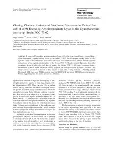
MCP-4 in these extracts are below 1 ng/75 mg of tissue protein. Localization of MCP-4 in Human Vascular Tissues—MCP-1 is known to be expressed in human atherosclerotic lesions, mainly by macrophage-foam cells but also in intimal smooth muscle cells and endothelial cells (30 –32). We studied the expression of MCP-4 in atherosclerosis using coronary artery specimens from heart transplant recipients and carotid endarterectomy specimens from patients undergoing vascular reconstructive surgery for arterial occlusive disease. MCP-4 expression was found to be associated with the luminal endothelium of human atherosclerotic coronary vessels (Fig. 10). It was also found on the endothelium of small adventitial blood vessels in all of the coronary vessels examined regardless of the degree of atherosclerosis. Four of the coronaries examined did not contain severe lesions (noncalcified, slightly thickened intima) but had a discrete macrophage infiltrate. We did not find MCP-4 to be strongly associated with these macrophages, although some areas of very weak staining could be seen. However, two of the coronary vessels were almost totally occluded and contained large areas of calcification. In these specimens, MCP-4 was found to be expressed by a subset of macrophages deep in the intimal lesion near areas of calcification. We also found MCP-4 strongly associated with macrophages in the two carotid endarterectomy specimens examined (Fig. 11). In these specimens, the lack of staining for Von Willebrand factor revealed loss of the luminal endothelium. Numerous intimal microvessels could be seen, but MCP-4 did not appear to be expressed by the endothelial cells lining these vessels. Using primers designed to flank the open reading frame of MCP-4 cDNA, we carried out reverse transcriptase-PCR on RNA extracted from eight carotid endarterectomy specimens. mRNA expression was found in seven of eight specimens (results not shown). DISCUSSION
In the present work we describe the identification and characterization of a novel human CC chemokine of the MCP family, MCP-4. This 75-amino acid residue chemokine was shown to induce chemotaxis in monocytes with nanomolar potency and to provoke a transient rise in cytosolic Ca21 concentrations in these cells. Similar to other members of the CC chemokine family, MCP-4 was found to be inactive toward human peripheral blood neutrophils, but in a concurrent study by Uguccioni and co-workers (33), MCP-4 was shown to induce functional responses in T cells, eosinophils, and monocytes. Using chemokine binding studies as well as functional response data, we have investigated which receptors on monocytes are implicated in MCP-4 binding and signaling. In monocytes MCP-4 as well as MCP-3 completely displaced binding of 125 I-labeled MCP-1. Since MCP-1 and MCP-3 are known ligands of the CC-CKR-2B receptor, while only MCP-1 seems to interact with the CC-CKR-2A splicing variant (10, 34, 35), we concluded that at least part of the high affinity binding of MCP-4 to monocytes is due to the CC-CKR-2B receptor. This conclusion was further supported by binding studies to CHO cells, stably transfected with the human CC-CKR-2B receptor. Displacement of 125I-MCP-1 binding in these cells by MCP-4, MCP-3, and other chemokines was very similar to that seen in monocytes, although affinities of all members of the MCP family were somewhat lower for the recombinant CC-CKR-2B receptor in CHO cells as compared with monocytes. It is possible that the latter effect is due to differences in the microenvironment for the receptor between monocytes and the recombinant CHO cells. Functional response studies and desensitization studies in monocytes with MCP-4 and other b-chemokines are largely in agreement with the binding studies. MCP-1, MCP-3, and
Characterization of MCP-4
16411
FIG. 10. MCP-4 protein expression on the luminal endothelium of an atherosclerotic coronary vessel. Serial sections of a human coronary artery show MCP-4 protein expression by endothelial cells. A, staining for MCP-4 on the luminal endothelium; B, serial section stained with normal rabbit serum under the same conditions as for MCP-4; C, endothelial cells stained with Von Willebrand factor in a serial section. Bar, 50 mm.
MCP-4 all are potent chemotactic agents with EC50 values for MCP-4 about 5 times higher than for the other two chemokines. Calcium transient measurements showed the same pattern but with EC50 values for MCP-4 about 1 order of magnitude higher than seen with MCP-1 and MCP-3. Recombinant CKR-2B CHO cells (but not control CHO cells) also gave rise to a calcium transient following exposure to MCP-1, MCP-3, and MCP-4. Similar to the binding of MCP chemokines, dose-response curves were shifted somewhat to the right (EC50 for MCP-1 4.2 nM versus 1.48 nM in monocytes, EC50 for MCP-4 51.7 nM versus 22.6 nM in monocytes). A larger shift seen in the EC50 for MCP-3
(35.3 nM versus 1.39 nM in monocytes) is as yet not well explained. Chemokine [Ca21]i response desensitization studies also indicate that MCP-4, MCP-1, and MCP-3 share a common chemokine receptor. MCP-4 also showed a partial desensitization of RANTES and MIP-1a calcium transients, although our binding displacement studies did not show clear competition between MCP-4 and these chemokines (only at very high concentrations of MCP-4 were we able to displace 125I-labeled RANTES binding; see Fig. 5B). In addition to the CC-CKR-2A and -B receptors, monocytes are known to express mRNAs for the CC-CKR-1 (MIP-1a/RANTES receptor) (9, 36) and the pro-
16412
Characterization of MCP-4
FIG. 11. MCP-4 expression in macrophage-rich areas of a carotid plaque. Serial sections of a human carotid endarterectomy specimen show MCP-4 expression by macrophages in the lesion. A, staining for MCP-4 in the carotid plaque; B, serial section stained with normal rabbit serum; C, serial section stained with CD68 showing macrophages to be the source of MCP-4 protein expression. Bar, 50 mm.
miscuous basophil receptor CC-CKR-4 (15, 37). The CC-CKR-3 (eotaxin) receptor is reported not to be present on these cells (14). No data are as yet available on the presence or absence of the CC-CKR-5 receptor. All of these receptors (CC-CKR-1, -3, -4, and -5) have nanomolar affinity for RANTES (9, 13, 15, 16), a ligand whose binding in monocytes is not very well displaced by MCP-4. Based on this finding, we would suggest that this set of receptors is unlikely to possess a high affinity for MCP-4. Whether any of these receptors may accept MCP-4 with low affinity needs to be investigated further. At present, MCP-4 has not yet been purified from an authen-
tic human source. Therefore, the primary structure of the biologically relevant (mature) form has not yet been established. However, the alignment of its sequence with those of well characterized members of the b-chemokine family strongly suggests that the recombinant form synthesized by Drosophila cells (which starts with residue 24 at QPDAL) is the correct mature protein. The mature forms of all other members of the MCP family are known to start at this position. Preliminary data on mRNA expression for MCP-4 shows a wide tissue distribution. Small intestine, thymus, colon, and lung were all positive, while no transcript was seen in peripheral blood leu-
Characterization of MCP-4 kocytes, liver, heart, brain, skeletal muscle, and some other tissues. However, we have as yet been unable to demonstrate MCP-4 protein expression in any of the tissues tested. Using Western blot analysis, a procedure that could detect levels of MCP-4 as low as 1 ng/lane, no MCP-4 expression could be detected in blots containing 75 mg of tissue protein extract, indicating that the constitutive expression of MCP-4 protein in these tissues is very low. MCP-1 has been found in human atherosclerotic lesions (30 – 32) and is thought to play a central role in the recruitment of monocytes into the subendothelial space during atherogenesis (38). Given the fact that MCP-4 is also a potent chemotactic agent for monocytes, we investigated the expression of MCP-4 in human atherosclerotic carotid and coronary arteries. Using reverse transcriptase-PCR, we have established that human atherosclerotic lesions express MCP-4 mRNA. Furthermore, we have determined that the MCP-4 protein expression showed many similarities to previously published mRNA/protein expression data for MCP-1 in human atherosclerosis. We found variable levels of expression of MCP-4 on the luminal endothelium of atherosclerotic coronary arteries. Similar to our data for MCP-4, Takeya et al. (32) found MCP-1 to be expressed by luminal endothelial cells of human aortic atherosclerotic plaques. We also found MCP-4 protein expression on endothelial cells lining the vasa vasorum of coronary arteries regardless of the severity of the lesion, while Nelken et al. (31) failed to detect MCP-1 mRNA expression in these vessels. MCP-4 expression in adventitial vessels may be important in lesion development. It has been found that newly formed intimal vessels, thought to be important in plaque rupture, largely originate from the adventitial vasa vasorum (39). Of the six coronary vessels examined, four were found to contain only a small amount of intimal thickening. We did not find MCP-4 expression in the intima of these vessels, similar to results obtained by Nelken et al. (31) for MCP-1. In all reports on MCP-1 expression in advanced human atherosclerosis, macrophages have been identified as the main cell type expressing MCP-1 mRNA/protein (30 –32). Our data indicate that MCP-4 is expressed by macrophages in advanced lesions as found in both of the carotid plaques examined and also in two of the six coronary arteries. In the specimens studied, no clear indication for MCP-4 expression in (intimal) smooth muscle cells was found. Our in vitro studies with (pulmonary) smooth muscle cells have indicated that these cells express mRNA for MCP-4 when exposed to serum. Smooth muscle cell-associated MCP-1 has been seen in some studies on atherosclerotic lesions (31, 40) but not in others (30) and may relate to the type of lesion and its severity. More detailed studies on MCP-4 expression in atherosclerotic lesions are required to settle this point. Given the functional data for MCP-4 and its expression patterns in advanced atherosclerosis, we postulated that MCP-4, like MCP-1, may be involved in the recruitment of monocytes into the arterial wall during the disease process. The constitutive expression of MCP-4 mRNA in tissues such as lung, colon, and small intestine suggests that this chemokine may also play a role in the monocyte attraction in tissues chronically exposed to exogenous pathogens. In addition to a role in atherogenesis, MCP-4 may be implicated in other inflammatory conditions involving monocytes T-cells and/or eosinophils and associated pathophysiological conditions. Therefore, MCP-4 and/or its receptors may represent targets for pharmacological intervention. However, more studies are required before the origin and specific function of MCP-4 in relation to other CC chemokines can be established. Studies along this line are currently in progress.
16413
Acknowledgment—We are indebted to Dr. Haodong Li (Human Genome Sciences, Rockville, MD) for providing the full-length clone for human MCP-4. Drs. Adrian Chester and Karen Morrison (Harefield Hospital, Harefield, Middlesex, UK) are acknowledged for providing the specimen of human atherosclerotic coronary artery, and Drs. Peter Weisberg and Catherina Shanahan (Addebrooks Hospital, Cambridge) are thanked for the specimen of human carotid endarterectomy. REFERENCES 1. Miller, M. D., and Krangel, M. S. (1992) Crit. Rev. Immunol. 12, 17– 46 2. Baggiolini, M., Dewald, B., and Moser, B. (1994) Adv. Immunol. 55, 97–179 3. Oppenheimer, J. J., Zachariae, C. O. C., Mukaida, N., and Matsushima, K. (1991) Annu. Rev. Immunol. 9, 617– 648 4. Schall, T. J. (1991) Cytokine 3, 165–183 5. Horuk, R., and Peiper, S. C. (1995) Exp. Opin. Ther. Patents 5, 1185–1200 6. Holmes, W. E., Lee, J., Kuang, W. J., Rice, G. C., and Wood, W. I. (1991) Science 253, 1278 –1280 7. Murphy, P. M., and Tiffany, H. L. (1991) Science 253, 1280 –1283 8. Neote, K., Digregorio, D., Mak, J. K., Horuk, R., and Schall, T. J. (1993) Cell 72, 415– 425 9. Gao, B. J-L., Kuhns, D. B., Tiffany, H. L., McDermott, D., Li, X., Francke, U., and Murphy, P. M. (1993) J. Exp. Med. 177, 1421–1427 10. Charo, I. F., Myers, S. J., Herman, A., Franci, C., Connolly, A. J., and Coughlin, S. R. (1994) Proc. Natl. Acad. Sci. U. S. A. 91, 2752–2756 11. Yamagami, S., Tokuda, Y., Ishii, K., Tanaka, T. and Endo, N. (1994) Biochem. Biophys. Res. Commun. 202, 1156 –1162 12. Combadiere, C., Ahuja, S. K., and Murphy, P. M. (1995) J. Biol. Chem. 270, 16491–16494 13. Daugherry, D. L., Siciliano, S. J., DeMartino, J. A., Malkowitz, L., Sirotina, A., and Springer, M. S. (1996) J. Exp. Med. 183, 2349 –2354 14. Kitaura, M., Nakajima, T., Imai, T., Harada, S., Combadiere, C., Tiffany, H. L., Murphy, P. M., and Yoshie, O. (1996) J. Biol. Chem. 271, 7725–7730 15. Power, C. A., Meyer, A., Nemeth, K., Bacon, K. B., Hoogewerf, A. J., Proudfoot, A. E. I., and Wells, T. N. C. (1995) J. Biol. Chem. 270, 19495–19500 16. Samson, M., Labbe, O., Mollereau, C., Vassart, G., and Parmentier, M. (1996) Biochemistry 35, 3362–3367 17. Van der Straten, A., Johansen, H., Rosenberg, M., and Sweet, R. W. (1989) Curr. Methods Mol. Biol. 1, 1– 8 18. Angelichio, M., Beck, J. A., Johansen, H., and Ivey-Hoyle, M. (1991) Nucleic Acids Res. 19, 5037–5043 19. Ramos, L., Oka, M., and Murname, A. (1992) Patent WO 92/05247 20. Boyum, A. (1984) Methods Enzymol. 108, 88 –102 21. Grynkiewicz, G., Poenie, M., and Tsien, R. Y. (1985) J. Biol. Chem. 260, 3440 –3450 22. Elshourbagy, N. A., Korman, D. R., Wu, H.-L., Sylvester, D. R., Lee, J. A., Nuthalaganti, P., Bergsma, D. J., Kumar, C. S., and Nambi, P. (1993) J. Biol. Chem. 268, 3873–3879 23. Johanson, K., Appelbaum, E., Doyle, M., Hensely, P., Zhao, B., Abdel-Meguid, S. S., Young, P., Cook, R., Carr, S., Matico, R., Cusimano, D., Dul, E., Angelichio, M., Brooks, I., Winborne, E., McDonnell, P., Morton, T., Bennett, D., Sokoloski, T., McNulty, D., Rosenberg, M., and Chaiken, I. (1995) J. Biol. Chem. 270, 9459 –9471 24. Von Heijne, G. (1983) Eur. J. Biochem. 133, 17–21 25. Sozzani, S., Luini, W., Molino, M., JiLek, P., Bottazzi, B., Cerletti, C., Matsushima, K., and Montovani, A. (1991) J. Immunol. 147, 2215–2221 26. Sozzani, S., Zhou, D., Locati, M., Rieppi, M., Proost, P., Magazin, M., Vita, N., VanDamme, J., and Montovani, A. (1994) J. Immunol. 152, 3615–3622 27. Sozzani, S., Molino, M., Locati, M., Luini, W., Cerletti, C., Vecchi, A., and Montovani, A. (1993) J. Immunol. 150, 1544 –1553 28. Yoshimura, T., and Leonard, E. J. (1990) J. Immunol. 145, 292–297 29. Newby, A. C., and George, S. J. (1993) Cardiovasc. Res. 27, 1173–1183 30. Yla¨-Herttuala, S., Lipton, B. A., Rosenfeld, M. E., Sa¨rkioja, T., Yoshimura, T., Leonard, E. J., Witztum, J. L., and Steinberg, D. (1991) Proc. Natl. Acad. Sci. U. S. A. 88, 5252–5256 31. Nelken, N. A., Coughlin, S., Gordon, D., and Wilcox, J. N. (1991) J. Clin. Invest. 88, 1121–1127 32. Takeya, M., Yoshimura, T., Leonard, E. J., and Takahashi, K. (1993) Hum. Pathol. 24, 534 –539 33. Uguccioni, M., Loetscher, P., Forssmann, U., Dewald, B., Li, H. D., Lima, S. H., Li, Y. L., Kreider, B., Garotta, G., Thelen, M., and Baggiolini, M. (1996) J. Exp. Med. 183, 2379 –2384 34. Myers, S. J., Wong, L. M., and Charo, I. F. (1995) J. Biol. Chem. 270, 5786 –5792 35. Combadiere C., Ahuja, S. K., Van Damme, J., Tiffany, H. L., Gao, J-L., and Murphy, P. M. (1995) J. Biol. Chem. 270, 29671–29675 36. Combadiere, C., Ahuja, S. K., and Murphy, P. M. (1995) DNA Cell Biol. 14, 673– 680 37. Hoogewerf, A. J., Black, D., Proudfoot, A. E. I., Wells, T. N. C., and Power, C. A. (1996) Biochem. Biophys. Res. Commun. 218, 337–343 38. Berliner, J. A., and Haberland, M. E. (1993) Curr. Opin. Lipidol. 4, 373–381 39. Kumamoto, M., Nakashima, Y., and Sueishi, K. ((1995) Hum. Pathol. 26, 450 – 456 40. Yu, X., Dluz, S., Graves, D. T., Zhang, L., Antoniades, H. N., Hollander, W., Prusty, S., Valente, A. J., Schwartz, C. J., and Sonensheim, G. E. (1992) Proc. Natl. Acad. Sci. U. S. A. 89, 6953– 6957