It was found that multiple copies of the phoS gene borne on pBR322 repressed enzyme production even in low-phosphate medium, whether it was introduced.
Vol. 152, No. 2
JOURNAL OF BACTERIOLOGY, Nov. 1982, P. 692-701
0021-9193/82/110692-10$02.00/0 Copyright C 1982, American Society for Microbiology
Cloning of and Complementation Tests with Alkaline Phosphatase Regulatory Genes (phoS and phoT) of Escherichia coli MITSUKO AMEMURA,1 HIDEO SHINAGAWA,1 KOZO MAKINO,1 NOZOMU OTSUJI,2t AND ATSUO NAKATAS* The Research Institute for Microbial Diseases, Osaka University, Suita, Osaka,1 and Faculty of Pharmaceutical Sciences, Kyushu University, Fukuoka,2 Japan Received 24 November 1981/Accepted 13 July 1982
The regulatory genes of alkaline phosphatase, phoS and phoT, of Escherichia coli were cloned on pBR322, initially as an 11.8-kilobase EcoRI fragment. A restriction map of the hybrid plasmid was established. Deletion plasmids of various sizes were constructed in vitro, and the presence ofphoS and phoT genes on the cloned DNA fragments was tested by introducing the plasmids into phoS64 and phoT9 strains for complementation tests. One set complemented only phoS64 but not phoT9; the other set complemented only phoT9 but not phoS64. We conclude that phoS64 and phoT9 mutations belong to different complementation groups and probably to different cistrons. The hybrid plasmid with the 11.8kilobase chromosomal fragment also complemented the phoT35 mutation. A smaller derivative of the hybrid plasmid was constructed in vitro which complemented phoT35 but did not complement phoS64, phoT9, or pst-2. Our results agree with the suggestion that phoT35 lies in a different complementation group from phoS, phoT, or pst-2 (Zuckier and Torriani, J. Bacteriol. 145:1249-1256, 1981). Therefore, we propose to designate phoT3S as phoU. The effect of amplification of phoS or phoT on alkaline phosphatase production was examined. It was found that multiple copies of the phoS gene borne on pBR322 repressed enzyme production even in low-phosphate medium, whether it was introduced into wild-type strains (partially repressed) or phoR (phoR68 or phoR17) strains (fully repressed), whereas the introduction of multicopy plasmids bearing the phoT gene did not affect the inducibility of the enzyme. The synthesis of alkaline phosphatase (AP) is the phosphate-binding protein which has been repressed by inorganic phosphate. The expres- shown to be the phoS gene product (10-12, 21, sion of its structural gene, phoA, is under com- 32). Furthermore, Zuckier and Torriani (33) sugplex genetic control. Three positive regulatory gested, on the basis of their genetic studies, that genes, phoB (4, 15), phoR (8, 9, 15), and phoM phoS and phoT belong to different cistrons. No (30), and three negative regulatory genes, phoR physiological difference was demonstrated. Our interest in the control of AP synthesis and (7-9, 15), phoS, and phoT (7, 10, 31) have been identified. It has been shown that the phoS and the related complex regulatory circuit motivated phoT genes are also responsible for highly spe- us to clone the relevant genes, which would cific phosphate transport (31). provide powerful tools and materials for elucidaRecently, Levitz et al. (17) reported that the tion of the molecular mechanisms. In this paper, phoS and phoT genes did not complement each we report the cloning and characterization of other in cis-trans tests and concluded that they phoS and phoT genes, and the results of complebelong to the same cistron. However, these mentation tests between them utilizing the investigators failed to give a satisfactory expla- cloned genes. nation for the fact that phoT mutants produce t After 2 years of heroic struggle against cancer, Nozomu Otsuji passed away on 2 July 1982, at the age of 50. Despite weakened health and debilitating therapy, his passion for science never weakened. He stimulated and participated in the work on the pho regulon, to which he made great contributions, until his last days. He was loved and admired by everybody who knew him.
MATERIALS AND METHODS Bacterial strains. The bacterial strains used in this work, other than the BC strains described below, are listed in Table 1. To compare the synthesis of AP, a set of strains isogenic except for the relevant mutation sites was 692
VOL. 152, 1982
CLONING OF phoS AND phoT GENES IN E. COLI
693
constructed. The phoR strains, C2, C3, and C5, were crossed by conjugation with strain CSH57. Colonies constitutive for AP synthesis were purified from the Ade+ and Strf recombinants (ANCC2, ANCC3, or S Strain J ource Genotype' ANCC5). The phoS64, phol9, or phoT35 mutation was transferred into CSH57 by Pt phage transduction ANC24 F- leu trp his argG rpsL Hfr cross: KtO (24). Cells of strain CSH57 were infected with P1 vir x CSH57 ilv met thi in strain C75, C90, or C4, and Ilv', APgrown ANCC2 F- leu phoR68 trp his Hfr cross: C2 x constitutive transductants were purified (ANCC75, argG rpsL ilv met thi CSH57 and ANCC4, respectively). ANCC3 F- leu phoR69 trp his Hfr cross: C3 x ANCC90, BC strains were isolated as follows: strain BE269 CSH57 argG rpsL ilv met thi ilv trp tna lac-2 Strr [1)) was mutagenized with N(FANCC4 F- leu purE trp his argG Pt transduction: methyl-N'-nitro-N-nitrosoguanidine ChemirpsL phoT35 met thi C4 x CSH57 cal Co.) as described by Nakata (Nakarai al. (26). APANCC5 F- leu phoR17 trp his Hfr cross: C5 x constitutive mutants were purified. et They were then argG rpsL ilv met thi CSH57 crossed, as with recipients, NS31-11 leu lacI ANCC75 F- leu purE trp his argG Pt transduction: proC thi [26]) as a donor. When no(HfrH recombinant rpsL phoS64 met thi C75 x colonies in which AP synthesis was repressed apCSH57 peared among the Trp+ Strf recombinants, the muANCC90 F- leu purE trp his argG Pt transduction: tants were classified as R2 groups by the old nomenrpsL phoT9 met thi C90 x clature (7). CSH57 Media. T-broth contained 10 g of tryptone (Difco C2 HfrC(PO2A) phoR68 (25) and 5 g of NaCl per liter. TG medium, a Laboratories) relAl pit-10 spoT) minimal salt solution buffered with Tris (pH 7.2) and tonA22 T2r 0.2% glucose, supplemented with either 6.4 C3 HfrC(PO2A) phoR69 CGSC 6318 (15) containing x 10-4 M (excess phosphate) or 6.4 x 10⁻⁵ M (limited relA1 pit-10 spoT) phosphate) KH2PO4, as described by Nakata et al. tonA22 T2r (26), was used. It was also supplemented, as needed, C4 F. G. Rothman with HfrC(PO2A) phoT35 amino acids or nucleosides at concentrations relAl pit-10 spoT) (10) previously described (6). Plates contained 1.5% agar. tonA22 T2r Tetracycline (10 Fg/ml) or ampicillin (40 Fg/ml) was CS F. G. Rothman added to the medium HfrC(PO2A) phoR17 for direct selection of transformrelA1 pit-10 spoTJ (6) ants or to ensure the presence of plasmids in liquid tonA22 T2 cultures used for enzyme assays. C75 (10) HfrC(PO2A) phoS64 We observed that growth inhibition by ampicillin spoT1 relA1 tonA22 was very weak in the TG medium we used. Therefore, T2r we used the Tcr gene on the plasmids to select against C90 (10) HfrC(PO2A) phoT9 the plasmid-cured cells which would produce AP spoT1 relA1 tonA22 constitutively. T2y Amay of AP actity. The AP phenotype was deterCSH57 F- ara leu lacYpurE gal Cold Spring mined by spraying colonies with a mixture of atrp his argG malA Harbor Labo- naphthylphosphate (Sigma Chemical Co.) and tetrazorpsL xyl mtl ilv ratory (24) tized o-dianisidine (Fast Blue B Salt; Sigma). With this metA(or B) thi spray, colonies synthesizing AP constitutively on TF- proC24 purE41 GS5 CGSC 5507 (29) broth plates or on TG plates with excess phosphate pyrF30 his-S3 thyA2S were stained dark brown (26, 28). pit-1 pst-2 metBl The assay of enzyme activity in liquid culture was nalA12 rpsL97 tsx-63? carried out as follows. One drop of toluene was added K10 HfrC(PO2A) pit-J spoTJ (25) to about 1 ml of culture, which was vigorously blended relAI tonA22 Tr in a Vortex mixer for 1 min and then incubated at 37C KLF48/ F148/ thi-J his4 aroD5 CGSC 4302 with shaking for 60 min, until the toluene evaporated KL159 proA2 recAI xyl-5 or A 0.1-ml sample of the toluene-treated completely. xyl-7 naUA12 tsx-J? or culture was added to 2.4 ml of 1 M Tris-hydrochloride tsx-29 A- supE44 (pH 8.0) containing 10-3 M p-nitrophenylphosphate KH693 F- thr-J leu-6 trp-J his-1 T. Miki (14) (Nakarai Chemical Co.). After incubation at 37°C, the argH-1 metE dnaA46 reaction was stopped with 0.5 ml of 0.5 M Na2HPO4, tna bglB bglR mtl-2 and the absorbance at 410 nm was measured in optical malA1 thi ara-13 gal6 cuvettes with a 1-cm lightpath, within 30 min after the lacY1 rpsL9 tonA2 addition of Na2HPO4. The optical density at 410 nm supE44 due to the disrupted cells was negligible at this diluKY7388 gal-1 gal-2 lac gImS X T. Miki (22) tion. The enzyme activity was expressed as microglmS-t (A i21 gImS) A moles of p-nitrophenol liberated per minute per millitna(A i2l tna) gram of cellular protein (measured with the Folin a For the strains constructed in our laboratory, only reaction (19]). Test of pst. For the test of the pst genotype, strain genotypes confirmed after isolation are described. GS5 (pit-) pst-2) was used. The cells to be tested were inoculated on a plate of TGly medium (TG medium in TABLE 1. Bacterial strains
694
AMEMURA ET AL.
which 0.2% glucose was replaced by 0.6% glycerol) supplemented with K2HPO4 (5 x 10⁻⁵ M) and DLglycerol-3-phosphate (0.4 mg/ml [29, 33]). Strain GS5 requires phosphoglycerol as a phosphate source, because it cannot transport inorganic phosphate due to its pit pst genotype. This strain becomes independent of added organic phosphate when either the pst+ or the pit' gene is introduced (29). Transformants of GS5 resulting from the introduction of hybrid plasmids carrying the phoS-phoT region, which could grow on plates without added phosphoglycerol, were scored as pst+, since the pit gene was mapped at a different locus on the chromosome (2, 29). Test of bgl. For the test of the bgl genotype, eosin methylene blue-salicin (1%) plates were used. Colonies of strain KH693 (bglB bglR) were white, and colonies of the bglB+ bglR strain were dark red on this plate (22, 27). When strain KH693 was transformed by a hybrid plasmid and the transformant formed dark red colonies on the plate, the plasmid was considered to carry the bglB+ gene. Purification of A glmS phage particles. Bacteriophage lambda was induced by the addition of mitomycin C (0.5 ,ug/ml) to a 200-ml culture of strain KY7388. Phage particles were collected by high-speed centrifugation (25,000 rpm in a no. 30 rotor for 90 min) and suspended in 10 mM Tris-0.1 mM Na2 EDTA (pH 8.0). They were then collected by CsCl blocl gradient centrifugation followed by CsCl equilibrium centrifugation (6). Two bands of phage particles were obtained after the equilibrium centrifugation, of which the lower, containing the A gimS phage, was withdrawn. Extraction of bacterial, phage, and plasmid DNA. Whole cell DNA was prepared from Escherichia coli K-12 strain KLF48/KL159 (no. 4302; Coli Genetic Stock Center, Yale University, New Haven, Conn.). Exponentially growing cells (absorbance at 600 nm, 0.6) were harvested and treated with solution I (lysozyme, 2 mg/ml; 10 mM EDTA; 25 mM Tris-hydrochloride [pH 8.0]) for 30 min at 0°C and lysed with 1% sodium dodecyl sulfate at 0°C for 60 min. After the cell debris was removed by centrifugation at 10,000 rpm in an SS34 rotor for 15 min, the supernatant was treated with solution II (phenol-chloroform-isoamyl alcohol 25:24:1 [vol/vol/vol]). The aqueous phase was withdrawn, and phenol was extracted with chloroformisoamyl alcohol 24:1 (vol/vol). DNA was precipitated twice with 2 volumes of ethanol and further purified by CsCl equilibrium centrifugation at 38,000 rpm in a no. 40 rotor for 40 h. Phage DNA was extracted from X glmS-1 phage particles with formamide (6). Plasmid DNA was prepared according to the method of Birnboim and Doly (3). The crude plasmid DNA solutions were further purified by cesium chlorideethidium bromide equilibrium centrifugation. Ethidium bromide was removed from the DNA solution with isopropanol saturated with aqueous 5 M NaCl-10 mM Tris-1 mM Na3-EDTA (pH 8.5) (6). Cloning of E. coli genes. Purified chromosomal and pBR322 DNA were digested separately with restriction endonuclease EcoRI. The vector DNA was treated with E. coli AP to remove 5'-phosphate to prevent self-ligation (13). Recombinant plasmids were constructed in vitro by ligation of the E. coli DNA fragments and the pBR322 vector by using bacteriophage T4 ligase. T4 ligase was kindly supplied by S.
J. BACTERIOL.
Harashima, Osaka University. Ligation was performed at 8°C overnight in ligation buffer containing 66 mM Tris-hydrochloride (pH 7.6), 6.6 mM MgCI2, 10 mM dithiothreitol, and 0.5 mM ATP. The DNA concentrations were about 30 pLg/ml for vector DNA and 100 Fg/ml for passenger DNA in a total volume of 50 to 100 1J. One unit of T4 ligase per 1lg of DNA to be ligated was used. Transformation was performed according to the method of Lederberg and Cohen (16) by using an appropriate recipient with a mutation in the gene to be cloned. To check the efficiency of these processes, we picked eight transformant colonies, and the plasmids were prepared by the rapid method (3). If more than 20%o of the transformants contained hybrid plasmids, the transformants were transferred to duplicate plates (TG with antibiotic), one for a master plate and the other for the AP phenotype test. When more than 80% of the transformants possessed independent hybrid plasmids, the mixture of plasmids prepared from such transformants was stored as a gene bank. Restriction map. The restriction maps of recombinant plasmids were constructed by digesting the DNA with combinations of restriction endonuclease and subsequent electrophoresis on agarose or polyacrylamide gels. The procedures employed have been described by Davis et al. (6). Most of the restriction enzymes were purchased from Takara Shuzo Co. Ltd. RESULTS
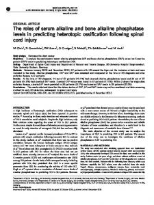
Cloning of a chromosomal fragment complementing the phoS and phoT mutations. The chromosomal fragment complementing the phoS64 and phoT9 mutations was isolated from two different sources, X glmS-1 phage and E. coli cells. The transducing X glmS-1 phage contains the E. coli chromosomal fragment covering the region from tna to glmS, including bgl (14, 23). The phoS gene is located between bgl and glmS (14). We thought it likely that the phoT gene is located on this fragment, since' the phoT gene is closely linked to the phoS gene (10). The DNA isolated from A glmS particles was digested with EcoRI and ligated to the EcoRI site on plasmid pBR325. This DNA was used to transform either phoS or phoT mutant cells. Several ampicillinresistant transformants that are repressed for AP synthesis, that is, they show complementation of the phoS or phoT mutation, were isolated. Plasmids were extracted from several colonies, and they were able to transform and to complement either the phoS or phoT mutation. The cloned EcoRI fragments of these plasmids were compared by digestion with restriction enzymes EcoRI, HindIII, MluI and PstI, and all the fragments showed the same restriction pattern (pSN400, Fig. 1). DNA was also prepared from E. coli strain KLF48/KL159, digested with EcoRI, and ligated into plasmid pBR322, as described in Materials and Methods. Transformation experiments were performed with a phoR mutant (strain ANCC2) as a recipient. Transformants resistant to tetra-
VOL. 152, 1982
CLONING OF phoS AND phoT GENES IN E. COLI Eco R I
uhl
FIG. 1. Restriction map of pSN400. An EcoRI fragment of A gImS-1 phage DNA was cloned on plasmid pBR325. The plasmid complemented phoS, phoT, pst-2, and bglB mutations.
cycline were selected on a T-broth plate containing tetracycline, and AP- transformants (white or slightly stained colonies by chromogenic substrate) were purified. The plasmid DNA was extracted from these transformants, and their sizes and the pattern of DNA fragments generated by digestion with restriction endonucleases were compared. They were classified into four groups according to the restriction patterns. The EcoRI fragment cloned in one of them (pSN401) was found to show the same restriction map for HindIII, MluI, and PstI as the DNA fragment isolated from transducing phage A glmS (Fig. 2). Strains ANCC75 (phoS64) and ANCC90 (pho79) were transformed by this plasmid (pSN401). It was found that this plasmid could complement both of these mutations. Although pSN401 was originally isolated as a plasmid complementing the phoR mutation, strains C2 (phoR68) and C5 (phoR17), harboring pSN401, were unable to synthesize AP not only on TG plates with excess phosphate but also on plates with limited phosphate. This phenomenon also occurred with pSN400.
695
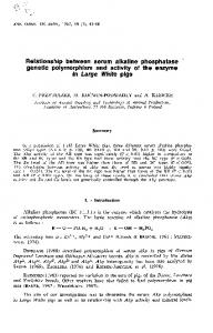
The identity of the EcoRI fragment on the hybrid plasmid with fragment derived from glmS was also supported by the genetic markers borne on the plasmids. Strains KH693 (bglB bglR) and GS5 (pit-i pst-2) were transformed with either pSN400 or pSN401. All the tetracycline-resistant transformant colonies from the former strain were dark red on eosin methylene blue-salicin plates (BglB+ phenotype), and those from the latter strain could grow on TGly (5 x 1i-0 M K2HPO4) plates (Pst+ phenotype). We have not examined whether pSN400 and pSN401 carry the bglR allele. The similarity of the two hybrid plasmids (pSN400 and pSN401) derived from different sources and screened in different genetic backgrounds is summarized in Table 2. Both of them showed the same phenotypes in each genetic background and thus are likely to carry the same alleles. Restriction map of the plasmids and complementation tests on plates. A restriction map of the cloned chromosomal fragment (EcoRI fragment) was constructed (Fig. 2). There were four HpaI, two Hindlll, two MluI, and two PstI cleavage sites. A variety of deletion plasmids were constructed by partial or complete digestion with an appropriate enzyme followed by self-ligation. Their properties were examined by complementation tests. We also examined the sizes and restriction patterns of the deletion plasmids, and they were confirmed to be the ones shown in Fig. 2. A plasmid from which the HindIII1-HindIII2 fragment was deleted (pSN507) complemented both phoS and phoT mutations, but not bglB. The following deletion plasmids were constructed from pSN507. A plasmid lacking the HpaIl-HpaI2 fragment (pSN517) complemented phoT9 but not phoS64. Plasmid pSN537, which lacks both HpaI1-HpaI2 and HpaI3-HpaI4 fragments, complemented only phoT9. Plasmids lacking the fragment between HpaI2 and HpaI4 (pSN557), and between HpaIj and HpaI4 (pSN567) did not complement either phoS64 or phoT9. In plasmid pSN577, the MluI1-MluI2 segment of pSN517 was excised and religated in the
gImS-1
TABLE 2. AP activity of various strains transformed by hybrid plasmids derived from A DNA (pSN400) and from E. coli chromosomal DNA (pSN401) AP activity' BglB Pst GSS KH693 ANC24 Plasmid (pit-I ANCC2 ANCC75 ANCC90 GS5 (wild pst-2) (bglB type) (phoR68) (phoS64) bgIR) (phol9) (pit-1 pst-2) + + pSN400 0.005 ± 0.001 0.004 ± 0.001 0.008 ± 0.001 0.004 ± 0.0004 0.005 ± 0.001 + + pSN401 0.005 ± 0.001 0.006 ± 0.001 0.006 ± 0.001 0.003 ± 0.001 0.006 ± 0.001 pBR322 0.009 ± 0.001 0.41 ± 0.18 3.86 ± 0.19 3.86 + 0.52 1.86 ± 0.072 a In the medium supplemented with excess phosphate, except GS5, for which the medium was supplemented with DL-glycerol-3-phosphate (0.04%) (29, 33). Activity was measured as micromoles of p-nitrophenol liberated per minute per milligram of cellular protein ± standard deviation.
J. BACTERIOL.
AMEMURA ET AL.
6%
/
p SN 507
/
/
,/
E,l/HIP1 P2 H:2
Ml
-I-I-Ii
H3
517H7Z 537
1-rzz-
547
'-C
557 567 577
a
I I
}1
I
I
t
1L
-i
1-
1
I
II
I-I I
J
H3
M2 I 1I
508 518
I
I
I
,I
a
1 I
I
AI
c
phoT , pst-2
phoS
O
I
---i~~~~~~~
2 I
3 I
pho U
4 I
5 I
6 I
7 a
8 kb
FIG. 2. Restriction map of pSN401 containing an EcoRI fragment covering the phoS phoT region of the E. coli chromosome and of deletion plasmids derived from it. Plasmid pSN507 was formed by deletion of a HindIII fragment, measuring 3.6 kb, located between HpaI4 and EcoRI2 sites on pSN401. Other deletion plasmids were constructed from pSN507. Cleavage sites: E, EcoRI; H, HpaI; Hi, HindlIl; M, MluI; P, PstI.
Plasmid pSN508, lacking the MluII-MluI2 opposite orientation. This plasmid did not complement the phoT9 mutation. Thus, it is clear fragment, complemented phoS64 but not phoT9. that the phoT gene lies within the 2.6-kilobase When the PstI fragment of pSN508 (from the (kb) HpaI2-HpaI3 segment covering site MluI1. PstI2 site to the PstI site on the Apr gene in
CLONING OF phoS AND phoT GENES IN E. COLI
VOL. 152, 1982
TABLE 3. Classification of AP constitutive mutants by complementation tests on platesa
Strain
pSN518 (phoS+)
pSN537 (pholr)
pSN547 (phoU+)
697
ment the phoT35 mutation. These results indicate that a wild-type allele which complements phoT35, but not phoT9, lies on the DNA fragment between the HpaI3 and HindIII cleavage
sites on pSN507. + BC68 We isolated nine AP-constitutive mutants + BC69 which belong to the R2 group by the old classifi+ BC72(phoT [1]) + cation (7). Plasmids pSN518 (phoS+), pSN537 BC73 + BC74 (phoT+), and pSN547 (phoT35+) were trans+ BC76 ferred into these strains and tested for AP syn+ BC77 thesis on plates. As shown in Table 3, all of + BC78 these mutants could be assigned to either the + BC79 phoS or phoT group, except one which was a Ten tetracycline-resistant transformed colonies complemented only by pSN547 (BC76). were transferred to TG plates with either excess or These results indicate that the mutations limited phosphate. These plates were also supple- pho79 and phoT35 belong to different complemented with the required amino acids and tetracycline mentation groups, which was initially suggested (20 i.g/ml). +, White colonies (complementation took by Zuckier and Torriani (33). Therefore, we place); -, colored colonies (complementation did not take place) after spraying with chromogenic substrate. propose to designate the phoT35 mutation as phoU instead of phoT Complementation tests were also performed pBR322) was removed, the deletion plasmid with the pst-2 mutant, which was isolated as a (pSN518) still complemented the phoS64 muta- mutation deficient for highly specific phosphate tion. It is obvious that the phoS gene is located transport and was found to be partially represson the 1.3-kb fragment between the PstI2 and covers site HpaI2. Therefore, we conclude that the phoS and phoT genes can be independently complemented by the wild-type alleles, that is, the wild-type allele is transdominant over each mutant. Plasmid pSN547, which lacks the HpaI1HpaI3 fragment (Fig. 2) was unable to complement either phoS64 or phoT9. However, when it was introduced into another phoT mutant (strain C4; phoT35), the synthesis of AP was repressed, which meant that complementation took place. The plasmids pSN517, pSN537, and pSN547 were transferred into strain ANCC4 (phoT35). AP synthesis was repressed in transformants carrying either pSN517 or pSN547 but not in those carrying pSN537. Plasmid pSN577, in which the MluI fragment of pSN517 was religated in the opposite orientation, did not comple-
MluIj cleavage sites and
ible for AP synthesis (29, 31, 32). The plasmids mentioned above were transferred into the pit pst mutant (strain GS5) and tested for Pst phenotype. It was found that the plasmids which complemented phol9 also complemented pst-2, and none of those that failed to complement phoT9 complemented pst-2. Thus, pho79 and pst-2 are as yet inseparable by complementation tests, and they belong to a different complementation group from phoS or phoU. Effect of hybrid plasmids on AP synthesis. The complementation tests were also performed by measuring AP activities of cells grown in either excess-phosphate or limited-phosphate medium. Although pSN401 was originally isolated as a plasmid complementing the phoR mutation, its derivative pSN507 (phoS+ phoPT pst' phoU'), which carries a deletion of the HindIII fragment, provokes a negative AP phenotype even in low-
TABLE 4. AP levels of strains carrying pSN507 and its derivatives in various genetic backgrounds AP levelb ANCC57 ANCC2 Plasmid Mediuma ANC24 ANCC90 (wild type)
(phoR68)
(phoS64)
(phol9)
0.005 ± 0.001 0.005 ± 0.002 0.006 ± 0.001 0.006 t 0.001 0.127 ± 0.04 0.032 t 0.01 0.007 ± 0.001 0.094 ± 0.037 NDC 0.007 ± 0.002 0.004 ± 0.001 0.015 ± 0.006 pSN518 1.73 ± 0.02 ND 0.18 ± 0.11 0.007 ± 0.002 0.57 ± 0.036 2.52 ± 0.35 0.012 ± 0.001 0.016 ± 0.002 pSN537 3.63 ± 0.31 3.03 t 0.21 3.63 ± 0.21 0.323 ± 0.011 1.80 ± 0.20 2.68 ± 0.31 0.008 ± 0.002 0.65 ± 0.02 pSN567 1.65 t 0.7 4.18 ± 0.28 5.42 ± 0.32 0.20 ± 0.02 a TG medium supplemented with excess phosphate (HP) or limited phosphate (LP). b Micromoles of p-nitrophenol liberated per minute per milligram of cellular protein ± standard deviation. I ND, Not determined.
pSN507
HP LP HP LP HP LP HP LP
698
J. BACTERIOL.
AMEMURA ET AL.
TABLE 5. AP levels of strains carrying the phoS gene on a low-copy-number plasmid AP level' Plasmid
Mediuma
ANC24 (wild type) 0.008 ± 0.001 6.99 ± 0.5 0.008 ± 0.001 5.57 ± 0.1
ANCC2 (phoR68)
ANCC3 (phoR69)
ANCC75 (phoS64) 0.015 ± 0.001 3.18 ± 0.44 2.02 ± 0.35 6.19 ± 0.92
ANCC90 (phoT9) 2.29 ± 0.14 4.74 ± 0.21 1.28 ± 0.24 4.62 ± 0.65
ANCC4 (phoT3S) 2.22 ± 0.19 3.10 ± 0.45 1.28 ± 0.01 3.55 ± 0.07
0.84 ± 0.08 1.52 ± 0.03 HP 0.22 ± 0.01 1.82 ± 0.05 LP 1.39 ± 0.01 2.94 ± 0.08 HP pMF3 0.45 ± 0.07 1.65 ± 0.17 LP a TG medium supplemented with excess phosphate (HP) or limited phosphate (LP). b Micromoles of p-nitrophenol liberated per minute per milligram of cellular protein ± standard deviation.
pSN5083
phosphate medium (Table 4). This effect is due to phoS, since a phoS+ (phoT) plasmid (pSN518) prevents full induction of AP synthesis in wild-type E. coli and totally prevents synthesis in the phoR68 mutant.
When pSN518 was introduced into ANCC90 (phoT9), all the transformant colonies showed the AP constitutive phenotype on T-broth plates. In TG medium, the transformants grew at a growth rate one-third that of the strain carrying only pSN567, and they frequently segregated AP-repressible colonies. The level of AP activity in ANCC90 carrying pSN518 varied considerably from experiment to experiment, which we consider to reflect the degree of the segregation (data not shown). We interpret this phenomenon as follows. Overproduction of the phoS gene product in phoT cells has an adverse effect on cell growth, and rare PhoT+ revertants resulting from mutation or recombination between the phoT gene on the chromosome and a portion of the phoT gene borne on the plasmid eventually comprise a considerable portion of the cell population after growth in TG medium, in which PhoT+ cells grow more rapidly than PhoTcells. To test this assumption, we introduced the phoS gene into a low-copy-number plasmid. Plasmid DNA of pSN508 was digested with EcoRI and inserted in the EcoRI site of the lowcopy-number plasmid pMF3, which possesses the F replicon (20). ANCC75 was transformed with this DNA. The cells were screened for transformants resistant to ampicillin but sensitive to tetracycline and showing repressed synthesis of AP. Plasmid DNA was isolated and was confirmed to be the result of insertion of the EcoRI fragment of pSN508, containing the phoS
into pMF3 (pSN5083). This plasmid was introduced into strains with various genetic backgrounds, and the levels of AP were measured in liquid medium (Table 5). The results of AP synthesis under repressed or derepressed conditions were as expected: no inhibition of AP synthesis by pSN5083 in phoR mutants and full derepression in a wild-type strain carrying the plasmid. When pSN5083 was introduced into ANCC90, no growth inhibition of the transformed cells was observed, and the transformants did not segregate AP-repressible colonies after they were grown in TG medium, in contrast to the case of the multicdpy plasmid, pSN518. Complementation of phoS64 but not phoT9 or phoT35, by the plasmid was observed, as expected. The deletion plasmid pSN547 repressed AP synthesis in the phoT35 mutant but not in phoT9 and phoS64 mutants (Table 6). It did not prevent full derepression in the wild-type strain or the phoR68 mutant. AP synthesis in the phoT35 mutant was not repressed by pSN537 (phoT) (data not shown) or by pSN5083 (phoS+) (Table 5). All of these results are consistent with those obtained in the plate tests with the chromogenic substrate. DISCUSSION We have cloned an E. coli chromosomal fragment which complements the phoS and phoT mutations. This fragment was also found to contain the bglB and pst-2 genes. A series of deletion plasmids were constructed in vitro, including one which complemented only the phoS64 mutation but not phoT9, and one which complemented only phoT9 but not phoS64.
gene,
TABLE 6. AP levels of strains carrying pAN547 AP levelb Mediuma ANCC4 (phoT35) ANCC90 (phoT9) ANCC75 (phoS64) ANCC2 (phoR68) ANC24 (wild type) ± ± ± ± 0.01 ± 0.001 2.28 0.09 2.50 0.18 0.46 0.03 HP 0.007 0.002 2.39 ± 0.10 3.56 ± 0.11 1.61 ± 0.22 0.22 ± 0.02 LP 5.57 ± 0.18 a TG medium supplemented with excess phosphate (HP) or limited phosphate (LP). b Micromoles of p-nitrophenol liberated per minute per milligram of cellular protein ± standard deviation.
CLONING OF phoS AND phoT GENES IN E. COLI
VOL. 152, 1982
Therefore, we conclude that the phoS and phoT mutations are located in different genes. Our results are contradictory to the conclusion drawn by Levitz et al. that phoS and phoT are not separate cistrons (17). Their conclusion was based on their result that F'111 phoS and F'111 phoT did not complement phoT and phoS recipients, respectively. Although these workers used recA strains for recipients, they could not rule out some genetic rearrangements, such as deletion or insertion involving transposable genetic elements, since these processes have been shown to be recA independent. Their conclusion was also based on several assumptions, some of which were not substantiated by experimental evidence. We have also experienced difficulties in complementation tests with large F' factors such as F'111 due to instability of the plasmid state. We tried to test complementation between phoS and phoT alleles by isolating F' factors as described by Low (18). The strain Hfr P13(P0104) ilv+ phoS phoTP (or phoS+ phoT) tna+ cys was crossed with F- ilv- phoS+ phol7 (or phoS phoT) tna cys+ and recA, and colonies Pho- (white after spraying with a chromogenic AP substrate) were observed among Ilv+ Cys+ recombinants. Some of them were found to be also Tna+. However, we could not confirm the fertility of the recombinants, apparently because of their instability. Cox et al. (5) reported that, based on complementation tests with partial diploids, there are three complementation groups in addition to the phoS group, represented by the alleles pstA2, phoT32, and pstB401, and all four genes are part of an operon. The gene order was reported to be uncDC, with the pstA-(pstB phoT)-phoS pstA gene being promoter proximal. Our present work indicates that the gene order in this region is bglB phoU-(pst-2 phoT)-phoS. The pst-2 mutation was refered to as pstA2 by Cox et al. (5). We have not tested the pstB401 mutation. We believe that the promoter of the phoS gene is not located near pstA but instead is on the PstI2MluI1 fragment which contains the phoS gene (Fig. 2). Plasmid DNA of pSN518 (Fig. 2) was digested with EcoRl and MluI, and the resulting single-stranded DNA ends were filled in by using the four deoxyribonucleoside triphos...
...
699
phates and T4 DNA polymerase. The blunt ends of the DNA were ligated with T4 DNA ligase. This plasmid (pSN5182) complemented a phoS mutation, as judged by the repression of AP synthesis and the production of the phosphatebinding protein (Morita et al., unpublished data). This suggests that a promoter and signals for initiation of translation must be located within the 1.3-kb PstI2-MluI1 segment containing the phoS gene. Furthermore, it is likely that the pstA (pst-2) region and the phoU (phoT35) gene comprise independent operons, since deletion of the region containing pstA or phoU did not prevent the remaining gene from complementing the appropriate mutation (Fig. 2 and Table 7). The restriction map of the cloned fragment and complementation tests revealed that the phoS gene is located on a 1.3-kb PstI2-MluI1 segment and the phoT gene on a 2.6-kb HpaI2HpaI3 segment. Since the phoT gene overlapped the MluI1 cleavage site, the length of the phoS gene is presumed to be less than 1.3 kb. The maximal length of phoS identified in the present studies, is only slightly more than the calculated size of the gene coding for the phoS precursor protein of molecular weight 39,000 (estimated by sodium dodecyl sulfate-polyacrylamide gel electrophoresis; Morita et al., manuscript in preparation). The phoT35 mutation is constitutive for AP synthesis and produces phoS protein (10, 33) and therefore was classified as phoT. Zuckier and Torriani (33) reported that phoS and pst-2 mutants are deficient in the phosphate-specific transport system, whereas the phoT35 mutant is not. Because of its distinct phenotype and separate position on the chromosome, they have suggested that the phoT35 represents a gene different either from phoTor pst-2. We could not separate the phoT gene from the pst-2 gene physically, since all deletion plasmids which complemented phoT also complemented the pst2 mutation. We have shown in this paper that the DNA fragment which complements phoT35 is separable from those that complement phoT9 or pst-2. Therefore, from the work of Zuckier and Torriani and our present results, we proposed to designate phoT35 as a new gene, phoU. Cells containing multicopy phoS plasmids
TABLE 7. AP levels of GS5(pit-l pst-2) carrying various hybrid plasmids AP level' Mediuma pSN547 (phoU+) pSN567 pSN537 (phoTr) pSN507 (phoS+ phol phoU+) 2.44 ± 0.02 1.03 ± 0.14 0.147 ± 0.011 HP 0.006 ± 0.001 1.34 ± 0.08 2.68 ± 0.15 LP 0.013 ± 0.001 0.165 ± 0.013 a TG medium supplemented with DL-glycerol-3-phosphate (0.04%) and with either excess (HP) or limited (LP) phosphate. b Micromoles of p-nitrophenol liberated per minute per milligram of cellular protein ± standard deviation.
700
AMEMURA ET AL.
J. BACTERIOL.
LITERATURE CITED showed decreased levels of AP even under derepressed conditions. The strong inhibition by 1. Aono, H., and N. Otsusil. 1968. Genetic mapping of regulator gene phoS for alkaline phosphatase in Escherichia phoS plasmids was more evident when the host coli. J. Bacteriol. 95:1182-1183. was a phoR strain. When phoS+ plasmids were 2. Badnmann, B. J., and K. B. Low. 1980. Linkage map of introduced into a phoT strain, the generation Escherichia coli K-12, edition 6. Microbiol. Rev. 44:1-56. time was increased three to four times relative to 3. Blnbohn, H. C., and J. Doly. 1979. A rapid alkaline extraction procedure for screening recombinant plasmid the phol* strain carrying the same plasmids. Nucleic Acids Res. 7:1513-1523. The prolonged generation time was not observed 4. DNA. Bracha, M., and E. Yagil. 1973. A new type of alkaline when the plasmids contained both phoS and phosphatase-negative mutants in E. coli K12. Mol. Gen. Genet. 122:53-60. phoT genes. Our preliminary analysis of the G. B., H. Rosenberg, J. A. Dowue, and S. Silver. cellular proteins by sodium dodecyl sulfate- 5. Cox, 1981. Genetic analysis of mutants affected in the Pst gel electrophoresis polyacrylamide suggested inorganic phosphate transport system. J. Bacteriol. 148:1that a large amount of phoS protein was pro9. duced in the phoS mutant carrying the phoS+ 6. Davis, R. W., D. Botsteln, and J. R. Roth. 1980. Advanced bacterial genetics. Cold Spring Harbor Laboratory, Cold plasmid. Excess phosphate-binding protein proHarbor, N.Y. duced by the multicopy phoS gene seems to 7. Spring Echols, H., A. Garen, S. Garen, and A. Torriani. 1961. cause adverse physiological effects. The phoT Genetic control of repression of alkaline phosphatase in E. coli. J. Mol. Biol. 3:425-438. gene might play a regulatory role in phoS gene expression, or the phoT gene product might 8. Garen, A., and H. Echols. 1962. Genetic control of induction of alkaline phosphatase synthesis in E. coli. Proc. work cooperatively with phoS protein. Natl. Acad. Sci. U.S.A. We cloned the phoS gene while we were 9. Garen, A., and H. Echols.48:1398-1402. 1962. Properties of two regulattrying to clone the phoR gene on pBR322. During genes for alkaline phosphatase. J. Bacteriol. 83:297300. ing the course of the attempts, we have cloned A., and N. Otsuji. 1964. Isolation of a protein four different chromosomal DNAs on pBR322 10. Garen, specified by a regulator gene. J. Mol. Biol. 8:841-852. which repress AP synthesis in the phoR68 mu- 11. Gerdes, R. G., and H. Rosenberg. 1974. The relationship tant grown on TG plates with excess phosphate. between the phosphate-binding protein and a regulator gene product from Escherichia coli. Biochim. Biophys. One of them was found to contain phoS gene. 351:77486. The remaining three showed different restriction 12. Acta Gerdes, R. G., K. P. Strickland, and H. Rooenberg. 1977. cleavage patterns, and we have not yet characRestoration of phosphate transport by the phosphateterized them. They might carry one of the genes binding protein in spheroplasts of Escherichia coli. J. Bacteriol. 131:512-528. similar to phoS or phoA which are regulated by Goodman, H. M., and R. J. MacDonald. 1979. Cloning of phosphate and by the regulatory genes, such as 13. hormone genes from a mixture of cDNA molecules. compete might genes Such phoR. with and phoB Methods Enzymol. 68:75-90. phoA for a positive regulatory factor, such as 14. Kanazawa, H., F. Tamura, K. Mabuchl, T. Mid, and M. Futal. 1980. Organization of unc gene cluster of EschephoB gene product. Their gene products might richia coli coding for proton-translocating ATPase of compete for processing machinery with phoA if oxidative phosphorylation. Proc. Natl. Acad. Sci. U.S.A. they are genes coding for secretory proteins. 77:7005-7009. The effect of these genes on AP synthesis in the 15. Kreuzer, K., C. Pratt, and A. Torrlanl. 1975. Genetic analysis of regulatory mutants of alkaline phosphatase of phoR68 mutant is likely to be due to the incoli. Genetics 81:459-468. creased gene dosage or amount of the gene 16. E. Lederberg, E. M., and S. N. Cohen. 1974. Transformation product as the result of their presence on the of Salmonella typhimurium by plasmid deoxyribonucleic acid. J. Bacteriol. 119:1072-1074. high-copy-number plasmid pBR322. Since all the E. coli strains used in the present 17. Levitz, R., R. Bittan, and E. Yagi. 1981. Complementation tests between alkaline phosphatase-constitutive muwork were Rec+, the complementation experitants (phoS and phoT) of Escherichia coli. J. Bacteriol. ments might reflect genetic recombination rather 145:1432-1435. than true complementation. We think this is 18. Low, B. 1968. Formation of merodiploids in matings with a class of Rec- recipient strains of Escherichia coli K12. unlikely. The phoR68 mutant was complementNatl. Acad. Sci. U.S.A. 60:160-167. ed by the phoS plasmid (pSN518). This strain 19. Proc. Lowry, 0. H., N. J. Rosebrough, A. L. Farr, and R. L. was cured it after AP constitutivity of recovered Randall. 1951. Protein measurement with the Folin phenol the plasmid. The plasmid prepared from the reagent. J. Biol. Chem. 193:265-275. Kline. 1977. Restriction endonutransformant was introduced into the phoS and 20. Manis, J. J., and B. C.mutagenesis of the F sex factor clease mapping and phoT mutants; it still complemented the phoS replication region. Mol. Gen. Genet. 152:175-182. mutation but not the phoT mutation. 21. Medveczky, N., and H. Rosenberg. 1970. The phosphateACKNOWLEDGMENTS We thank Frank G. Rothman, Barbara J. Bachmann, and Toru Miki for generously supplying the bacterial strains and Satoshi Harashima for kindly supplying T4 DNA ligase. We are grateful to Tadao Horiuchi and Junichi Kawamata for encouragement.
binding protein of Escherichia coli. Biochim. Biophys. Acta 211:158-168. 22. MIki, T. 1978. Genetic and physical analyses of the replication origin and dnaA gene of Escherichia coli chromosome by molecular cloning and cleavage mapping with restriction endonucleases. Annu. Rep. Inst. Virus Res. Kyoto Univ. 21:1-26. 23. Miki, T., S. Hiraga, T. Nagata, and T. Yura. 1978. Bacteriophage A carrying the Escherichia coli chromo-
VOL. 152, 1982
24. 25.
26.
27. 28.
CLONING OF phoS AND phoT GENES IN E. COLI
somal region of the replication origin. Proc. Nati. Acad. Sci. U.S.A. 75:5099-5103. Miller, J. H. 1972. Experiments in molecular genetics. Cold Spring Harbor Laboratory, Cold Spring Harbor, N.Y. Morris, H., M. J. Schlesnger, M. Bracha, and E. Yagil. 1974. Plciotropic effects of mutations involved in the regulation of Escherichia coli K-12 alkaline phosphatase. J. Bacteriol. 119:583-592. Nakata, A., G. R. Peterson, E. L. Brooks, and F. G. Rodman. 1971. Location and orientation of the phoA locus on the Escherichia coli K-12 linkage map. J. Bacteriol. 107:683-689. Prasd, I., and S. Schaeler. 1974. Regulation of the glucoside system in Escherichia coli K-12. J. Bacteriol. 120:638-650. Rothman, F., and J. R. Coleman. 1968. Kinetics of transcription and translation of a repressed gene. J. Mol. Biol. 33:527-531.
701
29. Sprague, G. F., Jr., R. M. Bell, and J. E. Cronan, Jr. 1975. A mutant of Escherichia coli auxotrophic for organic phosphates: evidence for two defects in inorganic phosphate transport. Mol. Gen. Genet. 143:71-77. 30. Wanner, B. L., and P. Lattereil. 1980. Mutants affected in alkaline phosphatase expression: evidence for multiple positive regulators of the phosphate regulon in Escherichia coli. Genetics 96:353-366. 31. WUlsky, G. R., R. L. Bennett, and M. H. Malamy. 1973. Inorganic phosphate transport in Escherichia coli: involvement of two genes which play a role in alkaline phosphatase regulation. J. Bacteriol. 113:529-539. 32. Willsky, G. R., and M. H. Malamy. 1976. Control of the synthesis of alkaline phosphatase and the phosphatebinding protein in Escherichia coli. J. Bacteriol. 127:595609. 33. Zucker, G., and A. Torriani. 1981. Genetic and physiological tests of three phosphate-specific transport mutants of Escherichia coli. J. Bacteriol. 145:1249-1256.