The Fab region of an IgG2b antibody (AM7B2.1) reactive to the herbicide atrazine was cloned into a plasmid vector using the polymerase chain reaction and two ...
Protein Engineering vol.6 no.8 pp.981-988, 1993
Cloning, sequencing and expression of the Fab fragment of a monoclonal antibody to the herbicide atrazine
Vernon K.Ward1, Peter G.Schneider2, Sabine B.Kreissig1, Bruce D.Hammock2*3 and Prabhakara V.Choudary1-4 'UC Davis Antibody Engineering Laboratory and Department of Entomology, 2Department of Entomology and 3Department of Environmental Toxicology, University of California, Davis, CA 95616, USA 4
To whom correspondence should be addressed
The Fab region of an IgG2b antibody (AM7B2.1) reactive to the herbicide atrazine was cloned into a plasmid vector using the polymerase chain reaction and two sets of degenerate oligonucleotide primers designed to mimic the amino acid variation at the N-termini of xL-chains and TH-chains. These primers also provide a secretion signal fused precisely to the antibody gene sequence for secretion of the mature antibody. A further set of universal oligonucleotide primers was developed for the direct sequencing of the VH and C m regions of TH-chains and the VL and C L regions of xL-chains without subcloning and were used to determine the sequence of this antibody. The xL-chain was found to not possess a conserved Cys residue at position 23 and the implications of this observation are discussed. The cloned genes were expressed in Escherichia coli using a commercially available T7 RNA polymerase-based plasmid. The clones were also expressed in a 17 RNA polymerasebased system containing an attenuated version of the T7 RNA polymerase promoter, plus a lac promoter placed in an antisense orientation, to enhance plasmid stability. The expressed products were confirmed as atrazine reactive by binding to an atrazine derivative conjugated with alkaline phosphatase. Key words: antibody cloning/antibody expression/atrazine/pesticide analysis/recombinant antibody
Introduction In recent years, much interest has arisen in the use of antibodies for determining environmental contaminants with some notable successes in pesticide residue analysis (Kaufman and Clower, 1991). One of the problems with this type of environmental assay is the development and production of suitable antibodies. The production of antibodies to small molecules requires conjugation to large molecules to elicit an immune response (Harrison et al., 1988; Jung et al., 1989) and often involves large scale immunization and screening strategies. As the molecule of interest becomes smaller, it becomes increasingly difficult to develop antibodies with the desired properties and there is often a fine line between developing a useful antibody and production of antibodies which lack the required sensitivity and specificity or which exhibit handle recognition (Harrison et al., 1991). This renders the antibody unusable for detection of environmental contaminants by inhibition-based ELISA assays. To overcome this problem, it is often necessary to immunize many animals and the production of useful monoclonal antibodies can involve © Oxford University Press
large-scale screening strategies. In addition, although polyclonal and monoclonal technologies produce sufficient antibody for most analytical applications, it is expensive to produce large amounts of antibody for applications including widespread monitoring programs, remediation or therapy following toxic exposure. As the field of small molecular immunoassay matures, these problems will have to be addressed and the recent development of recombinant antibody systems offers many advantages in respect of alleviating some of these problems, particularly for large-scale production and development of screening strategies (Huse etal., 1989; Marks et al., 1991; Pluckthun, 1991a, b; Winter and Milstein, 1991). Another potential advantage of recombinant antibodies is the ability to engineer novel domains such as reporter enzymes (Ducancel et al., 1993), metal binding sites (Pessi et al., 1993), and affinity tags (Schmidt and Skerra, 1993) directly into antibody molecules. One potential limitation, however, is the very fine tolerance in the affinity of an antibody to its small target. This means that if recombinant antibodies are to be useful, it is important to produce a recombinant antibody that mimics the original antibody as closely as possible. As a step towards achieving this goal, we have developed a set of degenerate oligonucleotide primers for the cloning of antibody genes which mimic as closely as possible the amino acid sequences found at the N-termini of antibody heavy (H> and light (L)-chains and used these to clone the Fab region of a monoclonal antibody (AM7B2.1) reactive to the herbicide atrazine (Karu et al., 1991). The derived Fab antibody was capable of binding atrazine and the binding could be inhibited by the free analyte. Additional universal primers were synthesized for the direct sequencing of antibody clones and used to sequence the recombinant AM7B2.1 clones. Analysis of the light chain sequence revealed an uncommon amino acid sequence which has been associated with poor expression of recombinant antibodies in Escherichia coli (Glockshuber et al., 1992). Two different T7 RNA polymerase-based systems were used to express the AM7B2.1 clones as Fab antibody fragments. The first system was the commercially available vector pET3d (Studier et al., 1990) and the second system is described in this manuscript. This system was developed in an attempt to overcome the stability problem which can be associated with recombinant antibodies. Materials and methods Cloning strategy The cloning strategy used here involved an initial reverse transcriptase reaction of poly(A) + RNA extracted from hybridoma cells followed by two polymerase chain reactions (PCR). The first PCR reaction used the same C-terminal primer used for the reverse transcriptase reaction and degenerate Nterminal primers which contain homology to the antibody sequences and most of a prokaryotic leader sequence, pelB (Lei et al., 1987) for the H-chain and ompA (Mowa et al., 1980) for the L-chain. The second PCR used the same C-terminal primer and new overlapping N-terminal primers which completed the leader sequence and provided cloning and expression infor981
V.K.Ward et al.
Table I. N-terminal PCR primers H-chain primers petB primer
(K) M V Y L L P T A A A G L L L L A ACGTCATATGTCC47TGCTATACCTATTGCCTACCK3CAGCCGCTGGATTGTTATTACTCGCT Ncol (AAG)
HV1 primer G L L
L
L
A
A
Q
P
A
V
A
E / Q V Q L K E S G
GGATTGTTATTACTCGCTGCCCAACCAGCCGTGGCCG/CAGGTGCAGCTGCAGCAGC/TCTGG
HV2 primer G L L L L A A Q P A V A Q V Q L K E S G GGATTGTTATTACTCGCTGCCCAACCAGCCGTGGCCCAGGTACAGCTGAAGGAGTCAGG HV3 primer G L L L L S G A Q A V A E V Q/K L V E GGATTGTTATTACTCGCTGCCCAACCAGCCGTGGCCGAGGTGC/AAGCTGGTGGAGTCTGG L-chain primers ompA primer EcoRl RBS M K K T A I A I A TGGC ATGAA 77TXTGCAC AAGGAGAAAAATAAAATGAAAAAGAC AGCTATCGCGATTGCA V A L A G F GTGGCACTGGCTGGTTTC VL1 primer V A L A G F A T V A Q A D I Q/K M T Q GTGGCACTGGCTGGTTTCGCTACCGTAGCGCAGGCCGACATCC/AAGATGACCCAG VL2 primer D V A L A G F A V A I V/L M/L T Q GTGGCACTGGCTGGTTTCGCrACCGTAGCGCAGGCCGACATT G / T TG A /cTGACTCAG The primers used in the two step PCR protocol are listed 5'—3'. For the H-chain, the three HV primers, representing seven possible N-termmi, were used in an initial PCR reaction and this PCR product was used as the template for a second PCR reaction using the pelB primer. The HV primers contain homology to antibody sequences (bold face type) and ~50% of the leader sequence information. The pelB primer contains homology to the 5'-end of the HV primers for amplification purposes and the remainder of the pelB leader sequence as well as an Ncol restriction endonuclease site for cloning purposes. In order to improve secretion of the H-chain, a Lys residue (K) was incorporated into the pelB leader sequence by site-directed mutagenesis as described in Materials and methods. The L-chain primers were used in a similar manner to the H-chain primers. The LV primers, representing six possible N-termini, contain homology to the N^rmini of xL-chains (bold face type) and contain half of the ompA leader sequence including homology to the ompA primer. The ompA primer contains homology to the LV primers, a ribosome-binding site (RBS) for polycistronic message capabilities and an £coRI restriction site for cloning purposes. Extra nucleotides were incorporated at the 5'-end of the ompA and pelB primers to enhance restriction endonuclease digestion for cloning purposes.
mation as described below. The PCR products were cloned to allow production of a polycistronic message in plasmid vectors. Development of N-terminal primers Using amino acid variability as the primary criterion, three gamma heavy (-y^-chain primers (designated HV1, HV2 and HV3) with 7-fold total degeneracy and two kappa light (*L)chain primers (designated LV1 and LV2) with 6-fold total degeneracy, were synthesized (Table I). These primers contained homology to the mature N-termini of antibody coding sequences and leader sequence information. For the H-chain, the second PCR primer provided the remainder of the pelB leader sequence and an Ncol restriction endonuclease site for cloning. The Ncol recognition sequence contains an ATG start codon which allows precise placement of the start codon for expression purposes. The second L-chain primer contained the remainder of the ompA leader sequence, a ribosome binding site and an £coRI cloning site. C-terminal primers The H-chain C-terminal primer was designed to be homologous to the hinge region of the H-chain to allow cloning of the Fd (VH -f C m ) region. The primer also contained stop codons and an EcoRl cloning site to allow ligation to the N-terminus of the L-chain construct for polycistronic message production (Table II). The L-chain C-terrninal primer (Table U) was designed to 982
recognize the 3' end of the xL-chain and to provide a translational stop codon as well as a BamHi cloning site. Vector construction A vector (pGEMEB) with blue/white selection and appropriate restriction endonuclease sites for cloning purposes, was constructed from the existing vector pGEM5Zf(-) (Promega Corp) as follows. The restriction endonuclease recognition sequences for EcoN and BamHI were introduced into the pGEM5Zf(-) vector by subcloning the Spel to £coRV multicloning site (MCS) region of pBluescriptSK" (Stratagene) into the Spel and EcoRV sites of pGEM5Zf(-). This retained the reading frame in the MCS and, thus, blue/white selection, while transferring the EcoRl and BamlU sites from pSK~ to pGEM5Zf(-) to create the vector pGEMEB. This vector already contained an Ncol site and was suitable for cloning the antibody PCR products. In order to express the antibody clones, a further vector was constructed based upon pGEMEB. The region of pGEMEB including the T7 RNA polymerase promoter to the Ncol site was replaced with a ribosome-binding site and the immediate 5' upstream sequences of bacteriophage T7 gene 10 by site-directed mutagenesis using the method of Kunkel (1985) as described by Ward et al. (1992). The mutagenesis primer was 5' - ATCCCGCGGCC ATGGT AG ATCTCCTTCTT AAAGTTAAACAATTCGCCT-3'. This created the vector pGTWl (see
Expression of an atrazine reactive antibody
Table n . Constant region (C-terminal) primers HCG1 primer
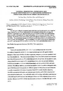
EcoRl 3'-AACACGGGTCCCTAACAACTACTTTCACCGTAC7T,4,4GGACGTG-5' HCG2 primer EcoRl 3'-GGGACAGGAGGTACGTTTACTACTTTCACCGTAC7T^/ 70 mM KC1, 1 mM each dNTP, 5 U reverse transcriptase and 1 mM vanadyl ribonuclease complexes, in a 20 /xl reaction volume at 42°C for 30 min. PCR amplification was performed directly upon the first strand cDNA products generated above. The quantity of each cDNA reaction material placed into each PCR reaction and the reaction conditions were optimized empirically. The L-chain PCR contained 5 y.\ of the reverse transcriptase reaction, 0.5 /*g for each primer, 0.2 mM dNTPs, 1.5 mM MgCl2) 50 mM KC1, 10 mM Tris-Cl, pH 9.0, 0.1% Triton X-100 in a reaction volume of 100 y\. The reactions were overlaid with 100 /tl of mineral oil and the PCR performed in a Hybaid thermal cycler. The initial cycle of amplification was 95°C for 2 min, 55°C for 2 min and 72°C for 5 min, followed by 30 cycles of 95°C/30 s, 55°C/30s, 72°C/1 min and a final cycle identical to the initial cycle. An aliquot of each PCR reaction was analyzed by agarose gel electrophoresis to confirm amplification of the correct size products. The H-chain PCR was identical to the L-chain reaction except for the primers and reverse transcriptase reaction used. Cloning and sequencing of PCR products The PCR products were cloned separately into the pGEMEB vector using the appropriate restriction endonucleases and five H-chain clones and five L-chain clones were isolated. The sequences of the cloned Fd and x-antibody cDNAs were determined by double-stranded dideoxy sequencing using a Sequenase kit (USB). In addition to using the universal forward and reverse primers, primers were synthesized which are homologous to conserved regions within xL~chains a n d th e Fd region of yn~ chains. These primers are described in Figure 2 (Results and
B H M
2030
560
M L
^2320 1930\ 1371 \ 1260^ 700\
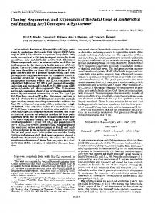
Fig. 1. Agarose gd dectrophorcsis of (A) PCR reaction products and (B) cloned products. PCR reactions were performed as described in Materials and methods using the primer sets described in Tables I and D~. (A) Lane h is the first H-chain PCR reaction, lane H is the second H-chain reaction containing the pelB leader sequence, lane 1 is the first L-chain PCR reaction, lane L is the second L-chain PCR reaction containing the ompA leader sequence and ribosome binding site, lane M is a marker lane containing a Hindm digest of lambda DNA. The size of the mol. wt markers is shown in base pairs. (B) Lane H is the H-chain PCR product cloned into the pGEMEB vector, lane L is the L-chain PCR product cloned into the pGEMEB vector, lane M is a marker lane containing a BsiEU digest of lambda DNA. The size of the molecular size markers is shown in base pairs. The cloned products are indicated by arrowheads ( > ) .
discussion). Both strands of the antibody clones were sequenced and confirmation of the sequence was obtained by repeating the sequencing in one direction using single-strand rescued DNA as a template. Expression of AM7B2.1 Fab fragment For expression of the AM7B2.1 sequences, the H- and L-chain clones were subcloned into the T7 RNA polymerase-based expression vector pET3d (Studier et al., 1990). Screening of the subcloning reactions was performed using the Escherichia coli strain XL 1-Blue, as this does not contain T7 RNA polymerase, hence expression should not occur and thus select against the desired clones. In order to express the Fab fragment of the AM7B2.1 antibody, the pET3d/AM7B2.1 was transformed into E.coli strain JM109(DE3) (Promega) which contains a gene for T7 RNA polymerase under the control of a lac promoter and is thus inducible by IPTG. As a control, pET3d/AM7B2.1 was also transformed into E.coli strain JM109 which does not contain the T7 RNA polymerase gene. Expression was carried out by inoculation of a single colony into LB media and growth of the 983
V.K.Ward et al. —^-
II
\rt-*eqF—&-
Lmef
•
cu.^-*-
^
C4. —
-VLsaqR
VH
VUaqF U«jf CU«f CW)H Ua*