Neurology Asia 2015; 20(3) : 305 – 307
Cluster-tic syndrome and bilateral internuclear ophthalmoplegia as the manifestation of multiple sclerosis Ali Ulvi Uca
MD,
Hasan Hüseyin Kozak
MD
Department of Neurology, Meram Faculty of Medicine, Necmettin Erbakan University, Konya, Turkey Abstract This report describes a 35-year-old female suffering from a cluster-tic syndrome and bilateral internuclear ophthalmoplegia as the initial manifestation of multiple sclerosis. Magnetic resonance imaging of the brain revealed multiple pontine hyperintense lesions. To our knowledge, there is no previous report of multiple sclerosis presenting as cluster-tic syndrome and bilateral internuclear ophthalmoplegia in the literature. The cluster headache attacks and peri-ocular neuralgiform pain resolved after treatment with intravenous methyl-prednisolone and oral carbamazepine. INTRODUCTION Cluster-tic syndrome is a rare headache syndrome characterized by the coexistence of three types of pain attacksin which trigeminal neuralgia co-occurs with cluster headache.1,2 We report the case of a patient diagnosed as having cluster-tic syndrome and bilateral internuclear ophthalmoplegia (INO) as the manifestation of multiple sclerosis (MS). We are not aware of previous similar report in the literature. CASE REPORT A thirty-five year-old female patient presented to our outpatient clinic with 3 years history of cluster headaches. The headache during the attacks occurred 3-5 times a day, lasting 0.5-1 hours for 3-4 weeks, and she was pain-free for one year afterwards. Her headaches were localized to the left orbital and peri-orbital region. They were severe and throbbing; associated with ipsilateral conjunctival hyperemia, lacrimation, nasal congestion, rhinorrhea, and palpebral fissure narrowing due to eyelid edema. During the attacks, the patient received subcutaneous sumatriptan and had methylprednisolone as prophylactic treatment. The patient experienced a change in the character in her headaches a week before her recent visit, with a “trigeminal neuralgia” like pain immediately precede the cluster headache. This was described as severe left periocular sharp shock-like pain. The pain may occur spontaneously or when triggered by innocuous
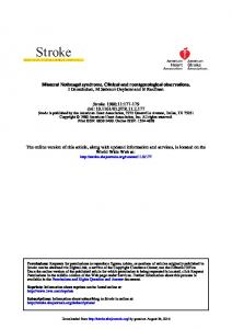
stimuli in the facial and intraoral areas. It lasted for 2-5 minutes just before the onset of headache. The patient also complained of diplopia, which started three days after the onset of pain. Neurological examination revealed horizontal diplopia during lateral gaze in both eyes. The patient had adduction deficit in one eye and nystagmus in the other eye on lateral gaze bilaterally. Vertical gazes and the eyes at primary position were unremarkable. Laboratory tests, including blood chemistry, rheumatologic and immunologic screening tests were normal. The cerebrospinal fluid (CSF) IgG-index was 1.69 and CSF isoelectric focusing was positive for oligoclonal bands in the gamma regions (Pattern 2). Serum and CSF aquaporin-4 antibodies were negative. Magnetic resonance imaging (MRI) showed multiple pontine, cervical and periventricular hyperintense lesions, with some of the lesions showing contrast enhancement. (Figure 1, 2). The patient was diagnosed with MS.3,4 In particular, the presence of both contrast-enhanced and non-contrast enhanced lesion would satisfy the 2010 revision to the McDonald Criteria for dissemination of time as well as space.4 The patient was first treated with intravenous methylprednisolone at 1g/day for 5 days. Her headaches remitted but she had persistence of the left periocular neuralgic pain. Carbamazepine was then added at the starting dose of 200 mg daily, and gradually increased to 1,200 mg a day. Patient was followed up for three months with a daily dosage of 1200 mg of carbamazepine and showed good clinical response. She remained free of headache
Address correspondence to: Ali UlviUca, Necmettin Erbakan Üniversitesi Meram Tıp Fakültesi, Nöroloji Anabilim Dalı. Meram, 42080, Konya, Turkey. Tel: +90 332 223 7981, E-mail:
[email protected]
305
Neurology Asia
Figure 1: T2 weighted axial brain MRI showing bilateral multiple pontine demyelinating lesions, where the left trigeminal root inlet and main sensory nucleus show hyperintense lesions.
and the left peri-ocular neuralgic pain. DISCUSSION Cluster-tic syndrome is characterized by the coexistence of three types of pain attacks.1 Pain resembling trigeminal neuralgia may be the first to occur during or after throbbing clustertype headaches. The second is a bout of cluster
September 2015
headaches, and the third, a mixture of both.5 These three types of pain could be provoked by a trigger manoeuver (e.g. washing, smoking, chewing, shaving, talking, touching). Although there is no proof of a common pathophysiology, both types of pain always occur in the same side of the face, often involving the same trigeminal territory, and could be elicited by the same maneuver.6,7 MS patients have a 20-fold increased risk for developing trigeminal neuralgia.8 The association with cluster headache is rare, with only a few cases reported in the literature.9,10 INO is characterized by impaired horizontal eye movement that is caused by a lesion in the medial longitudinal fasciculus (MLF). Lesions in the MLF result in the failure of adduction on attempted lateral gaze. We report here a patient suffering from cluster-tic syndrome. This rare headache variant and bilateral INO was the presenting symptom of her MS. MRI scans showed bilateral pontine hyperintense lesions, where the left trigeminal root inlet and main sensory nucleus also showed hyperintense lesions. The association between cluster-tic syndrome and MS is rare, and the mechanism of the pain is still unknown. The acute demyelination may give rise to clustertic syndrome through the trigeminovascular system activation, and the associated autonomic signs could be mediated by central autonomic reflexes, also involved in cluster-tic syndrome pathogenesis.10 Carbamazepine was found to be effective in our patient and may be the preferred medication for prophylactic treatment in MS with cluster-tic syndrome. This case also suggests that in patients with cluster-tic syndrome, neuroimaging may be useful to demonstrate the structural lesions. REFERENCES
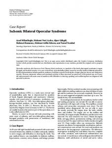
Figure 2: T2 weighted sagittal cervical MRI showing multiple pontine and cervical multiple hyperintense lesions.
306
1. González-Quintanilla V, Oterino A, Toriello M, et al. Cluster-tic syndrome as the initial manifestation of multiple sclerosis. J Headache Pain 2012; 13:425-9. 2. Wilbrink LA, Weller CM, Cheung C, et al. Cluster-tic syndrome: a cross-sectional study of cluster headache patients. Headache 2013; 53:1334-40. 3. McDonald WI, Compston A, Edan G, et al. Recommended diagnostic criteria for multiple sclerosis: guidelines from the International Panel on the Diagnosis of Multiple Sclerosis. Ann Neurol 2001; 50:121-7. 4. Polman CH, Reingold SC, Bandwell B, et al. Diagnostic criteria for multiple sclerosis: 2010 revisions to the McDonald criteria. Ann Neurol 2011; 69:292-302.
5. Monzillo PH, Sanvito WL, Da Costa AR. Clustertic syndrome: report of five new cases. Arq Neuropsiquiatr 2000; 58:518-21. 6. Watson P, Evans R. Cluster-tic syndrome. Headache 1985; 25:123-6. 7. Alberca R, Ochoa JJ. Cluster-tic syndrome. Neurology 1994; 44:996-9. 8. Cruccu G, Biasiotta A, Di Rezze S, et al. Trigeminal neuralgia and pain related to multiple sclerosis. Pain 2009; 143:186-91. 9. Leandri M, Cruccu G, Gottlieb A. Cluster headache like pain in multiple sclerosis. Cephalalgia 1999; 19:732-4. 10. Gentile S, Ferrero M, Vaula G, et al. Cluster headache attacks and multiple sclerosis. J Headache Pain 2007; 8:245-7.
307