[Downloaded free from http://www.jiaomr.in on Friday, March 17, 2017, IP: 168.151.137.14]
Original Article
Co-relation of variables as determined from panoramic radiograph and evaluating their significance in eruption of permanent mandibular third molar Kushal Amin, K. Vasavi, Sonal Vahanwala, C. D. Nayak, S. S. Pagare, S. S. Ramdev Department of Oral Medicine and Radiology, Padmashree Dr. D. Y. Patil Dental College and Hospital, Nerul, Navi Mumbai, India
ABSTRACT Purpose of the Study: Purpose of the study is to investigate whether the variables associated with the permanent mandibular third molar (PMM3) and arch dimensions could be co-related and significantly differentiated between a fully erupted and mesially impacted PMM3 among a set of Indian population. Study Design: A standardized panoramic radiograph was taken of subjects of age 21 years and above. Patients with missing tooth from mandibular arch, subjects undergoing or having history of orthodontic treatment, subjects having disto-angular, horizontal or vertical impacted PMM3 were excluded from the study. Subjects were divided into 2 groups: (1) mesially impacted PMM3 and (2) vertically erupted PMM3. Following measurements were taken from acetate paper tracing of standardized panoramic radiograph: (1) Angulation of long axis of PMM3 to permanent mandibular second molar (theta) (2) Angulation of PMM 3 to base of mandible (theta 2) (3) Gonial angle (theta 3) (4) Mesio-distal width of PMM 3 (5) Retro molar space. From these measurements Ganss ratio (retro molar space /PMM3 crown width.) was calculated. Results and Conclusion: Results revealed that angle theta 1, angle theta 2, retro molar space and Ganss ratio were positively co-related and highly significant variables associated with the mesially and vertically erupted teeth as measured on panoramic radiograph. Using these variables a long-term study can be carried out to predict the ultimate position of lower third molar in the arch so that if there is a probability of the tooth being impacted at a later age, a prophylactic germectomy can be performed at an early age. Key words: Angulation of the third molar, mandibular third molar and panoramic radiograph
INTRODUCTION Evolutionists have taught us that humans have evolved from ape like ancestors that possessed larger jaws and teeth than us. In the process of evolution the jaw has become smaller, allowing smaller room for third molars and resulting in numerous dental problems. The third molars (commonly called as wisdom teeth) account for 98% of all impacted teeth and permanent mandibular third molar (PMM3) is the most commonly impacted tooth after maxillary third molar.[1,2]
great variation in size, shape, position, root formation, time of development and path of eruption which is initially in horizontal direction and as the jaw grows the angulation changes to mesio-angular to vertical.[4] The prevalence of mandibular third molar impaction is variable in different populations ranging from nil in Nigerians[2] to 39% in Finns.[5] Third molar impaction is the most important clinical issue because it is involved in number of pathologies such as pericoronitis, caries, periodontitis, pathological resorption of mandibular second molar, cyst formation, benign and malignant odontogenic tumors and incisor crowding.[6,7]
A tooth is said to be impacted when it is completely or partially unerupted and is positioned against another tooth, bone or soft tissue so that its further eruption is unlikely.[3]
The aim of the study was to investigate variables associated with mesially impacted and vertically erupted group in a set of Indian population.
The average age of eruption of lower third molar is 20 years although it may continue till 25 years.[4] Third molars exhibit
MATERIALS AND METHODS
Correspondence Dr. Kushal Amin, 11/4 Hari Smruti, Vishwabharti society, Correspondence: Juhu lane, Andheri (w), Mumbai – 400058. E-mail:
[email protected] 14
The study population consisted of 60 patients seen in the out patient department of Padmashree Dr. D. Y. Patil Dental college and Hospital, Nerul, Navi Mumbai. Patients
Journal of Indian Academy of Oral Medicine and Radiology / January - March 2008 / Volume 20 / Issue 1
[Downloaded free from http://www.jiaomr.in on Friday, March 17, 2017, IP: 168.151.137.14]
Amin, et al.: Variables in eruption of permanent mandibular 3rd molar undergoing orthodontic treatment/or having history of orthodontic treatment, missing teeth in mandibular arch and having horizontal or vertical, disto-angular impaction status were excluded from the study. Only subjects of 21 years and above were included in the study. The subjects were divided into 2 groups: 1) Vertically erupted PMM3 2) Mesially impacted PMM3. A standardized panoramic radiograph of each subject was taken following a standardized technique by means of orthopantomogram machine (Xtropan 2000). Out of 60 subjects 30 had mesially impacted teeth and remaining had vertically erupted teeth. Measurements The radiographs were then traced on overlying acetate paper and analyzed by a single observer and the following measurements were taken [Figures 1 and 2]: 1) Inclination of third molar (theta 1) Measured as the angle formed between the long axis of PMM3 and permanent mandibular second molar (PMM2). 2) Angle made by PMM3 with the base of the mandible (theta - 2) Measured as the angle formed between the long axis of PMM3 and the tangent drawn to the inferior border of the mandible. 3) Gonial angle (theta - 3) Measured as the angle formed between the tangent drawn to the inferior border of the mandible and posterior border of the mandible.
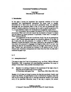
Figure 1: Shows an orthopantograph of a mesio-angular impacted tooth with an overlying lead acetate tracing paper
4) Mesio-distal crown width (m-d) Measured as the greatest distance between mesial and distal extent of the crown. 5) Retro-molar space Measured as the distance between the distal contact point of PMM2 and the junction of the anterior border of the mandible with the occlusal plane (line joining the highest point on the 1st premolar cusp with the highest point on mesio-buccal cusp of PMM2). 6) Ganss ratio Ratio of retro molar space to m-d width of PMM3. Statistical analysis A statistical analysis of the measurements was carried out. The results were expressed as means and standard deviation of means. Chi square test and paired t tests were also carried out to find the significance of the test. Further, Pearson’s and Spearson’s co-efficient analysis were carried out to find the positive co-relation between the variables.
RESULTS 1) Table 1 shows the frequency distribution of angle theta 1 which ranged from minimum of 6 to maximum of 72 degrees. The highest frequency (around 53.3%) was seen in the range of 1–20 degrees which accounted for vertically erupted teeth. There was an equal distribution of frequency (18.3 %) in the range of 20–60 degrees which accounted for most of the mesially impacted teeth. 2) Table 2 shows the frequency distribution of angle theta 2 which ranged from minimum of 24 to maximum of
Figure 2: Shows the tracing of the mesially impacted tooth where: Theta 1-angulation of PMM3 to PMM2, theta 2 - angulation of PMM3 to base of mandible, theta 3 - gonial angle, M-D - mesio-distal crown width, A-B - retro molar spacer
Journal of Indian Academy of Oral Medicine and Radiology / January - March 2008 / Volume 20 / Issue 1
15
[Downloaded free from http://www.jiaomr.in on Friday, March 17, 2017, IP: 168.151.137.14]
Amin, et al.: Variables in eruption of permanent mandibular 3rd molar 91 degrees. The highest frequency (48.3%) was seen in the range of 60–80 degrees which accounted for most of the vertically erupted teeth. 26.7% of the frequencies were distributed in the range of 40–60 degrees which accounted for mesially impacted teeth. 3) Table 3 shows the frequency distribution of gonial angle which ranged from minimum of 109 degrees to maximum of 139 degrees. Most of the frequencies were distributed between the range of 110 and 130 degrees for both mesially and erupted teeth. 4) Table 4 shows frequency distribution of mesio-distal crown width ranged from minimum of 10 mm to maximum of 17 mm. The highest frequency (61.70%) was in the range of 14–16 mm and around 26.7% of frequencies were distributed in the range of 12–14 mm. Mesio-distal crown width of both mesially impacted and erupted teeth was more or less within same range. 5) Table 5 shows frequency distribution of retro molar space Table 1: Frequency distribution of data θ1 (angulations of long axis PMM3 to long axis of PMM2) Degrees 1-20° 20-40° 40-60° 60-80° Total
Frequencies
Percentage
32 11 11 6 60
53.3 18.3 18.3 10 100
Table 2: Frequency distribution of data θ2 (angulation of PMM3 to base of mandible) Degrees
Frequencies
Percentages
1-20° 20-40° 40-60° 60-80° 80-100° Total
0 10 13 29 8 60
0 16.7 21.7 48.3 13.3 100
Table 3: Frequency distribution of data θ3 (gonial angulations) Degrees
Frequencies
Percentages
100-110° 110-120° 120-130° 130-140° Total
3 22 28 7 60
5 36.7 46.7 11.7 100
Table 4: The frequency distribution of m-d (PMM3) mm 10-12 12-14 14-16 16-18 Total 16
Frequencies
Percentages
3 16 37 4 60
5 26.7 61.7 6.7 100
which ranged from minimum of 4 mm to maximum of 24 mm. The highest frequency (55%) was in the range of 10–15 mm which accounted for most of the mesially impacted teeth and around 30% of the frequencies were distributed in the range of 15–20 mm which accounted for most of the vertically erupted teeth. 6) Table 6 shows the frequency distribution of Ganss ratio which ranged from minimum of 0.35 to maximum of 1.54. 48.3% of the frequencies were distributed between the range of 0.5 and 1 which accounted for most of the mesially impacted teeth. Similarly, around 41.7% of the frequencies were distributed in the range of 1–1.5 which accounted for most of the vertically erupted teeth. 7) Table 7 shows mean, standard deviation, difference between means and standard error of difference of mesially impacted and fully erupted teeth. As seen in the table a significant difference in angle theta 1, theta 2, retromolar space and Ganss ratio is observed between the two groups, which will be discussed later. Chi square test and paired t test revealed that the variables theta 1, theta 2, retromolar space and Ganss ratio were highly significant as P value was less than 0.05. Pearson’s Table 5: Frequency distribution of retro molar space mm 5-10 10-15 15-20 20-25 Total
Frequencies
Percentages
6 33 18 3 60
10 55 30 5 100
Table 6: Frequency distribution of Ganss ratio Ratio 0.1-0.5 0.5-1 1-1.5 1.5-2 Total
Frequencies
Percentages
1 29 25 5 60
1.7 48.3 41.7 8.3 100
Table 7: Mean, standard deviation, difference between means and standard error of difference of mesially impacted and fully erupted PMM3. Variable
Vertically erupted Mean sd
Theta-1 11.20 4.72 Theta-2 73.9 8.16 Theta-3 120.60 6.22 M-d (PMM3) 13.60 1.33 Retro molar 15.80 3.04 space Ganss ratio 1.22 0.19
Mesially impacted Mean sd
Difference between mean
Standard error of difference
43.06 17.70 50.10 15.99 123.93 7.41 14.50 1.14 11.50 2.19
-31.86 23.80 -3.33 -0.9 4.3
9.53 7.26 2.04 2.82 6.29
0.78 0.14
0.44
10.23
Theta in degrees; m-d in mm; retro molar space in mm
Journal of Indian Academy of Oral Medicine and Radiology / January - March 2008 / Volume 20 / Issue 1
[Downloaded free from http://www.jiaomr.in on Friday, March 17, 2017, IP: 168.151.137.14]
Amin, et al.: Variables in eruption of permanent mandibular 3rd molar and Spearman’s co-relation analysis proved that the variables were positively co-related.
DISCUSSION Third molar removal is the most frequent oral surgical intervention.[8] Due to the pathologies associated with the impacted wisdom teeth, assessment of the germ position and prognosis of its eruption is of general interest for the dentist. A school of thought states that removal of third molars at an early age seems to have less surgical and postoperative complications[9] and hence their early prophylactic removal is frequently advised.[8,10] A study was done by Ricketts et al. which described germectomy as a simple, short and relatively atraumatic surgical intervention[9] However, prophylactic removal of third molar is not justified for the fear of associated pathologies since the risk of the development of the pathologies is negligible (1–12% of problematic eruptions).[11] Our study enlists the variables associated with the impacted and erupted third molar as measured from the panoramic radiograph. In our study, a considerable difference in the mean of angle theta 1 between the two groups was observed. Mean of mesially impacted teeth was 43.06 (±17.7) degrees and vertically erupted was 11.20 (±4.72) degrees. Thus, we can conclude that greater the angulation of the tooth greater the chances of impaction. Hence, the tooth angulation is an important variable. Similarly, a significant difference was observed for angle theta 2. The mean in vertically erupted teeth was 73.9 (±8.16) degrees and 50.10 (±15.99) degrees in mesially impacted teeth hence, greater the angle made by the third molar with the base of the mandible greater the chances of eruption. Skeletal growth pattern is one of the factors governing eruption of third molar.[12] Studies have suggested that gonial angle is smaller in impacted group.[13] In our study no such finding was observed. Studies have indicated that mesio-distal crown width differs between the two groups. In our study, a difference of 0.9 mm was observed which is not a significant difference, so we can conclude that crown width is not an important variable. Numerous studies were done to measure retromolar space. Earlier technique for predicting lower third molar eruption
was done by Ricketts et al. The study was conducted on 74 orthodontic treated cases which showed that the distance from a point Xi (centre of the ramus) to the most distal point of second molar had a threshold value of 25 mm,[9,14] 20 mm was regarded as inadequate space whereas 30 mm was regarded as adequate space for eruption of the wisdom tooth. In our study the mean retro molar space in mesially impacted teeth was 11.50 mm and in vertically erupted teeth was 15.80 mm which is a significant difference between the two groups. Thus, lesser the space available greater the chances of impaction. Ganss et al.[15] used rotational tomograms and concluded that when the ratio is greater than 1,around 70% of teeth would erupt in the arch. However, lower values indicate impaction of the lower third molar. Ganss ratio remained constant between 16 and 20 years in impacted teeth whereas there was an increase by 0.6 between the age of 13–20 years in erupted teeth. In our study, the mean of Ganss ratio for mesially impacted teeth was less than 1 and in vertically erupted group was greater than 1 [Table 7]. Thus, it was concluded that when the Ganss ratio is less than 1 there is a higher probability of teeth being impacted thus making Ganss ratio an important variable. Thus, the present study enlists the variables: Angle theta 1, angle theta 2, retromolar space and Ganss ratio as the most important variables associated with the mesially impacted and vertically erupted teeth as measured on the panoramic radiograph.
CONCLUSION Based on the variables i.e., angle theta 1, angle theta 2, retromolar space and Ganss ratio, in our study it is possible to carry out a long-term study so as to predict the ultimate position of lower third molar in the arch, so that if there is a probability of the tooth being impacted at a later age, a prophylactic germectomy can be performed at an early age.
REFERENCES 1. 2. 3. 4. 5.
Alling CC, Helfrick JF, Alling RD. Impacted teeth. Philadelphia: WB Saunders; 1993. Hattab FN, Rawashdeh MA, Fahmy MS. Impaction status of third molars in Jordanian students. Oral Surg Oral Med Oral Path Oral Radio Endod 1995;79;24-9. Oral and maxillofacial surgery. 5th ed. By W. Harry. Archer. Peterson LJ. Principles of management of impacted teeth. In: Peterson LJ,Ellis III E, Hupp JR, Tucker MR, editors. Contemporary oral and maxillofacial surgery. 3rd ed. 1998. p. 215-48. Aitasalo K, Lehtinen R, Oksala E. An orthopantomographic study prevalence of impacted teeth. Int J Oral Surg 1972;1:117-20.
Journal of Indian Academy of Oral Medicine and Radiology / January - March 2008 / Volume 20 / Issue 1
17
[Downloaded free from http://www.jiaomr.in on Friday, March 17, 2017, IP: 168.151.137.14]
Amin, et al.: Variables in eruption of permanent mandibular 3rd molar 6.
Daley TD. Third molar prophylactic extraction: A review and analysis of literature. Gen Dent 1996;44:310-20. 7. Vasir NS, Robinson RJ. The mandibular third molar and late crowding of mandibular incisors: A review. Br J Orthod 1991;18:59-66. 8. Bruce RA, Frederideson GC, Small GS. Age of patients and morbidity associated with mandibular third molar surgery. J Am Dent Assos 1980;101:240-5. 9. Ricketts RM, Turley S, Chaconas S, Schulhof RJ. Third molar enucleation: Diagnosis and technique. J Calif Dent Assoc 1976;4:52-7. 10. Fielding AF, Douglass AF, Whitley RD. Reasons for early removal of impacted third molars. Clin Prev Dent 1981;3:19-23. 11. National Institute of Health. Concensus development conference for removal of third molars. Int Dent J 1980;38:2 12. Bjork A Jensen E, Palling M. Mandibular growth and third molar
18
impaction. Acta Odontol Scand 1956;14:231-2.35-6. 13. Richardson ME. The etiology and prediction of mandibular third molar impaction. Angle Orthod 1977;47:165-72. 14. Forsberg CM, Vingren B, Wesslen U. Mandibular third molar eruption in relation to available space as assessed on lateral cephalograms. Swed Dent J 1989;13:23-31. 15. Ganss C, Hoehban W, Kielbassa AM, Umstadt HE. Prognosis of third molar impaction. Oral Surg Oral Med Oral Path Oral Radiol Endod 1993;76:688-93.
Source of Support: Nil, Conflict of Interest: Nil
Journal of Indian Academy of Oral Medicine and Radiology / January - March 2008 / Volume 20 / Issue 1