[Downloaded free from http://www.ijo.in on Tuesday, November 28, 2017, IP: 195.16.121.41]
Brief Communications Co‑existing ligneous conjunctivitis and IgG4‑related disease Wei‑Yu Chiang, Ting‑Ting Liu1, Wan‑Ting Huang1, Ming‑Tse Kuo Herein, we elucidate that ligneous conjunctivitis (LC) was proved as an IgG4‑related disease (IgG4‑RD) by a series of pathologic studies from primary and recurrent episodes of an LC patient. LC was diagnosed based on clinical presentation and pathological appearance; furthermore, combined with serological examination and immunohistochemical study, the case also conformed to the diagnosis of IgG4‑RD. The IgG4‑RD, broadly discussed in recent times, is an idiopathic disease entity with tissue fibrosis possibly involving multiple organs. To the best of our knowledge, IgG4‑RD has never been reported with LC. By reporting the clinical course and literature review, we should pay attention to the association between these two diseases. Key words: IgG4, IgG4‑related disease, ligneous conjunctivitis
Ligneous conjunctivitis (LC) is a rare form of chronic conjunctivitis characterized by a fibrinous pseudomembrane on the conjunctiva and the development of firm fibrin‑rich, woody pseudomembranous lesions mainly on the tarsal conjunctivae.[1] Similar lesions may also involve extra‑ocular mucosa such as the gingiva, ear, upper and lower respiratory tracts, renal collecting system, or female genital tract.[1] The IgG4‑related disease (IgG4‑RD) is an idiopathic disease entity with tissue fibrosis possibly involving multiple organs. It is characterized by elevated serum IgG4 concentrations and tissue infiltration by IgG4‑positive plasma cells, accompanied by tissue fibrosis and sclerosis.[2] IgG4‑RD has been broadly discussed in recent times, with a rapidly increasing understanding of the disease and its causes. Access this article online Quick Response Code:
Website: www.ijo.in DOI: 10.4103/0301-4738.190154 PMID: ***
Departments of Ophthalmology and Pathology, Kaohsiung Chang Gung Memorial Hospital and Chang Gung University College of Medicine, Kaohsiung, Taiwan, R.O.C. 1
Correspondence to: Dr. Ming‑Tse Kuo, Department of Ophthalmology, Kaohsiung Chang Gung Memorial Hospital and Chang Gung University College of Medicine, No. 123, Dapi Road, Niaosong Dist., Kaohsiung 83301, Taiwan, R.O.C. E‑mail:
[email protected] Manuscript received: 10.10.14; Revision accepted: 14.02.16
In this case report, we show that LC may be an IgG4‑RD based on the ocular pathological findings, clinical presentation, and serological tests.
Case Report A 34‑year‑old woman presented with bilateral itching, redness, and mucinous discharge. Several bilateral woody hyalinized masses were identified on the tarsal conjunctiva [Fig. 1]. She also had mucinous saliva; besides, a cyst partially limiting the vocal cord movement was identified over the right posterior cricoids. Neither did the patient have any contributory history of ocular trauma nor did any of her family members. Initially, unusual, varied‑size giant papillae were found over the tarsal conjunctiva, and received mass excision 3 times, cryotherapy, triamcinolone injection (40 mg/ml), antiproliferative soaking with mitomycin C, and amniotic membrane transplantation because of medically refractory symptoms. Although symptoms were dramatically relieved after each surgery, the patient was always followed‑up for tumor regrowth with associated discomfort for up to 1–2 years after each surgery [Fig. 2]. Recurrent conjunctival lesions were assessed by immunohistochemical microscopy for serial samples of conjunctival biopsies. Pathological analysis confirmed the diagnosis of LC [Fig. 3] and revealed the characteristics of IgG4‑RD [Fig. 4]. Combined with clinical presentation and elevated serum IgG4 level (274 mg/dl), IgG4‑RD was further diagnosed. There was no obvious lymphadenopathy in physical examination and chest X‑ray. Laboratory examination revealed that eosinophil (0.7%), rheumatoid factor (nonreactive), anti‑nuclear antibody (negative), erythrocyte sedimentation rate (3 mm/h), C3 (98.4 mg/dl), C4 (15.3 mg/ dl), IgG (1500 mg/dl), and IgM (230 mg/dl) were all within normal limit, but IgE (133 IU/ml) and IgA (531 mg/dl) were elevated.
Discussion In this case, we found LC may be an IgG4‑RD. To the best of our knowledge, IgG4‑RD has never been associated with LC. In this case, LC was diagnosed by the patient’s clinical and pathological manifestations. Her condition was also diagnosed as a definite case of IgG4‑RD [Table 1][3] because she met the clinical criteria (diffuse/localized swelling and masses in single or multiple organs), hematological criteria (serum IgG4 level: 274 mg/dl), and the histopathological criteria (marked lymphocyte and plasmacyte infiltration This is an open access article distributed under the terms of the Creative Commons Attribution‑NonCommercial‑ShareAlike 3.0 License, which allows others to remix, tweak, and build upon the work non‑commercially, as long as the author is credited and the new creations are licensed under the identical terms. For reprints contact:
[email protected]
Cite this article as: Chiang WY, Liu TT, Huang WT, Kuo MT. Co-existing ligneous conjunctivitis and IgG4-related disease. Indian J Ophthalmol 2016;64:532-4.
© 2016 Indian Journal of Ophthalmology | Published by Wolters Kluwer - Medknow
[Downloaded free from http://www.ijo.in on Tuesday, November 28, 2017, IP: 195.16.121.41] July 2016
a
b
Figure 1: Conjunctival lesions of ligneous conjunctivitis. Woody hyalinized masses were identified on both the right (a) and left (b) tarsal conjunctiva
a
533
Brief Communications
b
a
Figure 2: Recurrent conjunctival lesions of ligneous conjunctivitis. Refractory recurrence was noted after surgical excision on both the right (a) and left (b) sides
a
b
c
d
e
f
b
Figure 3: Microscopy with H and E stain revealed background fibrotic stroma with prominent inflammatory cell infiltration, especially in plasma cells, which demonstrated a typical histological finding of ligneous conjunctivitis (a: ×40, bar = 200 μm; b: ×400, bar = 20 μm)
and fibrosis; infiltration of IgG4‑positive plasma cells; ratio of IgG4‑positive cells/IgG‑positive cells >40% and >10 IgG4‑positive plasma cells/high‑power field). However, we cannot exclude the possibility that our patient had two unrelated diseases. A future study of more LC cases will provide more supportive evidence. The clinical characteristics of female bilateral sides and palpebral conjunctiva are prone to be affected by LC.[1] The possible etiologies of LC include deficiency of plasmin‑mediated extracellular fibrinolysis, tranexamic acid drug exposure, and plasminogen deficiency. [1] However, the diagnosis of LC is mainly based on typical clinical and histological findings. LC is chronic conjunctivitis typically with woody‑like pseudomembranous lesions on the tarsal conjunctivae. Recurrence is common after surgical excision alone, but combination with antiproliferative agent mitomycin C and amniotic membrane transplantation may be beneficial.[4,5] In one case report, medication including topical frozen plasma, heparin, 0.1% dexamethasone, and
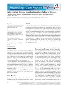
Figure 4: Immunohistochemical studies support that ligneous conjunctivitis is an IgG4‑related disease. The immunohistochemical study of CD3 (a: bar = 100 μm) and CD20 (b: bar = 100 μm) showed T lymphocyte predominance, which was highlighted by CD3. The immunohistochemical study revealed increased percentage of IgG4/IgG (c: bar = 100 μm) and the number of IgG4‑positive plasma cells per high‑power field (d: bar = 100 μm). Both light chains of κ (e: bar = 100 μm) and λ (f: bar = 100 μm) were stained, indicating polyclonal hypergammaglobulinemia
Table 1: Comprehensive clinical diagnostic criteria for IgG4‑related disease 1. Clinical examination showing characteristic diffuse/localized swelling or masses in single or multiple organs 2. Hematological examination shows elevated serum IgG4 concentrations (135 mg/dl) 3. Histopathologic examination shows (1) Marked lymphocyte and plasmacyte infiltration and fibrosis (2) Infiltration of IgG4‑positive plasma cells: Ratio of IgG4‑positive/ IgG‑positive cells >40% and >10 IgG4‑positive plasma cells/HPF Definite: 1+2+3 Probable: 1+3 Possible: 1+2 HPF: High‑power field
[Downloaded free from http://www.ijo.in on Tuesday, November 28, 2017, IP: 195.16.121.41]
534
Indian Journal of Ophthalmology
tobramycin was used for successful treatment.[6] Cyclosporin A and azathioprine were also reported to be effective.[7,8] Typical histological findings of LC include thinned or eroded epithelium with superficial or subepithelial deposits of amorphous hyaline‑like eosinophilic materials, and multiple foci of persisting granulation tissue with accompanying inflammatory cells, mainly lymphocytes, plasma cells, and granulocytes.[1] IgG4‑RD mainly affects middle‑aged to elderly men, and it can occur in various organs, including the central nervous system, salivary glands, thyroid gland, lungs, pancreas, biliary duct, liver, gastrointestinal tract, kidneys, prostate gland, retroperitoneum, and lymph nodes, but that clinical symptoms depend on the location of the lesion.[2] The elevated serum IgG4 concentration and tissue infiltration of IgG4‑positive plasma cells are characteristic features of IgG4‑RD, while there is no definite pathogenetic mechanism and underlying immunological abnormalities.[2] Overexpression of specific cytokines including interleukin 10 (IL‑10) and transforming growth factor β (TGF‑β) has been reported.[9] IL‑10 and TGF‑β have potent effects in directing B cells to produce IgG4 and induce fibroplasia, respectively.[2] Umehara et al. proposed a comprehensive diagnostic criterion for IgG4‑RD [Table 1].[3] At present, no specific treatment guideline is available for IgG4‑RD. Although it responds well to steroid therapy, recurrence and relapse occur following the early reduction or withdrawal of medication.[2]
Vol. 64 No. 7
determination of serum IgG4 levels in a larger sample of LC patients might conclusively elucidate whether LC is an IgG4‑RD. Financial support and sponsorship Nil. Conflicts of interest There are no conflicts of interest.
References 1. Schuster V, Seregard S. Ligneous conjunctivitis. Surv Ophthalmol 2003;48:369‑88. 2. Umehara H, Okazaki K, Masaki Y, Kawano M, Yamamoto M, Saeki T, et al. A novel clinical entity, IgG4‑related disease (IgG4RD): General concept and details. Mod Rheumatol 2012;22:1‑14. 3. Umehara H, Okazaki K, Masaki Y, Kawano M, Yamamoto M, Saeki T, et al. Comprehensive diagnostic criteria for IgG4‑related disease (IgG4‑RD), 2011. Mod Rheumatol 2012;22:21‑30. 4. Meire FM, Claerhout I, Kestelyn PH. Use of mitomycin C and r‑tPA for the management of conjunctival membrane and cataracts in a child with conjunctivitis lignosa. Br J Ophthalmol 2000;84:1204‑5. 5. Barabino S, Rolando M. Amniotic membrane transplantation in a case of ligneous conjunctivitis. Am J Ophthalmol 2004;137:752‑3. 6. Ku JY, Lichtinger A, Yeung SN, Kim P, Cserti‑Gazdewich C, Slomovic AR. Topical fresh frozen plasma and heparin treatment of ligneous conjunctivitis in a Canadian hospital setting. Can J Ophthalmol 2012;47:e27‑8.
As for the pathophysiologic correlation between LC and IgG4‑RD, there is no literature available to illustrate the common features. In LC, inflammatory cells, mainly lymphocytes and plasma cells, are typical histological findings;[10] on the other hand, lymphocyte and IgG4 plasma cell infiltration are the histopathological features in IgG4‑RD.[3] Thus, we postulate that lymphocyte and plasma cell may be the key roles for both diseases.
7. Hidayat AA, Riddle PJ. Ligneous conjunctivitis. A clinicopathologic study of 17 cases. Ophthalmology 1987;94:949‑59.
Conclusion
10. R o d r í g u e z ‑ A r e s M T , A b d u l k a d e r I , B l a n c o A , Touriño‑Peralba R, Ruiz‑Ponte C, Vega A, et al. Ligneous conjunctivitis: A clinicopathological, immunohistochemical, and genetic study including the treatment of two sisters with multiorgan involvement. Virchows Arch 2007;451:815‑21.
LC may be an IgG4‑RD with ocular mucosal (conjunctival) change, which has never been reported. Future studies with conjunctival biopsy for immunohistochemistry and
8. Shimabukuro M, Iwasaki N, Nagae Y, Nakagawa Y, Ohtori Y, Inoue Y, et al. Ligneous conjunctivitis: A case report. Jpn J Ophthalmol 2001;45:375‑7. 9. Nakashima H, Miyake K, Moriyama M, Tanaka A, Watanabe M, Abe Y, et al. An amplification of IL‑10 and TGF‑beta in patients with IgG4‑related tubulointerstitial nephritis. Clin Nephrol 2010;73:385‑91.