2876 • The Journal of Neuroscience, March 14, 2007 • 27(11):2876 –2882
Cellular/Molecular
Cocaine- and Amphetamine-Regulated Transcript: Stimulation of Expression in Rat Vagal Afferent Neurons by Cholecystokinin and Suppression by Ghrelin Guillaume de Lartigue, Rod Dimaline, Andrea Varro, and Graham J. Dockray Physiological Laboratory, School of Biomedical Sciences, University of Liverpool, Liverpool L69 3BX, United Kingdom
The neuropeptide transmitter cocaine- and amphetamine-regulated transcript (CART) inhibits food intake and is expressed by both vagal afferent and hypothalamic neurons. Here we report that cholecystokinin (CCK) regulates CART expression in rat vagal afferent neurons. Thus, CART was virtually undetectable after energy restriction for 24 h, but administration of CCK to fasted rats increased CART immunoreactivity, and refeeding of fasted animals promptly increased CART by a mechanism sensitive to a CCK-1 receptor antagonist. In vagal afferent neurons incubated in serum-free medium, CART was virtually undetectable, whereas the orexigenic peptide melaninconcentrating hormone (MCH) was readily detected. The addition of CCK rapidly induced CART expression and downregulated MCH. Using a CART promoter–luciferase reporter vector transfected into cultured vagal afferent neurons, we showed that CCK stimulation of CART transcription was mediated by activation of protein kinase C and cAMP response element-binding protein (CREB). The action of CCK on CART expression was inhibited by the orexigenic peptide ghrelin, through a mechanism that involved exclusion of phosphorylated CREB from the nucleus. Thus, CCK reciprocally regulates expression of CART and MCH within the same vagal afferent neuron; ghrelin inhibits the effect of CCK at least in part through control of the nuclear localization of phosphoCREB, revealing previously unsuspected modulation of gut– brain signals implicated in control of food intake. Key words: satiety; CREB; melanin-concentrating hormone; ghrelin; vagus; food
Introduction The mechanisms controlling food intake are attracting considerable attention, not the least because of their significance in understanding the recent dramatic increase in obesity (Schwartz et al., 2000; Woods, 2004). Termination of feeding involves gastrointestinal signals (Dockray, 2004; Murphy and Bloom, 2004; Woods, 2004) that include non-nutrient distension of the stomach and nutrient-dependent release of gastrointestinal satiety hormones, of which cholecystokinin (CCK) is one of the beststudied examples (Liddle, 1997; Dockray, 2004). Both gastric distension and gut hormones such as CCK inhibit food intake via vagal afferent neurons (Gonzalez and Deutsch, 1981; Smith et al., 1985; Moran and Kinzig, 2004). Transmission at the central terminals of these neurons in the nucleus tractus solitarius activates ascending pathways to the hypothalamus where there is integration of a variety of different peripheral signals including those from adipose tissue (e.g., leptin) and the endocrine pancreas (e.g., insulin) (Schwartz et al., 2000; Luckman and Lawrence, 2003).
Received Dec. 20, 2006; revised Jan. 26, 2007; accepted Jan. 26, 2007. We thank the Wellcome Trust and the Medical Research Council for financial support. We are grateful to Drs. Michael Kuhar, Perry Barrett, Joseph Nevins, and David Ginty for the supply of plasmids and to Rotta Pharmaceuticals for the gift of lorglumide. Correspondence should be addressed to Dr. Graham J. Dockray, Physiological Laboratory, School of Biomedical Sciences, University of Liverpool, Crown Street, Liverpool L69 3BX, UK. E-mail:
[email protected]. DOI:10.1523/JNEUROSCI.5508-06.2007 Copyright © 2007 Society for Neuroscience 0270-6474/07/272876-07$15.00/0
The neuropeptide transmitter systems implicated in the control of feeding behavior within the hypothalamus include those associated with the stimulation of food intake [e.g., neuropeptide Y, melanin-concentrating hormone (MCH), agouti-related peptide] or with inhibition of food intake [e.g., cocaine- and amphetamine-regulated transcript (CART)] (Kristensen et al., 1998; Lambert et al., 1998; Schwartz et al., 2000; Ellacott and Cone, 2004; Hunter et al., 2004). The latter was discovered by differential mRNA display as a transcript exhibiting increased expression in rats treated with amphetamine or cocaine (Douglass et al., 1995). Interestingly, CART is expressed in vagal afferent neurons that also express CCK-1 receptors (Broberger et al., 1999), and there is evidence that it can inhibit ingestive behavior from both hindbrain and hypothalamic sites (Aja et al., 2001; Zheng et al., 2001; Smedh and Moran, 2003). Recent studies suggest that in addition to expression of CCK-1 receptors, vagal afferent neurons also express the leptin receptor (Ob-R) and receptors associated with stimulation of food intake including the ghrelin (GHS-1), cannabinoid (CB1), orexin (OXR1), and MCH-1 receptors (Kirkup et al., 2001; Burdyga et al., 2002, 2003, 2004, 2006b; Dockray, 2003). Orexin A and ghrelin inhibit the discharge of vagal afferent neurons in response to CCK compatible with integration of both orexigenic and anorexigenic stimuli outside the CNS (Burdyga et al., 2003; Date et al., 2005). In addition, a growing body of evidence now indicates regulation by CCK of gene expression in these neurons. Thus, energy restriction of rats for 12–18 h or longer sharply increases
de Lartigue et al. • Control of CART Expression by CCK
J. Neurosci., March 14, 2007 • 27(11):2876 –2882 • 2877
expression of CB1 and MCH-1 receptors, and of MCH, and refeeding downregulates expression via CCK (Burdyga et al., 2004, 2006b). This is not a general response of all receptors associated with orexigenic function because OX-R1 and GHS-1 expression appears unchanged in these circumstances (Burdyga et al., 2003, 2006a). The phenotype of vagal afferent neurons therefore reflects energy ingestion over the previous 24 h. Because these neurons express CART, which has been linked to satiety effects (Kristensen et al., 1998; Lambert et al., 1998; Broberger et al., 1999), we hypothesized that CCK regulates CART expression in vagal afferent neurons, and we report here evidence consistent with the hypothesis.
Materials and Methods Materials. Unless stated otherwise, chemicals were obtained from Sigma (Poole, Dorset, UK); CCK8 and ghrelin were obtained from Bachem (St. Helens, UK), lorglumide was a gift from Rotta Pharmaceuticals (Milan, Italy), and Ro-32-0432 was obtained from Calbiochem (La Jolla, CA). Dr. M. Kuhar (Yerkes National Primate Research Center, Emory University, Atlanta, GA) and Dr. P. Barrett (Rowett Research Institute, Aberdeen Center for Energy Regulation and Obesity, Bucksburn, UK) provided CART promoter–luciferase reporter constructs of 120, 620, 3451, and 3100 bp upstream of the transcriptional start site (i.e., 120 CARTLuc, 620 CART-Luc, 3451 CART-Luc, and 3100 CART-Luc). Dr. J. Nevins (Duke University Medical Center, Durham, NC) kindly provided plasmids for the adenovirus product E1A and the inactive mutant ⌬2-36 E1A. Dr. D. Ginty (The Johns Hopkins University School of Medicine, Baltimore, MD) provided the cAMP response element-binding protein (CREB) dominant-negative mutant, ACREB plasmid. Animals. Studies were performed using adult male Wistar rats (250 – 400 g) housed at 22°C under a 12 h light/dark cycle with ad libitum access to food and water, unless stated otherwise. Fasting–feeding experiments. Rats were fasted for 24 h (water ad libitum) and subsequently refed for up to 2 h. Some fasted animals received CCK8 (10 nmol, i.p.) or saline and were killed 2 h later. In addition, some rats that were fasted for 24 h received the CCK-1 receptor antagonist lorglumide (10 mg/kg, i.p.) 15 min before refeeding and were killed 1 h after refeeding. Rats were killed by CO2 inhalation, and nodose ganglia were rapidly immersed in 4% paraformaldehyde for 1 h at 22°C, followed by 25% sucrose in 1 mol/L PBS overnight at 4°C. Cell culture. Nodose ganglia were dissected under aseptic conditions and digested for 90 min at 37°C in 3 ml of Ca 2⫹- and Mg 2⫹-free HBSS containing 50 l of 100 mM DTT and 1 mg/ml collagenase type Ia (Roche Diagnostics, Indianapolis, IN). Cells were dispersed by gentle trituration through siliconized Pasteur pipettes, washed twice with HEPES-buffered DMEM (HDMEM) containing 10% fetal calf serum (FCS) (Hyclone/ Perbio Science, Cramlington, UK) supplemented with 1% penicillin/ streptomycin solution and 2% antibiotic/antimycotic solution, plated onto four-well chamber slides, and maintained in HDMEM with 10% FCS at 37°C in 5% CO2, and the medium was changed every 48 h. For many experiments, cells were transferred to serum-free medium for 2–10 h before experimental treatments. Immunohistochemistry. Cryostat sections of fixed nodose ganglia (10 –15 m) were mounted on polysine-coated slides (Polysine; MenzelGla¨ser, Braunschweig, Germany) and processed for immunohistochemistry. Cultured neurons were fixed in 4% paraformaldehyde in PBS. The following antibodies were used: affinity-purified rabbit polyclonal antibody to CART (Phoenix Laboratories, San Antonio, TX); affinitypurified rabbit polyclonal antibody to phosphoCREB (Cell Signaling Technology, Beverly, MA); affinity-purified goat polyclonal antibody to MCH; rabbit polyclonal antibody to the CCK-1 receptor; rabbit polyclonal antibody to glial fibrillary acidic protein (GFAP; Santa Cruz Biotechnology, Calne, Wiltshire, UK); affinity-purified mouse monoclonal antibody to neuron-specific enolase (NSE; Abcam, Cambridge, UK); and affinity-purified mouse anti-Flag monoclonal antibody (Upstate Biotechnology, Hampshire, UK). Secondary antibodies were used as appropriate and included donkey anti-rabbit Ig conjugated to fluorescein isothiocyanate and donkey anti-mouse Ig labeled with Texas Red (Jackson
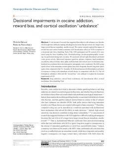
Figure 1. CCK-dependent expression of CART in vagal afferent neurons. Nodose ganglia from rats are shown. A, B, Rats fed ad libitum exhibiting CART-immunoreactive neurons. C, Reduced CART immunoreactivity after fasting for 24 h. D, Stimulation of CART immunoreactivity after fasting for 24 h and 1 h after an intraperitoneal injection of 10 nmol of CCK. E, F, Stimulation of CART after fasting for 24 h and refeeding for 1 h (E), which was inhibited by administration of the CCK-1 receptor antagonist lorglumide (F; 10 mg 䡠 kg ⫺1, i.p.) 15 min before refeeding. Representative images from 10 independent experiments are shown. Magnification: A, 20⫻; B–F, 40⫻.
ImmunoResearch, West Grove, PA). Specificity of immunostaining was determined by omitting the primary antibody and by preincubation with an excess of appropriate peptide where available. Samples were mounted in Vectashield with 4⬘,6-diamidino-2-phenylindole (DAPI; Vector Laboratories, Peterborough, UK) for nuclear localization and examined using an Axiopan Universal microscope (Zeiss, Oberkochen, Germany), and images were processed using the Axio Vision 3.0 Imaging system (Zeiss). Luciferase assay. Vagal afferent neurons were cultured in full medium for 2 d in 12-well plates. The medium was removed, and cells were transfected with firefly-luciferase constructs and a constitutively active Renilla luciferase plasmid as an internal control (Promega, Madison, WI); cells were transfected with 1 g of DNA per well, using CombiMag (OzBiosciences, Marseille, France) together with Transfast reagent (Promega), on a magnetic plate according to the manufacturer’s instructions. Cells were further incubated for 24 h before stimulation. Luciferase activity was measured by dual luciferase assay (Promega) according to the manufacturer’s instructions in a Lumat LB9507 luminometer (Berthold, Redbourne, Herts, UK). Results are presented as fold increase over unstimulated control, so 1.0 signifies no change in luciferase activity. Mutation of cAMP response element. To investigate the importance of the cAMP response element site for CCK-mediated CART transcription, this site was mutated from tgacgtca to gtcagtca in the 34510 CART-Luc construct using a QuikChange II XL site-directed mutagenesis kit (Stratagene Europe, Amsterdam, The Netherlands).
2878 • J. Neurosci., March 14, 2007 • 27(11):2876 –2882
de Lartigue et al. • Control of CART Expression by CCK
Results Energy restriction depresses CART in vagal afferent neurons in vivo In initial studies, we examined the localization of CART in vagal afferent neurons of rats fed ad libitum or fasted for 24 h. In the middle and caudal regions of the nodose ganglion from rats fed ad libitum, 45 ⫾ 2% of all neurons expressed CART, but after fasting for 24 h, CART immunoreactivity was detected in ⬍10% of neurons. Administration of CCK8 (10 nmol, i.p.) to fasted rats increased CARTexpressing cells within 1 h. In addition, 1 h after refeeding fasted rats, CART expression was restored, and this increase was inhibited by administration of the CCK-1 receptor antagonist lorglumide (Fig. 1). CART expression in vagal afferent neurons in culture The mechanisms controlling CART abundance in response to CCK were then examined in cultured vagal afferent neurons. After culture of dissociated rat nodose ganglia for 96 h, 79.1 ⫾ 1.8% of cells were identified as neurons on the basis of expression of NSE, and most of the remain- Figure 2. CART expression in cultured vagal afferent neurons. Vagal afferent neurons cultured for ⬎48 h showing CART der (12.7 ⫾ 1.3%) were identified as glial immunoreactivity (A, C, E, G, I, K ) or nuclei stained with DAPI in the respective fields (B, D, F, H, J, L). A, B, CART is undetectable cells on the basis of GFAP staining. Ap- after transfer of neurons to serum-free medium for 2 h. C, D, CART immunoreactivity is readily detected in cells in serum-free medium 2 h after the addition of 10 nM CCK. E, F, CART is also induced in cells in serum-free medium 2 h after the addition of 100 proximately 75% of neurons expressed nM PMA. G, H, The effect of CCK is reduced by the PKC inhibitor Ro-32-0432 (1 m). I–L, In serum-free medium, ghrelin (10 nM) CART (61.0 ⫾ 1.2% of all cells), and as has no effect on CART abundance (I, J ) but inhibits the effect of CCK (K, L). Arrows identify a representative neuron in each pair of expected, CART was expressed by ⬍1% of images; filled arrows show CART-positive neurons, and open arrows show CART-negative neurons. Representative images from GFAP-positive cells. Serum withdrawal for six independent experiments are shown. Scale bar, 50 m. 2 h reduced the population of cells expressing CART immunoreactivity to 6.2 ⫾ nuclear phosphoCREB (Fig. 3I ). However, when cells were 0.8% of the total (Fig. 2). However, the addition of CCK (10 nM) treated with ghrelin in the presence of CCK, there was a striking to serum-deprived vagal afferent neurons restored the proporexclusion of phosphoCREB from the nucleus that was associated tion of neurons expressing CART to that of control cultures (Fig. with localization to the cytosol (Fig. 3K ). In response to CCK 2 A, C). The addition of 12-myristate-13-acetate phorbol ester alone, antibody to total (i.e., phosphorylated and unphosphory(PMA) to neurons in serum-free conditions also increased CART lated) CREB revealed a predominantly nuclear localization, but immunoreactivity (Fig. 2 E); moreover, the PKC inhibitor Ro-32when CCK was combined with ghrelin, there was strong cytosolic 0432 blocked the effect of both CCK and PMA (data not shown), localization similar to that of phosphoCREB (Fig. 3 M, O). compatible with the idea that CCK induces CART via PKC (Fig. CCK stimulation of CART promoter–luciferase 2G). The orexigenic hormone ghrelin alone had no effect on reporter expression CART expression in vagal afferent neurons in serum-free meTo explore the mechanisms by which CCK regulates CART abundium (Fig. 2 I). However, the addition of ghrelin inhibited the dance in more detail, we then examined the expression of CART action of CCK, indicating an interplay between orexigenic and promoter–luciferase reporter constructs after transfection into anorexic peptides in regulating CART expression (Fig. 2 K). vagal afferent neurons (Fig. 4). In response to CCK, there was a CCK stimulation of CREB phosphorylation in vagal 2.3 ⫾ 0.1-fold increase in expression of a 3.45 kb sequence of the afferent neurons CART promoter region coupled to luciferase. Truncation of the Because it is known that CART expression is regulated by CREB sequence to 620 bp reduced the response to CCK, and further (Lakatos et al., 2002; Dominguez and Kuhar, 2004; Jones and truncation to 120 bp virtually abolished it (Fig. 4 A). The increase Kuhar, 2006), we asked whether CCK-1 receptor activation led to in luciferase activity in response to CCK was significantly reduced CREB phosphorylation in vagal afferent neurons. In vagal afferby the PKC inhibitor Ro-32-0432 (Fig. 4 B) and by ghrelin in ent neurons transferred to serum free-medium for 2 h, phosphocombination with CCK (Fig. 4C). Compatible with the idea that CREB was undetectable, but in the presence of 10 nM CCK for 30 CCK regulates CART expression via CREB, cotransfection with a min, there was abundant nuclear phosphoCREB (Fig. 3). The vector encoding a dominant-negative CREB protein, ACREB, PKC inhibitor Ro-32-0432 blocked the action of CCK in stimusignificantly inhibited the response to CCK. Moreover, cotranslating phosphoCREB in cultured vagal afferent neurons, and fection with a plasmid encoding E1A (which inhibits the CREB PMA stimulated it (Fig. 3 E, G), compatible with CCK phosphorcoactivator CBP) also significantly reduced the response to CCK, ylation of CREB via PKC. On its own, ghrelin did not increase whereas an inactive E1A mutant (⌬ 2-36 E1A) had no effect (Fig.
de Lartigue et al. • Control of CART Expression by CCK
J. Neurosci., March 14, 2007 • 27(11):2876 –2882 • 2879
Figure 3. CCK induces CREB phosphorylation through activation of PKC in vagal afferent neurons. Vagal afferent neurons cultured for ⬎48 h and stained with antibody to phosphoCREB (A, C, E, G, I, K ) or CREB (M, O) and DAPI (B, D, F, H, J, L, N, P), in the respective fields, are shown. A, B, Nuclear phosphoCREB staining was undetectable after transfer to serum-free medium for 2 h. C–F, Nuclear phosphoCREB was readily detectable after treatment with 10 nM CCK (C, D) and 100 nM PMA (E, F ) for 30 min. G, H, The effect of CCK was inhibited by pretreatment with 1 M Ro-32-0432. I, J, Nuclear phosphoCREB was undetectable after treatment with ghrelin (10 nM). K, L, Importantly, ghrelin (10 nM) in the presence of 10 nM CCK led to exclusion of phosphoCREB from the nucleus. M–P, Total CREB (M, N ) was unchanged by CCK and showed a cytoplasmic localization in the presence of ghrelin plus CCK (O, P). A–L, Arrows identify a representative neuron in each pair of images; filled arrows show phosphoCREB-positive neurons, and open arrows show phosphoCREB-negative neurons. M–O, Arrows indicate a representative neuron exhibiting nuclear localization of total CREB (M, N ) and a representative neuron exhibiting cytosolic localization of total CREB (M, O). Representative images from six independent experiments are shown. Scale bar, 50 m.
5). Finally, mutation of the Cre site in the 3.45 kb sequence virtually abolished responses to CCK. Together, the data are therefore compatible with a role for PKC, CREB, CBP, and the Cre element in mediating the effects of CCK. CREB-mediated CART–MCH switch Previous studies have shown that expression of the orexigenic peptide MCH in vagal afferent neurons is increased by fasting and decreased by CCK (Burdyga et al., 2006b). We therefore asked whether the loss of CART in serum-starved vagal afferent neurons was associated with increased MCH (Fig. 6). Serum withdrawal for 8 h was found to be associated with MCH expression in a substantial proportion of cells (53.0 ⫾ 0.9% of all cells); in parallel with the increased expression of CART on exposure to CCK, there was rapid loss of MCH expression so that after 120 min MCH was detectable in only 2.8 ⫾ 1.2% of cells (Fig. 6). At
⬃85 min after the addition of CCK, it was possible to identify separate populations of vesicles containing CART and MCH within the same neuron, indicating that these neurons switched between expression of CART and MCH depending on exposure to CCK (Fig. 6); in particular, at this time point, ⬍10% of cells expressed either CART or MCH alone, but 48.9 ⫾ 2.7% expressed both. To determine whether CREB regulated expression of wild-type CART and MCH in vagal afferent neurons, we then examined the consequences of transfecting cultured vagal afferent neurons with the dominant-negative mutant ACREB, identified by expression of the Flag epitope. Cells expressing ACREB made up 10.9% of the total population; in the same cultures, CCK-1 receptor expression was identified in 59.1% of all cells, and cells positive for both ACREB and CCK-1 receptor made up 10.2% of the total population. Thus, 93.1% of ACREB-transfected cells were CCK-1 receptor positive, reflecting a transfection efficiency for this population of 18%. Of the cells that expressed ACREB, only 15 ⫾ 2% expressed CART in response to CCK (data not shown), whereas untransfected cells in the same cultures exhibited CART expression in response to CCK similar to that described above. In control experiments in which cells were transfected with a vector encoding green fluorescent protein (GFP), the proportions exhibiting CART expression in response to CCK were again similar to controls (data not shown). Importantly, however, 96.6 ⫾ 1% of cells expressing ACREB also exhibited MCH after treatment with CCK, indicating that ACREB prevented CCK-induced downregulation of MCH.
Discussion
The present study shows that CCK regulates CART expression in vagal afferent neurons and that this depends on activation of PKC and CREB. The idea that CART might be a peptide mediator of the satiety effects of CCK has been recognized for some time (Broberger et al., 1999; Hokfelt et al., 2001). However, the regulation of CART expression by CCK in vagal afferent neurons adds an additional dimension to our understanding of this system. Although other examples of CCK-regulated expression of signaling molecules associated with feeding behavior have been described previously (Burdyga et al., 2004, 2006b), in each case CCK was found to depress expression of genes associated with orexigenic function (e.g., CB1 and MCH-1 receptors and MCH). The present findings suggest that activation of CCK-1 receptors on vagal afferent neurons is also associated with increased expression of a gene associated with satiety signaling. Two features of this system are noteworthy. First, within the same neuron, CCK acts as a switch that relatively rapidly depresses production of the
2880 • J. Neurosci., March 14, 2007 • 27(11):2876 –2882
Figure 4. CCK regulates CART transcription in luciferase promoter–reporter assays. A, Cultured vagal afferent neurons were transfected with empty vector (empty) and deletional mutants of the CART promoter of 3451, 3100, 620, and 120 bp upstream of the transcriptional start site in luciferase reporter vectors (empty, empty vector). Studies were performed 16 h after transfection when cells were transferred to serum-free medium and stimulated (filled bars) or not (open bars) with 10 nM CCK for 6 h. B, In vagal afferent neurons transfected with 3451CARTLuc, the response to 10 nM CCK was inhibited by 1 M Ro-32-0432. C, In vagal afferent neurons transfected with 3451 CART-Luc, the effect of 10 nM CCK was inhibited by preincubation for 30 min with 10 nM ghrelin (Gh). Responses to CCK are expressed relative to the appropriate control (1.00). *p ⬍ 0.05, **p ⬍ 0.01,*** p ⬍ 0.001 (t test; n ⫽ 6 –15). Error bars indicate SEM.
de Lartigue et al. • Control of CART Expression by CCK
tion that CCK regulates the expression of some receptors (CB1, OX-R1) and peptides (MCH, CART) controlling feeding behavior, the data indicate considerably more sophisticated patterns of integration of information at the level of vagal afferent neurons than previously supposed. Broberger et al. (1999) previously noted that CART expression in vagal afferent neurons was not changed by feeding different diets. The present data indicate that both food withdrawal in vivo and serum starvation in vitro lead to loss of CART immunoreactivity that is reversed by CCK. The loss of CART in serumdeprived cultured neurons is not attributable to a generalized depression of protein synthesis because MCH exhibits increased expression. Instead, it seems that CCK is able to switch the neurochemistry of vagal afferent neurons by decreasing the capacity for orexigenic signaling while enhancing the capacity for satiety signaling. The CART–MCH switch within these neurons is presumably part of a mechanism that allows control of meal size to be influenced by previous energy intake so that after energy restriction there is a transient and compensatory increase in food intake and a corresponding acceleration of gastric emptying to ensure accelerated delivery of food to the intestine. The observation that CCK regulates CART expression via CREB in vagal afferent neurons, and is blocked by ghrelin, is consistent with previous studies showing that CREB regulates CART expression (Lakatos et al., 2002; Dominguez and Kuhar, 2004; Jones and Kuhar, 2006), that CCK stimulates CREB phosphorylation (Burdyga et al., 2004; Sutton et al., 2004), and that ghrelin inhibits the effect of CCK. However, the observation that ghrelin inhibits the nuclear localization of phosphoCREB in the presence of CCK was unanticipated. Recent work in other systems has identified nuclear exclusion of CREB [e.g., in 6-hydroxydopamine-treated neurons (Chalovich et al., 2006)] as a novel regulatory event. The precise mechanism by which this is achieved is presently uncertain, and clearly additional work is required in this area. Until quite recently, the gastrointestinal mechanisms regulating feeding behavior were considered to depend on a relatively simple relay function on the part of vagal afferent neurons serving the gut with major sites of integration at the level of the brainstem and hypothalamus. It is now clear that this view should be revised. In particular, there appears to be two distinct neurochemical phenotypes adopted by vagal afferent neurons. In response to energy restriction, there is increased expression of
orexigenic peptide MCH and stimulates expression of the satiety peptide CART. Second, orexigenic peptides such as ghrelin inhibit the action of CCK and appear to do so by restricting the nuclear pool of phosphoCREB. Stimulation of CCK-1 receptors on vagal afferent neurons is well known to lead to inhibition of food intake, inhibition of gastric emptying, and stimulation of pancreatic secretion (Dockray, 2003; Moran and Kinzig, 2004), thereby limiting delivery of nutrient to the small intestine after a meal and ensuring optimal digestion of food that has already progressed beyond the pyloric sphincter. Recent evidence indicates that the effect of CCK on vagal afferent neurons is potentiated by leptin and by gastric distenFigure 5. CCK-stimulated CART transcription is mediated by Cre, CREB, and CBP in vagal afferent neurons. A, In cultured vagal sion (Schwartz et al., 1993; Wang et al., afferent neurons cotransfected with 3451CART-Luc and ACREB, stimulation of luciferase activity by CCK was inhibited compared 1997) and inhibited by ghrelin and with a control vector (EMPTY). B, An inhibitor of the CREB transcriptional coactivator CBP, E1A, inhibited the CART luciferase vector orexin A (Burdyga et al., 2003; Date et response to CCK compared with an appropriate E1A control vector (E1A⌬2-36). WT, Wild type. C, Mutation of the Cre site in the al., 2005). Together, with the observa- 3451CART-Luc vector (⌬CRE) abolished the response to CCK. ***p ⬍ 0.001 (n ⫽ 6). Error bars indicate SEM.
de Lartigue et al. • Control of CART Expression by CCK
J. Neurosci., March 14, 2007 • 27(11):2876 –2882 • 2881
Burdyga G, Lal S, Spiller D, Jiang W, Thompson DG, Attwood S, Saeed S, Grundy D, Varro A, Dimaline R, Dockray GJ (2003) Localization of orexin-1 receptors to vagal afferent neurons in the rat and humans. Gastroenterology 124:129–139. Burdyga G, Lal S, Varro A, Dimaline R, Thompson DG, Dockray GJ (2004) Expression of cannabinoid CB1 receptors by vagal afferent neurons is inhibited by cholecystokinin. J Neurosci 24:2708 –2715. Burdyga G, Varro A, Dimaline R, Thompson DG, Dockray GJ (2006a) Ghrelin receptors in rat and human nodose ganglia: putative role in regulating CB-1 and MCH receptor abundance. Am J Physiol Gastrointest Liver Physiol 290:G1289 –G1297. Burdyga G, Varro A, Dimaline R, Thompson DG, Dockray GJ (2006b) Feeding-dependent depression of melanin-concentrating hormone and melanin-concentrating hormone receptor-1 expression in vagal afferent neurones. Neuroscience 137:1405–1415. Chalovich EM, Zhu JH, Caltagarone J, Bowser R, Chu CT (2006) Functional repression of cAMP response element in 6-hydroxydopamine-treated neuronal cells. J Biol Chem 281:17870 –17881. Date Y, Toshinai K, Koda S, Miyazato M, Shimbara T, Tsuruta T, Niijima A, Kangawa K, Nakazato M (2005) Peripheral interaction of ghrelin with cholecystokinin on feeding regulation. Endocrinology 146:3518 –3525. Dockray GJ (2003) The brain-gut axis. In: Textbook of gastroenterology (Yamada T, Alpers DH, Kaplowitz L, Laine L, Owyang C, Powell DW, eds), pp 77–92. Philadelphia: Lipincott Williams and Wilkins. Figure 6. CCK switches between expression of CART and MCH in vagal afferent neurons. A–D, Immunocytochemical localization of CART and MCH in vagal afferent neurons exposed to serum-free medium for 8 h (A, DAPI; B, MCH; C, CART; D, overlay of B Dockray GJ (2004) Gut endocrine secretions and their relevance to satiety. Curr Opin Pharmaand C). Note the expression of MCH but not CART. E–H, In neurons incubated in serum-free medium with the addition of CCK (10 col 4:557–560. nM) for 85 min, coexpression of CART and MCH in the same neuron can be demonstrated (E, DAPI; F, MCH in neurite terminals; G, Dominguez G, Kuhar MJ (2004) Transcriptional CART in the cell soma and neurite terminals; H, overlay of F and G). I–L, In neurons incubated in serum-free medium (6 h) with the regulation of the CART promoter in CATH.a addition of CCK (10 nM) for 2 h, there is loss of MCH and stimulation of CART (I, DAPI; J, MCH; K, CART; L, overlay of J and K ). M–P, cells. Brain Res Mol Brain Res 126:22–29. In neurons transfected with ACREB, incubated in serum-free medium (6 h) with the addition of CCK (10 nM) for 2 h, the loss of MCH Douglass J, McKinzie AA, Couceyro P (1995) PCR is inhibited (M, DAPI; N, ACREB revealed by immunostaining for FLAG epitope; O, MCH; P, overlay of N and O). Filled arrows differential display identifies a rat brain mRNA indicate the position of nuclei for reference, and open arrows indicate the position of a terminal neurite containing both CART- and that is transcriptionally regulated by cocaine and MCH-immunoreactive vesicles. Representative images from six independent experiments are shown. Scale bar, 50 m. amphetamine. J Neurosci 15:2471–2481. Ellacott KL, Cone RD (2004) The central melanocortin system and the integration of shortand long-term regulators of energy homeostaCB1, MCH-1, and MCH and decreased expression of CART, sis. Recent Prog Horm Res 59:395– 408. whereas the converse pattern is characteristic of states of unGonzalez MF, Deutsch JA (1981) Vagotomy abolishes cues of satiety prorestricted energy supply. CCK is able, on its own, to switch duced by gastric distension. Science 212:1283–1284. neurons between the two states, and although ghrelin alone Hokfelt T, Holmberg K, Shi TJ, Broberger C (2001) CCK-ergic mechanisms in sensory systems. Scand J Clin Lab Invest Suppl 234:69 –74. appears not to determine the phenotype, it inhibits the action Hunter RG, Philpot K, Vicentic A, Dominguez G, Hubert GW, Kuhar MJ (2004) of CCK, at least in part by nuclear exclusion of phosphoCREB. CART in feeding and obesity. Trends Endocrinol Metab 15:454 – 459. A more detailed understanding of this system may well be Jones DC, Kuhar MJ (2006) Cocaine-amphetamine-regulated transcript exuseful in developing novel strategies for the therapeutic modpression in the rat nucleus accumbens is regulated by adenylyl cyclase and ulation of food intake. the cyclic adenosine 5⬘-monophosphate/protein kinase a second messenger system. J Pharmacol Exp Ther 317:454 – 461. References Kirkup AJ, Brunsden AM, Grundy D (2001) Receptors and transmission Aja S, Sahandy S, Ladenheim EE, Schwartz GJ, Moran TH (2001) Intracerein the brain-gut axis: potential for novel therapies. I. Receptors broventricular CART peptide reduces food intake and alters motor beon visceral afferents. Am J Physiol Gastrointest Liver Physiol havior at a hindbrain site. Am J Physiol Regul Integr Comp Physiol 280:G787–G794. 281:R1862–R1867. Kristensen P, Judge ME, Thim L, Ribel U, Christjansen KN, Wulff BS, Clausen Broberger C, Holmberg K, Kuhar MJ, Hokfelt T (1999) Cocaine- and JT, Jensen PB, Madsen OD, Vrang N, Larsen PJ, Hastrup S (1998) Hyamphetamine-regulated transcript in the rat vagus nerve: a putative mepothalamic CART is a new anorectic peptide regulated by leptin. Nature diator of cholecystokinin-induced satiety. Proc Natl Acad Sci USA 393:72–76. 96:13506 –13511. Lakatos A, Dominguez G, Kuhar MJ (2002) CART promoter CRE site binds Burdyga G, Spiller D, Morris R, Lal S, Thompson DG, Saeed S, Dimaline R, phosphorylated CREB. Brain Res Mol Brain Res 104:81– 85. Varro A, Dockray GJ (2002) Expression of the leptin receptor in rat and human nodose ganglion neurones. Neuroscience 109:339 –347. Lambert PD, Couceyro PR, McGirr KM, ll Vechia SE, Smith Y, Kuhar MJ
2882 • J. Neurosci., March 14, 2007 • 27(11):2876 –2882 (1998) CART peptides in the central control of feeding and interactions with neuropeptide Y. Synapse 29:293–298. Liddle RA (1997) Cholecystokinin cells. Annu Rev Physiol 59:221–242. Luckman SM, Lawrence CB (2003) Anorectic brainstem peptides: more pieces to the puzzle. Trends Endocrinol Metab 14:60 – 65. Moran TH, Kinzig KP (2004) Gastrointestinal satiety signals II. Cholecystokinin. Am J Physiol Gastrointest Liver Physiol 286:G183–G188. Murphy KG, Bloom SR (2004) Gut hormones in the control of appetite. Exp Physiol 89:507–516. Schwartz GJ, McHugh PR, Moran TH (1993) Gastric loads and cholecystokinin synergistically stimulate rat gastric vagal afferents. Am J Physiol 265:R872–R876. Schwartz MW, Woods SC, Porte Jr D, Seeley RJ, Baskin DG (2000) Central nervous system control of food intake. Nature 404:661– 671. Smedh U, Moran TH (2003) Peptides that regulate food intake: separable mechanisms for dorsal hindbrain CART peptide to inhibit gastric empty-
de Lartigue et al. • Control of CART Expression by CCK ing and food intake. Am J Physiol Regul Integr Comp Physiol 284:R1418 –R1426. Smith GP, Jerome C, Norgren R (1985) Afferent axons in abdominal vagus mediate satiety effect of cholecystokinin in rats. Am J Physiol 249:R638 –R641. Sutton GM, Patterson LM, Berthoud HR (2004) Extracellular signal-regulated kinase 1/2 signaling pathway in solitary nucleus mediates cholecystokinininduced suppression of food intake in rats. J Neurosci 24:10240 –10247. Wang YH, Tache Y, Sheibel AB, Go VL, Wei JY (1997) Two types of leptinresponsive gastric vagal afferent terminals: an in vitro single-unit study in rats. Am J Physiol 273:R833–R837. Woods SC (2004) Gastrointestinal satiety signals I. An overview of gastrointestinal signals that influence food intake. Am J Physiol Gastrointest Liver Physiol 286:G7–G13. Zheng H, Patterson C, Berthoud HR (2001) Fourth ventricular injection of CART peptide inhibits short-term sucrose intake in rats. Brain Res 896: 153–156.