Rotaviruses are a common cause of enteritis in the young of many, if not all, mammalian species (8, 10), including humans. Biochemical and biophysical studies ...
JOURNAL OF VIROLOGY, Mar. 1980, p. 976-982 0022-538X/80/03-0976/07$02.00/0
Vol. 33, No. 3
Coding Assignments of Double-Stranded RNA Segments of SA 11 Rotavirus Established by In Vitro Translation MICHAEL L. SMITH,* IEVA LAZDINS, AND IAN H. HOLMES Department ofMicrobiology, University of Melbourne, Parkville, Victoria 3052, Australia
The segmented double-stranded (ds) RNA genome of the simian rotavirus SA 11, after denaturation, can be translated in a cell-free protein synthesizing system. Of the 11 genome segments, 9 can be resolved on polyacrylamide gels and thus could be individually isolated and translated, providing a means of identifying the polypeptide encoded by each segment. On the basis of electrophoretic mobility of products in sodium dodecyl sulfate-polyacrylamide gels, the probable gene-coding assignments of dsRNA segments 1 to 6 were determined. RNA segments 1 to 4 code for polypeptides II, I2, I3, and I4, respectively; segment 5 codes for a polypeptide very similar in mobility to a minor polypeptide present in SA 11infected cells, 01A; and segment 6 codes for the major inner-capsid polypeptide Is. Rotaviruses are a common cause of enteritis in the young of many, if not all, mammalian species (8, 10), including humans. Biochemical and biophysical studies have shown that the rotavirus genome consists of 11 segments of double-stranded (ds) RNA (11, 21, 25, 32, 33). The simian rotavirus SA 11 (14, 22, 26) is readily cultivable in cell culture and is thus a useful model rotavirus for detailed laboratory study. Polyacrylamide gel electrophoresis of genomic RNA produces 10 bands, 1 band being composed of two RNA segments (segments 7 and 8) of equal mobility (20). Structural polypeptides of purified SA 11 virus have been well characterized and are similar to those of other rotaviruses (22, 32). Few studies of the virus-coded polypeptides in infected cells have so far emerged (16, 29); however, it appears that in addition to nine structural polypeptides, there are at least three nonstructural polypeptides. Recently, a method for assigning cognate RNA and polypeptide species for reovirus was published (17). Reovirus also contains a dsRNA segmented genome (24). By isolating individual dsRNA segments, denaturing them, and then adding them to a cell-free translation system, we could analyze the proteins produced, and compare them with true reovirus polypeptides. This enabled coding assignments to be made. With this method the polypeptides encoded by each of the first six dsRNA segments of SA 11 rotavirus have been determined. We regret that, due to the difficulty of separating segments 7, 8, and 9, and the apparent failure of our translation system to synthesize products related to rotavirus glycoproteins, we are unable to present assignments for the remaining segments at this stage.
MATERIALS AND METHODS Cells and virus. The fetal rhesus monkey kidney cell line MA104 was a gift from S. Matsuno. Cultures were grown in Eagle minimum essential medium containing nonessential amino acids, 10% fetal calf serum, and antibiotics (penicillin and streptomycin). SA 11 virus was kindly supplied by H. Malherbe and was plaque-purified twice in MA104 cells. A firstpassage stock (1.1 x 10⁷ PFU/ml) was used throughout this study. Virus stocks were infected cell lysates prepared by freeze-thawing and were stored at -70°C. Virus growth and purification. Confluent monolayers of MA104 cells were washed with phosphatebuffered saline and infected with a dilution of plaquepurified virus stock in virus diluent (Hanks balanced salt solution, 0.01 M N-2-hydroxyethylpiperazine-N'2-ethanesulfonic acid [HEPES], 0.02% gelatin) at a multiplicity of infection between 0.1 and 1 PFU/cell. After an adsorption period of 1 h at 37°C the inoculum was decanted and maintenance medium was added (minimal essential medium, 0.05% bovine serum albumin). The cells and culture fluid were harvested at 3 days postinfection, at which time gross cytopathic effects were evident. Initial fluorocarbon extraction was perfonned by the method of Rodger et al. (21). The resultant fluid was centrifuged at 24,000 rpm for 2.5 h at 4°C in a Spinco SW25.2 rotor, the virus being pelleted through 8 ml of 35% (wt/wt) sucrose in 0.002 M Tris-hydrochloride (pH 7.5). The concentrated virus was sonicated for 15 s and then banded in CsCl and concentrated by centrifugation as described by Rodger et al. (21). Plaque assay for SA 11. The method of Matsuno et al. (15) for the plaque assay of Nebraska calf rotavirus was adapted by S. Rodger in this laboratory for SA 11 rotavirus (unpublished data). Briefly, confluent monolayers of MA104 cells in plastic culture dishes were washed once with phosphate-buffered saline, the virus inoculum (suitably diluted in virus diluent) was added, and virus was allowed to absorb for 1 h at 37°C. Then the inoculum was removed and overlay medium (minimal essential medium, 0.05% bovine serum al976
VOL. 33, 1980
GENE CODING ASSIGNMENTS OF ROTAVIRUS
bumin, 5 ,ug of trypsin [1:250, Difco Laboratories] per ml, 0.5% agarose) was added. At 3 days postinfection the cells were fixed in formol-saline and stained with crystal violet, and the plaques were counted. DEAEdextran (15, 28) was not found to be necessary, but trypsin was essential. Plaques were clearly evident when observed at low magnification before fixing and staining. Preparation of [36Slmethionine-labeled SA 11 virus. Cells were infected as described above. At 6 h postinfection, the maintenance medium was replaced by medium containing only 10% of the usual methionine concentration and 5,uCi of [3S]methionine (1,300 Ci/mmol; Amersham Corp.) per ml. At 3 days postinfection the infected cultures were harvested, and the virus was purified as described. Preparation of labeled intracellular rotaviruscoded polypeptides. Confluent monolayers of MA104 cells in 35-mm plastic petri dishes were washed with phosphate-buffered saline and infected with SA 11 virus at a multiplicity of 10 PFU/cell. Virus was allowed to adsorb for 1 h at 37°C, and then the inoculum was removed and maintenance medium was added. At 10 or 12 h postinfection, the medium was changed to maintenance medium without methionine. After 30 min the medium was again changed to maintenance medium containing methionine at 10% of its normal concentration and 10 ,uCi of [3S]methionine per ml. Incubation was continued for 2 h, after which the cells were washed twice with ice-cold phosphatebuffered saline and dissolved in 200 pl of Laemmli sample buffer (12). DNA was sheared by passage through a 25-gauge needle. Extraction of dsRNA. Purified SA 11 virus in 0.002 M Tris-hydrochloride (pH 7.5) was made up to 1 ml with STE (0.15 M NaCl, 0.05 M Tris-hydrochloride, 1 mM EDTA, pH 7.5) buffer, and sodium dodecyl sulfate (SDS) was added to a final concentration of 1% (wt/vol). After 30 min at 37°C the solution was extracted three times at room temperature with an equal volume of water-saturated phenol. The aqueous phase was made 0.3 M with respect to sodium chloride, residual phenol was removed, and RNA was precipitated at -20°C by the addition of 3 volumes of ethanol. The precipitate was collected by centrifugation, washed three times with 90% ethanol, dried in air, and dissolved in 50 pl of distilled water. The amount of RNA was estimated by using the relation: one unit of optical density at 260 nm = 50 ,g of dsRNA per ml for reovirus (23). Convenient amounts (50 or 100 Ag) were stored as ethanol precipitates at -700C. Fractionation of individual dsRNA species. Preparative polyacrylamide slab gels (10%, 1.5 mm thick) were prepared by the method of Laemmli (12) but without a stacking gel. Before loading, the RNA was dissolved in Laemnli sample buffer and heated to 70°C for 2 min to resolve RNA aggregates (27). The gel was preelectrophoresed for 1 h at 40 mA with lower gel buffer, then the lower gel buffer in electrode reservoirs was replaced with Laenunli reservoir buffer, the RNA sample was loaded, and electrophoresis continued for 20 h (room temperature, 25 mA). The gel was stained with 0.005% ethidium bromide in 20 mM sodium acetate (pH 7.8) (27) for 15 min, and the bands were visualized under UV light. The bands were ex-
977
cised with a scalpel blade and eluted by diffusion as follows. The gel pieces were crushed through an 18gauge syringe needle into 2 ml of STE buffer. After being shaken at room temperature overnight, the gel pieces were extracted again with a further 1 ml of STE buffer. The buffer phases were pooled and processed as described below. Preparation of RNA segments for translation. The RNA segments eluted from gels were treated as described by McCrae and Joklik (17). Briefly, this involved extraction with isoamyl alcohol to remove ethidium bromide, then phenol extraction to remove monomeric acrylamide, and finally ethanol precipitation. The precipitate was washed twice in 80% ethanol, washed once in 100% ethanol, dried in air, and dissolved in 6 p1 of 90% (vol/vol) dimethyl sulfoxide (Me2SO) in water. The RNA was stored at -20°C until required. For translation, 2 pl of solution was removed and heated to 30°C for 5 min, and then the translation reaction mixture was added. In vitro translation. Denatured dsRNA was translated in a wheat germ system prepared essentially as described by Roberts and Patterson (19). The reaction mixture contained: 20 mM HEPES (pH 7.6), 39 mM KCI, 1 mM magnesium acetate, 2 mM dithiothreitol, 1 mM ATP, 20 ,uM GTP, 8 mM creatine phosphate, 30 Mg of creatine phosphokinase per ml, 0.2 mM Spermidine, to each of 19 unlabeled amino acids at 20 to 30 ,uM, 5 to 20 MuCi of [3S]methionine (1,300 Ci/ mmol Amersham), 10 pl of wheat germ (S-30) extract, and about 2 Mg of individual denatured dsRNA segments. The final volume was 50 p1. Reactions were incubated for 60 min at 30°C; then the reaction was terminated by chilling to 0°C. Samples of 5 01 were withdrawn, spotted onto glass fiber disks (Whatman, GF/A), and processed for trichloroacetic acid-precipitable radioactivity by the method of Roberts and Patterson (19). Analysis of translation products and viral polypeptides. Translation products and viral proteins were analyzed in SDS-polyacrylamide slab gels by using the Laemmli (12) Tris-glycine discontinuous buffer system. The acrylamide/bisacrylamide ratio was 30:0.8. SDS was not present in gels but only in reservoir buffer (0.6%) and loaded samples (1%) before commencement of electrophoresis, as suggested by Wyckoff et al. (34). Protein samples to be analyzed were treated with Laemmli sample buffer (12), (1% SDS, 2% 2-mercaptoethanol, final concentrations) and heated to 100°C for 5 min before being loaded onto the gel. Electrophoresis was performed at room temperature with a current of 25 mA per gel. Fluorographs were prepared as described by Bonner and Laskey (1) and Laskey and Mills (13). Molecular weight determinations. The molecular weights of SA 11 polypeptides were determined by comparison with standard protein molecular weight markers run on the same slab gel. The protein standards used were: ,B-galactosidase (Escherichia coli, molecular weight 130,000), phosphorylase a (rabbit muscle, molecular weight 94,000; Worthington Biochemicals Corp.), bovine serum albumin (molecular weight 68,000; Commonwealth Serum Laboratories, Australia), catalase (bovine liver, molecular weight 60,000), immunoglobulin G (heavy chain) (rabbit, mo-
978
SMITH, LAZDINS, AND HOLMES
J. VIROL.
lecular weight 53,000; D. Jackson), L-lactic dehydrogenase (bovine heart, molecular weight 36,000), carbonic anhydrase (bovine erythrocytes, molecular weight 29,500), a-chymotrypsinogen A (bovine pancreas, 25,700), myoglobin (whale skeletal muscle, 17,200). All except immunoglobulin G, phosphorylase a, and bovine serum albumin were obtained from the Sigma Chemical Co.
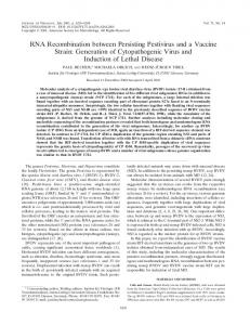
RESULTS Translation of unfractionated dsRNA. When suitably denatured, the dsRNA segments of the SA 11 genome stimulated the incorporation of [35S]methionine into trichloroacetic acidprecipitable material in a wheat germ cell-free protein synthesizing system. Figure 1 presents several parameters of this reaction. Addition of undenatured dsRNA up to 100 ,tg/ml did not inhibit endogenous activity (amino acid incorporation in the absence of any added mRNA) (9); however, no stimulation of [35S]methionine incorporation occurred either. Dimethyl sulfoxide (Me2SO) at the concentration used in this study (3.6%), did not inhibit endogenous activity (results not shown), in agreement with the findings of McCrae and Joklik (17).
Denaturation with 90% Me2SO did not require heating to 50°C, as was necessary for reovirus dsRNA (17). In fact, normal room temperatures were adequate, although equilibration to 30°C was chosen for convenience, because this was the temperature at which the wheat germ system was incubated. Once optimal concentrations of KCl and Mg were determined (39 and 1 mM, respectively) they were used throughout further experiments. An RNA concentration (80 ,tg/ml) sufficient to saturate the translation system was selected on the basis of preliminary studies (results not shown) for the optimization and time course experiments. Although high levels of incorporation were observed upon addition of rotavirus RNA, the specific stimulation of incorporation (i.e., the ratio of the incorporation in a system with added RNA to that in a system with no added RNA) was not as high as expected (maximum of about eightfold) due to significant endogenous incorporation by the particular wheat germ preparation used in this study. Polyacrylamide gel analysis of rotaviruscoded polypeptides. Since identification of
s0C
3}B
§m A 2125 IDD
2so
125
100
100
75
75
I I40
50
50
20
25
25
w 2
2 60 0
20
s0
60
40
100
1
2
3
4
0
20
40
60
M+ mM Time (inin) FIG. 1. KCI and Mg2+ concentration optima and time course of [35SJmethionine incorporation curves for the translation of denatured rotavirus dsRNA. Translation assays were as described in the text. The incubation time for the concentration optima curves was 60 min, after which 5-,ul samples were removed and the incorporation of [3SJmethionine into trichloroacetic acid-precipitable material was determined. The KCI concentration optimum was determined at a Mg2+ concentration of 1 mM (A), and the Mg2+ concentration optimum was determined at a KCI concentration at 39 mM (B). The time course incorporation curve (C) was performed at KCI and Mg2+ concentrations of 39 and 1 mM, respectively. For all three curves, the dsRNA concentration was 80 pg/ml, and the final Me2SO concentration was 3.6%. Symbols: 0, [35S]methionine incorporation in the presence of added denatured dsRNA; 0, [35S]methionine incorporation in the absence of added RNA, and in C this also represents the incorporation in the presence of undenatured rotavirus dsRNA (i.e., added to the translation mixture as an aqueous solution) at 50 or 100 pg/ml. Each of these three controls gave indistinguishable results. KCI
mM
GENE CODING ASSIGNMENTS OF ROTAVIRUS
VOL. 33, 1980
979
TABLE 1. Molecular weight estimates of SA 11 [35S]methionine-labeled translation products of rotavirus polypeptidesa denatured dsRNA was to be made on the basis first necessary it was of electrophoretic mobility, mol wt (x lo-3) Polypeptide to establish the migration patterns of rotavirus 130 II polypeptides in the SDS-polyacrylamide slab gel 93 12 system employed. 88 I3 This gel system gave good resolution of all 82 L4 62 known rotavirus polypeptides. Figure 2 shows O0 55 OIA typical patterns of SA 11 virion (structural) pro42 I5 teins, and also virus-specific proteins that appear 36 02 in [3S]methionine-labeled infected cells. No33 NS, menclature of these polypeptides is based upon 31 NS2 viral prowhich in Thouless (29), of the system 28 03 teins are designated by whether they are inner 26 04 (I) capsid structural, outer (0) capsid structural, a determined were estimates by Molecular weight or nonstructural (NO) proteins. They are numgel electrophoresis, using protein bered from highest to lowest molecular weight. polyacrylamide SA 11 virus possesses five inner (I, through 15) standards of known molecular weight. n 4Wu _= tnrougn ns anaA tour outer (Ul wJ4):I1 capsita poIypep- [Fig. 2]). A protein observed in SA 11-infected tides. In our modified systemrie14 and 15 are cells but not in pure virus was designated OIA. equivalent to I3 and 14, respectiively, in the sys- As first suggested by Thouless, this protein is tem of Thouless (29) (see Discussion). The num- possibly the precursor to however, since OlA ber and pattern of the proteins is a minor (see 2 and 4), the relaFig. product arations of purified virus are ess entiaryvthe same between these molecular two proteins will beofdifas described by Rodger et al. (2'2). In addition to tionship weights all structural proteins, virus-infeclted cells display ficult to elucidate. The two polypeptides not seen in piurified virus and SA 11 proteins are given in Table 1. A minor outer capsid protein, 03, was obthus considered nonstructural (NS1 and NS2 served only occasionally in virus-infected cells. This protein was also difficult to detect in [35S]methionine-labeled cells infected with other V V rotaviruses (29). However, it was always present in preparations of purified SA 11 virus (Fig. 2). S Virus infection of cells considerably reduced cell protein synthesis, allowing virus-spehost I,2 F~ . 1 lE cific including those-produced in small proteins, _ 14 ..... quantity, to be observed clearly (Fig. 2). Identification of the in vitro translation 01 oom _! products of unfractionated dsRNA. The translation products of denatured rotavirus G~A dsRNA were analyzed on polyacrylamide slab gels as described above, and the results are 4 15 4~t shown in Fig. 2. Products identical in mobility .,: to 11, I2, 13, I4, I5, and NS2 were observed. A *_ rA..,A-_-
O1;
IC UC
:
_05
_
*F'
.
.'
,.:M ";tK..
0:
I
E_
i_ __.
_
protein corresponding in electrophoretic mobil-
ity to NS, was only occasionally seen (not shown in Fig. 2). Proteins of equal mobility to the major (02) and minor (03 and 04) outer capsid proteins 03 WM were not produced. A major translation product (54,000 molecular weight) migrated slightly ahead of OIA. FIG. 2. SDS-polyacrylamide gel analysis of [35S]The fairly high endogenous incorporation of methionine-labeled, purified SA 1.I virus (V and V'); [3S]methionine in the wheat germ system provirus-infected (IC) and uninfected (UC) MAJO04 cells; near the gel front, and translation products of a wheat germ system duced densely labeled bands of low molecproducts translation made which without added RNA (-), or proga,rammed with unfractionated Me2SO-treated SA 11 dsRNA (+). V' is ular weight difficult to observe. Translation of individual species of dethe same as track Vexcept that it it a longer exposure to show the minor outer capsid pcglypeptides 03 and natured dsRNA. Total genomic dsRNA was 04. Analysis was on a 10% slab ge 1. fractionated by polyacrylamide gel electrophoS.
._
-
043
980
SMITH, LAZDINS, AND HOLMES
resis on a preparative scale as described in the text. A typical preparative scale gel is shown in Fig. 3. Not all RNA segments could be resolved, because segmnents 7 and 8 have identical mobilities in this gel system (20) and run together as a heavily staining band (Fig. 3). Bands were excised, and the RNA was eluted. The individual RNA segments (segments 1 to 6) were then denatured and translated as described in the text. Figure 4 shows the translation products of genome segments 1 to 4. The translation product of the denatured dsRNA segment 1 is identical in electrophoretic mobility to the virion structural polypeptide I,. Similarly, dsRNA segments 2, 3, and 4 produce labeled polypeptides corresponding to I2, I3, and L, respectively. Although there are endogenous bands in this region which migrate to positions close to the translation products of segments 2 and 4, they clearly do not interfere with the assignments. The translation of denatured dsRNA segment 5 produces a labeled protein of similar electrophoretic mobility to OIA (Fig. 5). However, whereas segment 5 appears to be translated very efficiently in vitro, 01A is quite difficult to observe in SA 11-infected cells. Genome segment 6 codes for the major inner capsid polypeptide 15. This segment appears to
J. VIROL.
0
1_
Si
S4
FIG. 4.
Wheat germ
cell-free protein synthesizing
system programmed with denatured dsRNA segments 1
through
4. Individual dsRNA segments
fr-om
were
re-
gel bands fr-om a preparative RNA gel and purified by organic solvent extraction and ethanol precipitation. After treatment with Me2SO they were added to a wheat germ translation system, and the [35S]methionine-labeled products were analyzed on a 10% SDS-polyacr-ylamide slab geL IC, SA 11 virus-infected MA1O4 cells labeled with [35S]methionine; 0, wheat-germ system with no covered
added
RNA.
excised
Translation
with dsRNA segment 1
(S3), and segment
UC
4
't,.
_
was
2
programmed
(S2),
segment
(S4).
S6
IC
...
system
(Si), segment
_.,
56-
S3
4~~~'
3
3 4-}
S2
S5
0
11
13-t 40= A
MO
,Z8-
9-
N06S, 02
10-
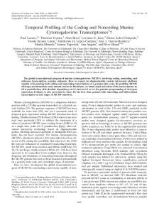
1I-FIG. 3. Preparative gel electrophoresis of SA 11 rotavirus dsRNA. RNA was extracted from purified virus and analyzed on 10%o SDS-polyacrylamide (Laemmli) slab gels. The gel was stained with ethidium bromide and photographed over a UV light box. The dsRNA segments are numbered in order of decreasing molecular weight.
4
FIG. 5. Wheat germ cell-free protein-synthesizing system programmed with denatured dsRNA segments 5 and 6. UC, Uninfected [36S]methionine-labeled MA104 cells; IC, SA 11 virus-infected, [35S]methionine-labeled MA104 cells; 0, no RNA added to the wheat-germ translation system; S5, denatured dsRNA segment 5 added; S6, denatured segment 6 added to the translation system. Products were analyzed on a 12%o slab gel.
VOL. 33, 1980
GENE CODING ASSIGNMENTS OF ROTAVIRUS
981
be translated efficiently both in vitro and in vivo, designated NCVP 1 by Matsuno and Mukoyama unlike segment 5. (16) may correspond to OIA in our system. In another recent study (5), translation of DISCUSSION transcription products of calf rotavirus appeared McCrae and Joklik (17) found that for the to yield only polypeptide I5 and possibly NS1 efficient translation of the denatured dsRNA and NS2. However, no comparison between lagenome of reovirus, a rapid shift in potassium beled translation products and virus-infected ion concentration, soon after the addition of cell proteins was done, so any lower-molecularRNA to their wheat germ system, was required. weight bands are of uncertain identity. Since infectivity and antigenic specificity of If this "salt jump" was not performed, none of the high-molecular-weight (lambda) polypep- rotaviruses depend on polypeptides of the outer tides were produced. This was not necessary for capsid (3, 30, 35), it is tantalizing that most of the translation of the rotavirus genome because the successful assignments so far are for inner all known high-molecular-weight proteins (I1 capsid proteins. From other studies in progress in this laboratory (I. Lazdins, unpublished data), through L) were formed. 02 is known to be a glycoprotein (22). No it appears probable that 04 is a glycoprotein, as product equal in mobility to this protein was well as 02, and in vitro translation of these may formed in vitro, which is consistent with other require variations in technique. These assignstudies (2, 4, 6) in which mRNA's of viral gly- ments, and the identification of the protein coproteins were used to program cell-free, pro- carrying type-specific antigenic determinants, tein-synthesizing systems. Often the unglyco- are of high priority to assist interpretation of sylated form of the viral protein was produced, epidemiological investigations based on electrowhich migrated significantly ahead of the gly- pherotypes (7, 11, 20; S. Rodger, manuscript in coprotein (i.e. of lower apparent molecular preparation). weight) on SDS-polyacrylamide gels. This posACKNOWLEDGMENIS sibility is currently under investigation in this We are very grateful to J. Phillips for helpful discussion on laboratory. in vitro translation and for supplying the wheat germ extract. This method of directly translating virion ge- We appreciate the advice of M. McCrae on the application of netic material clearly indicates primary gene his RNA denaturation technique to rotaviruses. We thank C. products, and clarifies the published data con- Adeney for technical assistance. This work was supported by the National Health and cerning viral polypeptides. Rodger et al. (22) Medical Research Council of Australia. M. Smith is the holder described four high-molecular-weight rotaviral of a Commonwealth Postgraduate Research award. polypeptides (e.g., p133, plO2, p99, and p92 for LITERATURE CITED SA 11), but others have observed only three (16, 18). Thouless (29) described an occasional split- 1. Bonner, W. M., and R. Laskey. 1974. A film detection method for tritium-labelled proteins and nucleic acid in ting of bands in the I2 or I3 region and suggested gels. Eur. J. Biochem. 46:83-88. this may be due to cleavage, similar to that of 2. polyacrylamide Both, G. W., S. A. Moyer, and A. K. Banerjee. 1975. reovirus (36). Our results demonstrate that there Translation and identification of the viral mRNA speare indeed four distinct high-molecular-weight cies isolated from subcellular fractions of vesicular stomatitis virus-infected cells. J. Virol. 15:1012-1019. polypeptides and that they are all primary gene J. C. 1978. Location of type-specific antigens in products. It is for this reason that we found it 3. Bridger, calf rotavirus. J. Clin. Microbiol. 8:625-628. necessary to change the designation of the major 4. Clegg, C., and I. Kennedy. 1975. Translation of semliki inner shell polypeptide from I4 (29) to 15. forest virus intracellular 26S RNA: characterization of the products synthesized in vitro. Eur. J. Biochem. 53: The assigmnent of dsRNA segment 5 to OlA 175-183. is quite firm, although the relationship between 5. Cohen, J., and P. Dobos. 1979. Cell free transcription and 01 has not been clearly established. OIA and translation of rotavirus RNA. Biochem. Biophys. Thouless (29) also showed a slight difference in Res. Commun. 88:791-796. migration between the 01 in infected cells and 6. Elder, K. T., J. M. Bye, J. J. Skehel, M. D. Waterfield, and A. E. Smith. 1979. In vitro synthesis, glycosylathe corresponding virus structural protein. Since and membrane insertion of influenza virus haetion, is produced in such small quantity in virusOlA magglutinin. Virology 95:343-350. infected cells, it will be difficult to compare it 7. Espejo, R. T., E. Calderon, N. Gonzalez, A. Salomon, A. Martuscelli, and P. Romero. 1979. Presence of two with the translation product of segment 5 and distinct types of rotavirus in infants and young children with 01 from virus particles, but the comparison hospitalized with acute gastroenteritis in Mexico City, (for example by limited proteolysis analysis) ap1977. J. Infect. Dis. 139:474-477. pears feasible. Unlike Matsuno and Mukoyama 8. Flewett, T. H., and G. N. Woode. 1978. The rotaviruses. Arch. Virol. 57:1-23. (16), neither Thouless (29) nor we were able to detect a polypeptide in infected cells which had 9. Grill, L. K., J. D. Sun, and J. Kandel. 1976. Effect of double stranded RNA on protein synthesis in an in the same mobility as the structural polypeptide vitro wheat germ embryo system. Biochem. Biophys. 01 of purified virus. We consider that the protein Res. Commun. 73:149-156.
982
SMITH, LAZDINS, AND HOLMES
10. Holmes, I. H. 1979. Viral gastroenteritis. Progr. Med. Virol. 25:1-36. 11. Kalica, A. R., M. M. Sereno, R. G. Wyatt, C. A. Mebus, R. M. Chanock, and A. Z. Kapikian. 1978. Comparison of human and animal rotavirus strains by gel electrophoresis of viral RNA. Virology 87:247-255. 12. Laemmli, U. K. 1970. Cleavage of structural proteins during the assembly of the head of bacteriophage T4. Nature (London) 227:680-685. 13. Laskey, R. A., and A. D. Mills. 1975. Quantitative film detection of 3H and "4C in polyacrylamide gels by fluorography. Eur. J. Biochem. 56:335-341. 14. Malherbe, H. H., and M. Strickland-Cholmley. 1967. Simian virus SAll and the related 0 agent. Arch. Gesamte Virusforsch. 22:235-245. 15. Matsuno, S., S. Inouye, and R. Kono. 1977. Plaque assay of neonatal calf diarrhea virus and the neutralizing antibody in human sera. J. Clin. Microbiol. 5:14. 16. Matauno, S., and A. Mukoyama. 1979. Polypeptides of bovine rotavirus. J. Gen. Virol. 43:309-316. 17. McCrae, M. A., and W. K. Joklik. 1978. The nature of the polypeptide encoded by each of the 10 doublestranded RNA segments of reovirus type 3. Virology 89:578-593. 18. Newman, J. F. F., F. Brown, J. C. Bridger, and G. N. Woode. 1975. Characterization of a rotavirus. Nature (London) 258:631-633. 19. Roberts, B. E., and B. M. Patterson. 1973. Efficient translation of tobacco mosaic virus RNA and rabbit globin as RNA in a cell-free system from commercial wheat germ. Proc. Natl. Acad. Sci. U.S.A. 70:2330-2334. 20. Rodger, S. M., and L. H. Holmes. 1979. Comparison of the genomes of simian, bovine and human rotaviruses by gel electrophoresis and detection of genomic variation among bovine isolates. J. Virol. 30:839-846. 21. Rodger, S. M., R. D. Schnagl, and L. H. Holmes. 1975. Biochemical and biophysical characteristics of diarrhea viruses of human and calf origin. J. Virol. 16:1229-1235. 22. Rodger, S. M., R. D. Schnagl, and L. H. Holmes. 1977. Further biochemical characterization, including the detection of surface glycoproteins, of human, calf, and simian rotaviruses. J. Virol. 24:91-98.
J. VIROL. 23. Shatkin, A. J. 1965. Inactivity of purified reovirus RNA as a template for E. coli polymerase in vitro. Proc. Natl. Acad. Sci. U.S.A. 54:1721-1728. 24. Shatkin, A. J., J. D. Sipe, and P. Loh. 1968. Separation of ten reovirus genome segments by polyacrylamide gel electrophoresis. J. Virol. 2:986-991. 25. Schnagl, R. D., and I. H. Holmes. 1976. Characteristics of the genome of human infantile enteritis virus (rotavirus). J. Virol. 19:267-270. 26. Schoub, B. D., G. Lecatsas, and 0. W. Prozesky. 1977. Antigenic relationship between human and simian rotaviruses. J. Med. Microbiol. 10:1-6. 27. Schuerch, A. R., W. R. Mitchell, and W. K. Joklik. 1975. Isolation of intact individual species of single and double-stranded RNA after fractionation by polyacrylamide gel electrophoresis. Anal. Biochem. 65:331-345. 28. Smith, E. M., M. K. Estes, D. Y. Graham, and C. P. Gerba. 1979. A plaque assay for the simian rotavirus SAl1. J. Gen. Virol. 43:513-519. 29. Thouless, M. E. 1979. Rotavirus polypeptides. J. Gen. Virol. 44:187-198. 30. Thouless, M. E., A. S. Bryden, and T. H. Flewett. 1978. Serotypes of human rotavirus. Lancet i:39. 31. Todd, D., and M. S. McNulty. 1976. Characterization of pig rotavirus RNA. J. Gen. Virol. 33:147-150. 32. Todd, D., and M. S. McNulty. 1977. Biochemical studies on a reovirus-like agent (rotavirus) from lambs. J. Virol. 21:1215-1218. 33. Verly, E., and J. Cohen. 1977. Demonstration of size variation of RNA segments between different isolates of calf rotavirus. J. Gen. Virol. 35:583-586. 34. Wyckoff, M., D. Rodbard, and A. Chrambach. 1977. Polyacrylamide gel electrophoresis in sodium dodecyl sulfate-containing buffers using multiphasic buffer systems: properties of the stack, valid Rf measurement, and optimized procedure. Anal. Biochem. 78:459-482. 35. Zi88i8, G., and J. P. Lambert. 1978. Different serotypes of human rotaviruses. Lancet i:38. 36. Zweerink, H. J., and W. K. Joklik. 1970. Studies on the intracellular synthesis of reovirus-specified proteins. Virology 41:501-518.