The FASEB Journal • FJ Express Full-Length Article
Coding region paraoxonase polymorphisms dictate accentuated neuronal reactions in chronic, sub-threshold pesticide exposure R. Orie Browne,* Liat Ben Moyal-Segal,† Dominik Zumsteg,‡ Yaron David,* Ora Kofman,§ Andrea Berger,§ Hermona Soreq,† and Alon Friedman*,1 *Departments of Physiology and Neurosurgery, Soroka University Medical Center, †The Department of Biological Chemistry, The Life Sciences Institute, The Hebrew University of Jerusalem, Jerusalem, Israel; ‡Krembil Neuroscience Centre, Toronto Western Hospital, University of Toronto, Toronto, Canada; and §Department of Behavioral Sciences, Zlotowski Center for Neurosciences, Ben-Gurion University of the Negev, Beersheva, Israel Organophosphate pesticides (OPs), known inhibitors of acetylcholinesterase (AChE), are used extensively throughout the world. Recent studies have focused on the ACHE/PON1 locus as a determinant of inherited susceptibility to environmental OP exposure. To explore the relationship of the corresponding geneenvironment interactions with brain activity, we integrated neurophysiologic, neuropsychological, biochemical, and genetic methods. Importantly, we found that subthreshold OP exposure leads to discernible physiological consequences that are significantly influenced by inherited factors. Cortical EEG analyses by LORETA revealed significantly decreased theta activity in the hippocampus, parahippocampal regions, and the cingulate cortex, as well as increased beta activity in the prefrontal cortex of exposed individuals—areas known to play a role in cholinergic-associated cognitive functions. Through neuropsychological testing, we identified an appreciable deficit in the visual recall in exposed individuals. Other neuropsychological tests revealed no significant differences between exposed and non-exposed individuals, attesting to the specificity of our findings. Biochemical analyses of blood samples revealed increases in paraoxonase and arylesterase activities and reduced serum acetylcholinesterase activity in chronically exposed individuals. Notably, specific paraoxonase genotypes were found to be associated with these exposure-related changes in blood enzyme activities and abnormal EEG patterns. Thus, geneenvironment interactions involving the ACHE/PON1 locus may be causally involved in determining the physiological response to OP exposure.—Browne, R. O., Ben Moyal-Segal, L., Zumsteg, D., David, Y., Kofman, O., Berger, A., Soreq, H., Friedman, A. Coding region paraoxonase polymorphisms dictate accentuated neuronal reactions in chronic, sub-threshold pesticide exposure. FASEB J. 20, E1103–E1113 (2006)
ABSTRACT
Organophosphate compounds (ops) are commonly used as agricultural pesticides and household insecticides throughout the world. Despite their common use and effectiveness in eradicating a wide range of harmful agricultural pests, their use poses a serious health hazard for humans. Surprisingly, their toxic properties have been known since the 1930s (1), and extensive research has verified that they act as inhibitors of the acetylcholine (ACh) hydrolyzing enzyme acetylcholinesterase (AChE) (2). Notably, acute poisoning by OPs leads to accumulation of ACh at cholinergic synapses in the peripheral and central nervous systems, potentially causing nausea, excessive salivation, incontinence, bradycardia, headache, fatigue, seizure, coma, and death (3). Nevertheless, although the short-term effects of acute OP poisoning are understood to a great extent, the long-term consequences of acute poisoning and chronic, subthreshold exposure are still not clear. Humans are capable of mounting a variety of responses to OP exposure. AChE and butyrylcholinesterase (BChE) in the circulation act as scavengers by irreversibly binding and consequentially inactivating the OP anticholinesterases (anti-AChEs) at their active sites. In experimental animals, the accumulation of ACh at cholinergic synapses in the brain and muscle following exposure was shown to initiate up-regulation of AChE mRNA, and protein (4). This protective response can be diminished as a consequence of a deletion in the AChE promoter, which induces constitutive overproduction of AChE, most likely exhausting the capacity for subsequent secondary overproduction in a reaction to exposure (5, 6). The paraoxonase gene (PON1), which maps close (5.5 Mb) to ACHE on the long arm of chromosome 7, codes for paraoxonase (PON), a plasma enzyme that can hydrolyze OPs. PON also possesses arylesterase and lactonase activity (7, 8) 1
Key Words: acetylcholinesterase 䡠 electroencephalography 䡠 LORETA 䡠 organophosphates 䡠 subthreshold exposure 0892-6638/06/0020-1103 © FASEB
Correspondence: Department of Physiology, Faculty for Health Sciences, Ben-Gurion University, Beer-Sheva 84105, Israel. E-mail:
[email protected] doi: 10.1096/fj.05-5576fje E1103
and is involved in inhibiting the oxidation of lowdensity lipoproteins (LDL) (9). PON1 is known to protect against OP exposure, as affirmed by the sensitivity of PON1-null mice to OP intoxication (10). However, the mechanism of this protection has not yet been fully elucidated. A variety of polymorphisms in the PON1 gene have been characterized (11). The PON1–108C/T promoter polymorphism affects enzyme levels by modulating expression. Another polymorphism affecting expression is the L55M substitution, which results in reduced PON enzyme levels (12). Substrate specificity is influenced by the Q192R substitution (13). Several studies have reported restlessness, forgetfulness, and other neuropsychiatric symptoms as common complaints in exposed human populations (14 –19). However, few significant changes in cognitive function have been detected by neuropsychological testing in populations exposed to low levels of OPs without any history of acute poisoning (17, 19 –23). Several factors may account for the apparent differences reported in these studies. It is not clear whether the concentration of exposure (which can be difficult to determine), the duration, type of exposure, or instead, individually inherited sensitivity is the most significant factor associated with exposure-related neuropsychological deficits. Variations in methods may also account for the apparent disparities in the literature. Furthermore, no specific phenomenological or mechanistic data exist regarding such changes. Despite the differing reports, accumulating data suggest that the function of the brain may be appreciably affected following exposure to OPs. These changes are often subtle and may be associated with genetic or environmental factors that have not always been considered in previous studies. Inherited, experiential, and environmental factors are all presumed to influence higher brain functions in humans. However, quantitative evaluation of each of these contributions is very difficult. We, therefore, combined biochemical, genetic, neurophysiologic, and neuropsychological methods in an effort to reveal the relation between environmental exposure and inherited sensitivity to OPs in an exposed population. Our data suggest the existence of a new underlying mechanism that explains the interactions between environmental and genetic factors in brain malfunction following low-concentration OP exposure.
MATERIALS AND METHODS Participants The local institutional review board approved the use of human subjects for this study. The study was conducted using volunteers from a rural, agricultural community where organophosphate pesticides (e.g., fenitrothion, chlorpyrifos, monocrotophos, ethion, and azinphos-methyl) are routinely used. Living in the community for an average of 26 yr, the residents are located 25–150 m from the fields. The exposed population consisted of 291 individuals with the following E1104
Vol. 20
August 2006
demographic profile: 149 males, 142 females, ages ranging from 4 to 90 yr (mean 36.5 yr, sd 21.7); 65 (22.3%) are agricultural workers. Twenty-eight individuals (9.6%) were excluded from the study because of a history of central nervous system (CNS)-related diseases (as obtained in medical questionnaire or medical records: meningioma, stroke, migraine, vertigo, anxiety, depression, attention deficit disorder, and schizophrenia). Sixty individuals gave written consent to participate in the study after reading an extensive consent form. The study participants had no history of acute pesticide intoxication. All control subjects for this study came from urban areas with no history of exposure to anticholinergic agents and had no known neurological disease or psychiatric problems. Control data for the biochemical analyses (n⫽91) were compiled in the same lab and with the same techniques as those in the present study. Control subjects for the EEG analysis (n⫽9) had an age and gender distribution similar to the exposed participants (exposed mean age: 46, control mean age: 42, exposed 58% male, 42% female, control 56% male, 44% female). Controls for the neuropsychological tests (n⫽24) were age (⫾5 yr), sex, and education matched. Airborne spread of pesticides The airborne spread of pesticides was evaluated using the Agri-Screen Ticket (Neogen, Lansing, MI, USA), which screens for the presence of cholinesterase inhibitors using a litmus-type disc impregnanted with cholinesterase enzyme. Inhibition of the enzyme results in a color change on testing. The test is sensitive to low parts per billion to ⬃6 parts per million. One test disc was placed in the field (positive control), and 5 more were placed at 20-meter intervals from the field prior to the time of spraying by a tractor-drawn sprayer. A negative control was placed inside a closed automobile at the same location. Test discs were collected after 12 h of exposure and tested for the presence of cholinesterase inhibitors. All discs except for the negative control tested positive. Blood samples Blood samples were drawn to BD Vacutainer® blood collection tubes (Becton-Dickinson, Franklin Lakes, NJ, USA) with citrate as an anticoagulant, centrifuged (1300 rcf, 4°C, 15 min) in a desktop centrifuge, and plasma was obtained. Whole blood and plasma were maintained at –70°C until use. Enzyme activity measurements Plasma paraoxonase activity was determined by adapting the spectrophotometric method (24) to a microtiter plate assay. Since several variations of the assay were published, the assay was calibrated for plasma dilution and substrate concentration. We found that a 1:10 dilution of plasma and a 1.2 mM paraoxon concentration were optimal, yielding high variability as reported for paraoxonase activity. Higher substrate concentrations (up to 6 mM) enabled higher hydrolysis rates but yielded lower variability, thus obscuring the population trends. Briefly, 10 l of plasma diluted 1:10 was placed in microtiter plate wells (Nunc, Roskilde, Denmark) in triplicate; the reaction was initiated by adding 190 l of the substrate, 1.2 mM paraoxon (Sigma, Jerusalem, Israel), in 0.26 mM Tris–HCl, pH 8.5, 25 mM CaCl2, and 0.5 M NaCl. Readings at 405 nm were repeated at minimal intervals for 10 min. Nonenzymatic breakdown of paraoxon was subtracted from the total rate of hydrolysis. Enzyme activity was calculated using the e405 for p-nitrophenol, 17 100 M/cm.
The FASEB Journal
BROWNE ET AL.
Plasma arylesterase activity was measured in 10 l of 1:40 diluted plasma mixed with 190 l of substrate (3.26 mM phenylacetate in 9 mM Tris–HCl, pH 8 and 0.9 mM CaCl2). Hydrolysis rates were determined at minimal intervals in UV-transparent 96-well plates (Greiner-Bio One GmbH, Frickenhausen, Germany), at 270 nm for 4 min. Enzyme activity was calculated using the e270 for phenol, 1310 M/cm. Plasma cholinesterase catalytic activity measurements involved adaptation of a spectrophotometric method (25) to a microtiter plate assay. ATCh (Sigma, 1 mM) or butyrylthiocholine (BTCh, Sigma, 10 mM) hydrolysis rates were measured following 20 min preincubation with 5 ⫻ 10⫺5 M iso-OMPA (tetraisopropylpyrophosphoramide) (Sigma), a specific BChE inhibitor, or 10⫺5 M 1,5-bis(4-allyldimethylammoniumphenyl)pentan-3-one dibromide (BW284C51, Sigma), a specific AChE inhibitor. Readings at 405 nm were repeated at 2-min intervals for 20 min. Nonenzymatic breakdown of substrate was subtracted from the total rate of hydrolysis. Enzyme activities were calculated using the e405 for 5-thio-2nitrobenzoate, 13 600 M/cm. Genotyping Genomic DNA was prepared from blood cells using the Gentra Whole Blood DNA Extraction Kit (Gentra, Minneapolis, MN, USA). Genotyping involved polymerase chain reaction (PCR) amplification of the corresponding gene regions, using Taq polymerase (Sigma) followed by agarose gel electrophoresis and Exo-Sap enzymatic purification (USB, Cleveland, OH, USA) of the PCR product. Standard automated sequencing utilized the BigDye Terminator cycle sequencing chemistry, ABI 3700 DNA Analyser and Data collection and Sequence Analysis software (Applied Biosystems, Foster City, CA, USA). PON1 Q192R and PON1 L55M polymorphisms were detected using the single nucleotide primer extension method (SNaPshot ddNTP Primer Extension kit, Applied Biosystems). Following PCR amplification and purification, the SNaPshot reaction was performed using primer 5⬘-GGCAGAAACTGG CTCTGAAGAC-3⬘ for the PON1 55 and 5⬘-GATCACTATTTTCTTGACCCCTACTTAC-3⬘ for PON1 192. Following extension and calf intestine phosphatase treatment (Amersham Biosciences, Freiburg, Germany), the products were subjected to electrophoresis on a 3700 ABI analyser and the results analyzed using Genescan software (ABI, Tel Aviv, Israel). Electroencephalography EEG was recorded from subjects using a clinical EEG unit (CEEGRAPH IV, Biological Systems Corp., Mundelein, IL, USA) with a sampling rate of 256 Hz (26,27). Twenty-five conventional Ag/AgCl surface electrodes were placed according to the international 10 –20 system, with additional electrodes for the detection of eye-movements and electrocardiogram activity. At least 25 suitable EEG segments (free of movement and eye artifacts, subjects with eyes closed) of 2 s duration were identified off-line by visual inspection for each subject. Power spectra (1–30 Hz, resolution of 0.5 Hz) were calculated using Fourier transform. The spectral power for discrete frequency bands (delta: 1.5– 6 Hz, theta: 6.5– 8 Hz, alpha 1: 8.5–10 Hz, alpha 2: 10.5–12 Hz, beta 1: 12.5–18 Hz, beta 2:18.5–21 Hz, beta 3: 21.5–30 Hz) was calculated for each subject. We also calculated the population averaged power of each electrode. Spectral power distribution maps were created using MATLAB (version 7.0). Source localization Low resolution brain electromagnetic tomography (LORETA) was used for the localization of presumptive cortical sources PARAOXONASE POLYMORPHISMS AND PESTICIDE EXPOSURE
of EEG activity (28). LORETA is based on the neurophysiologic assumption that neighboring cortical areas are likely to be coactivated and permits three-dimensional tomography of brain electrical data while requiring only simple constraints (smoothness of the solution) and no predetermined knowledge about the putative number of discernible source regions (29 –32). LORETA estimates the distribution of absolute current density (numerically, and ultimately visually using scaled color intensity) for brain electrical activity and displays it on a dense grid of 2,394 voxels based on the digitized Talairach Atlas, as provided by the Brain Imaging Centre, Montreal Neurological Institute (33). Calculation of LORETA is limited to cortical gray matter and hippocampi and has a spatial resolution of ⬃1–2 cm. The average current source densities for 23 distinct brain regions were calculated by averaging the LORETA values for each voxel within an anatomically defined region (34). These cortical regions were created by allocating the raw LORETA values of individual voxels to their corresponding Brodmann areas (BA) or cerebral gyri, based on the coordinates of the digitized Talairach Atlas, and subsequent grouping of these BAs into 23 arbitrary “functional” cortical regions (e.g., left mesial temporal, right lateral temporal, etc.). Neuropsychological tests We used a battery of neuropsychological tests related to attention, memory and executive functions, which included the Tower of Hanoi, a Serial Reaction Time (SRT), a Continuous Performance Test (CPT) in which digits from 1 to 9 were presented at a rate of 1/sec and the subject had to detect when the digit 9 followed the digit 1, the Digit Span (from the Wechsler Adult Intelligence Scale), a visual reproduction subtest of the Wechsler Memory Scale, and a long-term verbal memory from a Hebrew version of the RALT (Rey Auditory Learning Test). The two first tasks were computerized, and the others were paper and pencil. Details of the methods can be found in Lezak (35). Data analysis and statistics Student’s t test was used to calculate statistical significance for the differences among AChE, BChE, arylesterase, and paraoxonase activities, as well as differences in the area under the curve for each discrete frequency band of the EEG. Statistically significant differences between LORETA values were tested by using a nonparametric t test, on a voxel-by-voxel basis, with correction for repeated measure (36). We used three different thresholds for P values to determine statistical significance (P⬍0.01, P⬍0.05, and P⬍0.1).
RESULTS Neurophysiologic and neuropsychological assessment To determine the effect of exposure on brain activity, we performed EEG in 19 exposed and 9 control subjects with a similar age and gender distribution. Calculation of the averaged spectral power revealed a generally lower power in the exposed group, which reached statistical significant only in the theta band (ANOVA, P⫽0.03). When individual power was normalized, significantly lower power was found in the theta band and increased power in the beta 3 band (P⫽0.04 and E1105
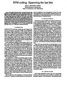
Figure 1. EEG analyses in OP-exposed group individuals show decreased power in the theta and increased power in the beta bands in specific brain regions: A) Average normalized power spectrum (1.5–30 Hz) of exposed (n⫽19) and non-exposed (n⫽9) groups. B) Area under the curve (AUC) of average normalized power by discrete frequency bands (delta: 1.5– 6 Hz, theta: 6.5– 8 Hz, alpha1: 8.5–10 Hz, alpha2: 10.5–12 Hz, beta1: 12.5–18 Hz, beta2: 18.5–21 Hz, beta3: 21.5–30 Hz) in exposed and non-exposed groups. In the exposed group, theta activity was significantly decreased (P⫽0.04) and beta3 activity was significantly increased (P⫽0.03). C–D) Percentage of increase in a normalized spectral power at the theta and beta3 bands is shown as a spectral power distribution map. Note the electrode-specific changes of spectral power. E–F) LORETA analyses showing cortical regions with statistically significant differences between exposed and non-exposed groups. E) Areas with significantly decreased current intensity (blue, P⬍0.05) in the theta band included bilaterally the amygdala, the hippocampus, the subcallosal area, the parahippocampal gyrus, and the anterior and posterior cingulate gyrus (Brodmann Areas 25, 30, 31, 34, 36, 37). F) Areas with significantly decreased (blue, P⬍0.05) current density in the beta 3 band included the hippocampus and parahippocampal gyrus bilaterally; increased sources (red, P⬍0.05) were found in the inferior, middle, and superior frontal gyri (areas 10, 11, 27, 46).
P⫽0.03, respectively) of exposed, compared with nonexposed groups (Fig. 1A–B). Topographically, the decrease of theta power was most prominent over the central region, whereas the increase of beta 3 power showed a more frontal distribution (Fig. 1C–D). These differences in surface spectral distribution are directly related to region-specific changes of their underlying cerebral sources, as evidenced by LORETA. Statistical nonparametric mapping of LORETA revealed significant differences in the localization of activity in all seven frequency bands of the exposed subjects, compared with controls. Most importantly, the exposed subjects exhibited markedly decreased theta activity in limbic cortical areas of both hemispheres, including mesial temporal structures, subcallosal areas, and posterior cingulate gyri. Cortical areas with significantly increased delta and beta3 activity were found in bilateral prefrontal areas, and alpha1 and alpha2 bands were localized to the occipital region (Fig. 1E–F and data not shown). To reveal the possible neuropsychological significance of the neurophysiologic findings, we conducted neuropsychological tests and compared the results for 23 of the exposed subjects and 23 controls, which were matched for age, sex, and education, using a t test for dependent measures. The only test that showed significant differences between the exposed and non-exposed groups was the delayed memory (20 min) portion of the visual reproduction subtest of the Wechsler E1106
Vol. 20
August 2006
Memory Scale (Fig. 2). An ANOVA for the effect of the group on the percentile score for immediate and delayed memory revealed a marginally significant omnibus effect for the group, F (1, 44) ⫽ 3.82, P ⫽ 0.056.
Figure 2. Neuropsychological tests revealed significantly decreased scores in delay memory: Exposed individuals showed decreased scores in the immediate and delayed memory portion of the visual reproduction subtest of the Wechsler Memory Scale. Only the delayed (20 min) portion, however, showed a statistically significant difference between the groups (see text for details).
The FASEB Journal
BROWNE ET AL.
Planned comparisons between groups for the immediate and delayed tests indicated that the exposed group was significantly impaired in the delayed memory task, F (1, 44) ⫽ 4.86, P ⬍ 0.05, but not for the immediate memory task, F (1,44) ⫽ 1.78, n.s. The mean and sd of the percentile scores for the exposed group were 25.04 ⫹ 12.6 for delayed recall and 28.56 ⫹ 17.6 for immediate recall. The matched control group had percentile scores of 37.43 ⫹ 23.7 and 36.13 ⫹ 20.7 for the delayed and immediate recall, respectively. To challenge our hypothesis that changes in current sources in mesial temporal lobe structures is associated with poor memory performance we analyzed separately mean current density in both right and left temporal lobe (medial part) for exposed individuals with low and high performance. Exposed individuals who performed poorly on memory tests (13.3 and 16% for immediate and delayed recall, respectively) also showed 10 times lower beta activity and 50% increase in theta compared to exposed individuals who showed better performance (32 and 38% for immediate and delayed recall, respectively). However due to the small sample size (n⫽6), these changes did not reach statistical significance (see Discussion). TABLE 1.
Exposure-related alterations in biochemical activity Biochemical assays for AChE, BChE, PON, and arylesterase activity were performed for 30 exposed subjects (see Table 1). Serum AChE activity was found to be significantly lower in exposed individuals compared with controls, compatible with the hypothesis of irreversible inhibition (36) (41% of control, P⬍0.001, Student’s t test). There was no significant difference in BChE activity. Both PON and arylesterase activity were significantly higher than predicted in the exposed subjects (447 and 441% of control, respectively, P ⬍ 0.001) (Fig 3A). Enzyme activities were not significantly different for agricultural workers (n⫽9) when compared to the local population (n⫽20) (Table 1). Alterations in biochemical activity reflect, besides exposure levels, also the individual’s reaction to the chemical stress, as manifested by modified gene expression (37). This, in turn, represents a composite effect of inheritance and environmental status (i.e., exposure). Therefore, as a follow-up to the biochemical investigation, we searched for genetic variation. Allele frequency in our study population was similar to that of the general Israeli population for PON1 55, PON1 192, and ACHE17130 ⌬HNF3. There was a higher frequency of the
Genetic and biochemical summary of exposed population
Subject
Age
1 2 3 4 5 6 7 8 9 10 11 12 13 14 15 16 17 18 19 20 21 22 23 24 25 26 27 28 29 30 Mean
44 48 51 53 57 47 49 31 26 51 31 46 60 43 53 20 50 16 47 51 70 52 42 46 54 40 60 62 28 53 46
Agricultural worker
yes
yes
yes yes yes yes
yes yes yes yes
PON -108
PON L55M
PON Q192R
TT CT CT CT CC CC TT CC CC TT CC CC CC CC CC CC CT CC CC CC CC CT TT CC CT CT CT CC CC TT
LL LM LM LL LL LM LL LL LL LL MM MM LL LL LM LL LL LL LL MM MM LL MM LM LL LM MM LL LM LL
QQ QQ QQ RR QQ QQ RR QQ QR QR QQ QQ QQ QR QR RR QQ QR QR QQ QQ QQ QQ QQ QQ QQ QQ QR QR QQ Mean SE
AChE Specific Activity (nmol/ min*ml)
BChE- Specific Activity (nmol/ min*ml)
PON-Specific Activity (nmol/ min*ml)
Arylesterase- Specific Activity (mol/min*ml)
101.97 137.22 160.87 168.87 171.97 172.70 177.18 181.89 187.16 189.58 190.66 195.21 195.79 195.93 196.80 196.87 198.33 213.55 214.58 216.88 217.79 221.26 226.53 230.46 232.28 238.62 252.54 274.06 279.39 201.27 6.92924
2231.17 3983.90 3763.94 4371.72 4528.69 4043.53 4915.15 4342.50 5077.09 4410.14 4665.69 5815.05 5901.26 4753.25 4338.76 4674.92 5847.56 4984.36 6387.74 4853.65 4607.45 6168.41 2109.98 3256.73 6336.29 6191.35 6602.36 6980.74 5098.75 4870.42 220.66556
189.66 108.89 132.19 517.65 150.83 125.63 477.95 224.60 466.97 425.09 105.19 90.00 188.39 210.81 229.04 545.46 119.79 478.24 496.75 111.87 211.56 187.32 144.96 101.23 197.59 157.57 165.83 452.71 312.63 252.64 28.40421
112.60 74.49 79.23 64.86 94.03 97.93 62.67 117.46 107.75 69.16 66.95 101.93 128.98 58.03 59.18 81.70 96.74 76.04 64.35 96.58 72.31 80.98 103.53 105.30 117.53 111.21 88.52 4.1075
PARAOXONASE POLYMORPHISMS AND PESTICIDE EXPOSURE
E1107
Figure 3. Biochemical analyses in OP-exposed individuals: A) Shown are the activities of AChE (nmol/min*ml) in exposed subjects (n⫽29) compared to the Israeli non-exposed population (n⫽91) (P⬍0.001); BChE (mmol*10/min*ml) (not significantly different); PON (nmol/min*ml) and arylesterase (mol/min*ml) (P⬍0.001). B) AChE and PON activities segregated by the PON1 55/192 genotype combinations: LLRR (n⫽3), LLQR (n⫽6), LLQQ (n⫽8), LMQR (n⫽2), LMQQ (n⫽5), MMQQ (n⫽6). In all graphs: open bars represent control, non-exposed group; filled bars, exposed group.
PON1 -108 C allele in our study than in the general Israeli population (0.70 and 0.40, respectively) (Table 2). Biochemical analysis by PON1 55, 192, and –108 genotypes revealed several significant trends. In general, AChE activity did not vary by PON1 genotype except for a slight decrease in LLRR compared with MMQQ individuals (180.97⫾8.30041 and 216.6⫾9.16877 nmole/min⫻ml, respectively, P⫽0.04) (Fig. 3B). In contrast, different PON1 genotypes correlated with significant alterations in paraoxonase activity. The presence of the PON1 L55M substitution (TTG to ATG) was consistent with a decrease in PON activity, presumably related to reduced PON1 mRNA and protein levels (12). We controlled for the effects on PON activity associated with the PON1 Q192R substitution by selecting individuals without the R allele. Interestingly, in the non-exposed population, MMQQ individuals (n⫽10) exhibited about one-third of the PON activity of LLQQ individuals (n⫽14) (MMQQ/LLQQ⫽0.36, P⬍0.0001). In contrast, in exposed subjects, MMQQ (n⫽6) and LLQQ (n⫽7) individuals displayed almost E1108
Vol. 20
August 2006
equal PON activity (MMQQ/LLQQ⫽1.3), suggesting that the M allele is associated with a greater increase in PON activity in exposed individuals. This is supported by our data that show 8.1 times more PON activity in exposed MM individuals than in controls but only 3.8 times more activity in exposed LL individuals than in controls. PON enzyme levels can be assessed by measuring the arylesterase activity (38). Indeed, arylesterase activity in individuals with the M allele was 6.0 times greater in exposed individuals than in non-exposed controls, whereas arylesterase activity in the exposed population without the M allele was only 3.5 times greater than in non-exposed individuals, consistent with the hypothesis of a more robust response to exposure in exposed individuals with the M allele. The PON1 Q192R (CAA to CGA) substitution, which affects catalytic efficiency (13), was associated with increased PON activity. In controls there was a genedependent increase in PON activity correlated with the presence of the R allele (PON-specific activity (nmol/ min*ml) QQ⫽29.6, QR⫽71.3, RR⫽99.9, slope⫽35.18, R2⫽0.9886) (Fig. 3B). Interestingly, changes in PON activity in exposed subjects were characterized by a much more robust change associated with the R allele (PON-specific activity (nmol/min*ml) QQ⫽150.7, QR⫽384.0, RR⫽513.7, slope⫽181.48, R2⫽0.9735). To control for the effects on PON activity contributed by the PON L55M substitution, we analyzed PON activity in individuals who lacked the M allele. Indeed, The R allele-associated changes in PON activity remained (Fig. 3B). In addition, we were able to identify an important difference between exposed and non-exposed groups. Non-exposed LLRR individuals (n⫽13) showed ⬃2 times more PON activity than non-exposed LLQQ individuals (n⫽10) (LLRR/LLQQ⫽2.09, P⬍0.0001), whereas in exposed subjects LLRR individuals (n⫽3) showed almost 3 times more PON activity than LLQQ individuals (n⫽7) (LLRR/LLQQ⫽2.86, P⬍0.0001), suggesting an exposure-dependent response associated with the PON1 Q192R substitution and a potential biosensor effect of the R allele in anti-AChE exposure. TABLE 2. PON1 and AChE allele frequency in this study and the general Israeli population Position, allele
PON1 –108 C T 162 (55) T (L) A (M) 575 (192) A (Q) G (R) ACHE -17130 ⌬HNF3 n a
This study
Israeli populationa
0.70 0.30
0.40 0.60
0.68 0.32
0.61 0.39
0.77 0.23
0.67 0.33
0.017 30
0.019 157
Bryk et al., 2005
The FASEB Journal
BROWNE ET AL.
Neurophysiologic alterations depend on PON genotype Our biochemical data revealed significant differences in the individual responses to exposure, associated with the presence or absence of the M (L55M) and R (Q192R) alleles in the PON1 gene. Thus, we hypothesized that the increase in beta3 activity observed in the EEG of exposed individuals may also differ among exposed individuals with different genetic profiles. We found significantly increased beta3 activity in the frontal cortical regions (decreased in temporal regions) of exposed individuals with the R allele compared with controls and with exposed individuals without the R allele. Interestingly, the M allele had no such effect (Fig. 4A). Comparing the LORETA values (representing the current source densities) in these brain regions revealed that exposed individuals with the R allele exhibited significantly increased frontal activity and
decreased temporal activity (Fig. 4B, P⬍0.03). Furthermore, non-exposed and exposed individuals without the R allele were more similar regarding the pattern of beta3 source activity and intensity.
DISCUSSION Our results support the following conclusions regarding the results of chronic subthreshold exposure to OPs: 1) The exposure leads to significant changes in the distribution of sources of brain activity in specific regions (mainly in limbic structures and frontal cortices); 2) These changes are associated with deficits in visual retention memory in exposed individuals; 3) No significant alterations were found in the biochemical activities of serum AChE and PON; and 4) Both biochemical and neurophysiologic changes are dependent on the relevant genetic makeup of an individual. Exposure to organophosphates leads to changes in brain activity
Figure 4. Enhanced beta activity in carriers of the R allele: A) LORETA values (i.e., current density) were calculated for each brain region in the beta3 frequency band and the % change compared to the control population is given. Robust changes were found only for exposed individuals with the R allele and in specific brain regions (Left temporal and Right frontal). B) Normalized current density values (arbitrary units) of beta3 activity in selected brain regions for nonexposed controls (white bars), exposed without the R allele (gray bars, n⫽14), and exposed with the R allele present (dark gray, n⫽8). Note that significant differences were found only for the exposed individuals carrying the R allele. PARAOXONASE POLYMORPHISMS AND PESTICIDE EXPOSURE
A previous study by Burchfiel and Duffy identified statistically significant increases in beta activity in individuals exposed to the AChE inhibitor, sarin (39). Our results, despite the small sample size, are consistent with these findings and add the observation of significant decreased theta activity in exposed individuals. The relevance of these changes is difficult to determine without more detailed information about the sources of the brain activity. This is especially true since the differences in power were not equally distributed between electrodes, which clearly suggest the involvement of specific brain regions (Fig. 1C–D). Using statistical analyses of LORETA current density distributions for the determination of OP-exposure related changes in cortical activity, we found markedly decreased theta activity in bilateral limbic structures (including the amygdala, the hippocampus, the parahippocampal regions, the subcallosal area and the cingulate cortex), and significantly increased beta3 activity in bilateral prefrontal cortical areas in the exposed group. These findings are consistent with research detailing the functional roles of these brain regions and the potential neuropsychological effects of OP exposure (see below). Notably, all of these regions are known to receive significant cholinergic input originating from the magnocellular basal forebrain cholinergic system (40, 41). This cholinergic system can be subdivided into a number of distinct cholinergic sites, including the nucleus basalis of Meynert, the medial septal nucleus, and the vertical and horizontal limb nuclei of the diagonal band of Broca. Efferents originating in the basal forebrain project diffusely throughout the cortex as well as in the hippocampus, amygdala, and olfactory bulb. Presumably, areas of the brain with significant cholinergic input may be particularly susceptible to OP exposure. The hippocampus and parahippocampal region were localized by LORETA as the sites of signifiE1109
cant differences in theta activity between exposed and control subjects. The theta frequency is extensively studied in the hippocampus and has been recognized in other areas of the limbic region as well (42, 43). Numerous reports have elucidated the role of cholinergic input in this region from the medial septum and diagonal band of Broca in generating the theta rhythm (42, 44 – 46), which is thought to be important in mnemonic function (43, 46, 47). Thus, the reduced theta rhythms in mesial temporal regions of chronically exposed individuals suggest disturbed cholinergic functions at rest. The observed changes in the beta activity of the prefrontal cortex may be associated with increased demands on attention and other cognitive functions following OP exposure. Several studies described the role of the cholinergic system in attentional processing (48 –55) and reported increased neuronal activity and ACh efflux in the prefrontal cortex of rats performing tasks that increase demands on attention (56 –58). Neuropsychological tests Of the neuropsychological tests performed in this study, the only one that showed a significant difference between the exposed and non-exposed groups was the delayed memory portion of the visual reproduction test. This outcome is consistent with the results of neuropsychological testing in other studies, where few, if any, detectable deficits in cognitive function were identified in individuals exposed to low levels of organophosphates (17, 19 –23). Interestingly, some correlation was found between decreased hippocampal theta and increased beta activity and poor performance in memory recall performance. While these strengthen the potential of source localization of resting EEG activity, future studies with increased sample size are awaited to confirm these results. No correlation was found between scores on the visual recall test and genotype or enzyme activities. The small sample size and the relative infrequency of specific genotypes in the present study make clear discrimination difficult. Future studies should seek to confirm genotypic variations in the OP susceptibility alluded to in this study. Alterations in biochemical activity On OP exposure, PON acts in a protective role by hydrolyzing OPs, thus preventing inhibition of AChE. In addition, PON exhibits peroxidase activity that protects AChE from oxidative damage (59 – 61). A reduction in PON levels or a functional defect is expected to leave AChE particularly susceptible to anti-AChE exposure. In the brains of experimental animals, it was shown that on AChE inhibition, there is a subsequent increase in ACh available to ACh receptors, thus activating a feedback response that results in overproduction of AChE while suppressing choline acetyltransferase (ChAT) and the vesicular ACh transporter (4). This feedback response may bring cholinergic activity E1110
Vol. 20
August 2006
into balance for the short term but may also be associated with cognitive deterioration in the long term as animal experiments suggest (62). Here we performed biochemical assays for AChE, BChE, PON, and arylesterase serum activity in 30 exposed subjects. Our results suggest a lasting reduction in serum AChE activity (41% of control) in exposed individuals, with a concomitant increase in PON (447%) and arylesterase (441%) activity. Previous studies reported an age-dependent increase in AChE activity (63, 64). Given that the average age of the exposed subjects in the present study was higher than our control population (46⫾0.95 vs. 34⫾2.28), this model would predict increased AChE activity in the exposed population. The fact that this was not the case supports the notion of persistent inhibition. The decrease of AChE activity and the increase of PON activity in exposed individuals could potentially reflect a dosedependent effect of exposure. In such a case we would expect a direct association between the extent of reduction in AChE activity and an increase in PON activity in exposed subjects. But this hypothesis was not supported by the data. An alternative hypothesis was that the increased PON activity is protective and results in increased AChE activity. This, however, was not supported either. Rather, our results support the conclusion that enzyme levels are a result of a complex interaction between inherited (genotype) and environmental (exposure) factors and that specific genotypes are associated with exposure-dependent changes in the biochemical activity of PON in the serum. Interestingly, this appears to be the first reported incidence of PON1 up-regulation in response to OP exposure, and it is consistent with a previous report from Hernandez et al., showing higher levels of PON activity in exposed than in control individuals during the period of lower exposure (65). Genetic influence on biochemistry and neurophysiology The increase in PON activity is most likely the result of multiple processes possibly acting concomitantly. The observed increase in PON activity was associated with increased arylesterase activity [arylesterase activity may be used as a measure of protein levels (37)], suggesting that higher PON1 protein levels contribute to increased PON activity. We have shown that the PON1 L55M substitution, which tends to result in reduced mRNA and protein levels (12), is associated with a much more robust up-regulated response in exposed individuals, suggesting an autoregulated mode of production. Another factor related to increased PON activity is the PON1 Q192R substitution, which significantly influences enzyme activity (Fig. 2B). This is compatible with the hypothesis that R PON1 possesses an insecticide biosensor activity that leads to subsequent increases in enzyme activity on exposure in a manner unrelated to enzyme levels (arylesterase activity in individuals with the R allele did not correlate with
The FASEB Journal
BROWNE ET AL.
increases in PON activity). Taken together, these findings suggest OP exposure-related changes in PON1 transcriptional and post-transcriptional processes that result in alterations of both PON1 protein levels and enzymatic functions. It is presently not known whether this effect is primarily peripheral or whether it occurs in other tissue types as well (e.g., brain). Our EEG data, showing increased beta3 in exposed individuals and uncharacteristically increased activity in the frontal regions of the cerebral cortex in individual carriers of the R allele, suggest that this effect may not be limited to the periphery. This notion is supported by the observation of the possible role of PON1 polymorphisms in the white matter lesions of the brain (66). Indeed, although PON is expressed in brain tissue (67), future research studies on animals and humans are needed to explore the role of altered PON expression on brain structure and function. The exposure-related decrease in AChE activity most likely reflects continued exposure to irreversibly inhibiting OPs in the exposed population. This finding is supported by a recent study showing decreased AChE activity in OP-exposed Parkinsonian (PD) patients compared with non-exposed PD patients (37). Notably, the physiological feedback response to anti-AChE exposure was described in the CNS and cannot be generalized to the periphery. Possibly, chronic exposure also leads to an “exhaustion” of the feedback pathway or that feedback is “limited” in the case of chronic exposure. In addition to the genetic influence on biochemistry, we found that markedly altered neurophysiologic function was correlated with paraoxonase promoter polymorphisms. The increased beta3 activity in frontal cortical regions of exposed individuals with the R allele, compared with controls and to exposed individuals without the R allele, suggests that the R allele may confer less of a “protective” effect on its carriers, thus allowing greater changes in brain activity following OP exposure. Another finding that links the presence of the R allele to changes in brain function is the observation that non-exposed individuals without the R allele were more similar in the intensity of their beta3 activity to exposed noncarriers of the R allele than to exposed carriers. The observations that the presence of the R allele is associated with both up-regulation of PON and alterations in brain activity following OP exposure may seem to contradict one another. Increased PON activity should be a protective factor enabling the carrier to hydrolyze OPs more effectively. Although this may be true in the short term, it is possible that this robust response either reflects or even plays a role in deleterious effects over the long term. Such a mechanism was suggested for the prolonged feedback response associated with AChE, as transgenic mice over-expressing AChE show cognitive deterioration over time (62). PON1 may also be involved in this process, since the two genes are so closely linked. We also mentioned that PON is expressed in brain tissue (especially blood vessels) and its over-expression may have a negative PARAOXONASE POLYMORPHISMS AND PESTICIDE EXPOSURE
influence on brain structure and function. Therefore, the increased PON1 activity that appears to be a potentially useful response to OP exposure may indeed be associated with neurophysiologic deficits following chronic exposure. A putative model of chronic, subthreshold organophosphate exposure On exposure to OPs, PON protects AChE by enzymatically degrading the poison and reducing the threat of oxidative damage (9, 61). OPs are stoichiometrically scavenged in the blood by BChE (67). The surplus OPs that remain active after these physiologically protective responses inhibit AChE and cause an increase of ACh available to muscarinic and nicotinic receptors. This relative overabundance of ACh activates a feedback response that overproduces AChE while suppressing choline acetyltransferase (ChAT) and the vesicular ACh transporter, in an attempt to balance cholinergic function in the short term (4). In the case of chronic exposure, this up-regulation fails to adequately meet the demands of excessive cholinergic stimulation, leading to up-regulation of PON and to consequent alterations in cholinergic neurotransmission, neuronal activity, and brain function. The entire biochemical and neurophysiological response to exposure depends on the genetic profile of the individual, (i.e., on PON1 polymorphism). Thus, although PON over-expression may protect against acute functional deficits associated with poisoning, it may lead to long-term alterations in higher brain functions. The authors are grateful to Ms. Carrie Zaga and Boris Bryk for assistance with experiments. This study was supported by the European Union (LSHM-computed tomography-2002– 503330), the German-Israeli-Foundation (Grant 673) and the Roestrees Foundation, UK, to H.S., the Israeli Ministry of Science to A.F. and H.S., and National Institute for Psychobiology in Israel grant 3– 02 to A.B. and O.K. R.O. Browne is a Kreitman Doctoral Fellow. L.B.M.S. is an Eshkol Doctoral Fellow.
REFERENCES 1. 2.
3.
4. 5. 6.
Beswick, F. W., and Maynard, R. L. (1987) Poisoning in conflict. In Oxford Textbook of Medicine (Weatherall, and colleagues, eds) pp. 6.59 –56.65, Oxford University Press, Oxford, UK DuBois, K. P., Doull, J., Salerno, P. R., and Coon, J. M. (1949) Studies on the toxicity and mechanisms of action of p-nitrophenyl diethyl thionosphosphate (parathion). J. Pharmaco. Exper. Therapeutics 95, 79 –91 Taylor, P. (2001) Anticholinesterase agents. In Goodman and Gilman’s The Pharmacological Basis of Therapeutics (Hardman, G., Limbird, L., and Gilman, A., eds) pp. 131–149, Pergamon, New York Kaufer, D., Friedman, A., Seidman, S., and Soreq, H. (1998) Acute stress facilitates long-lasting changes in cholinergic gene expression. Nature 393, 373–377 Soreq, H. (2005) Gulf War Syndrome, Psychological, and Chemical Stressors. In Encyclopedia of Stress (Fink, G., ed), Elsevier, in press Shapira, M., Tur-Kaspa, I., Bosgraaf, L., Livni, N., Grant, A. D., Grisaru, D., Korner, M., Ebstein, R. P., and Soreq, H. (2000) A
E1111
7.
8. 9.
10.
11.
12.
13.
14. 15.
16. 17. 18.
19.
20. 21. 22. 23. 24.
25.
transcription-activating polymorphism in the ACHE promoter associated with acute sensitivity to anti-acetylcholinesterases. Hum. Mol. Genet. 9, 1273–1281 Aharoni, A., Gaidukov, L., Yagur, S., Toker, L., Silman, I., and Tawfik, D. S. (2004) Directed evolution of mammalian paraoxonases PON1 and PON3 for bacterial expression and catalytic specialization. Proc. Natl. Acad. Sci. U. S. A. 101, 482– 487 Aharoni, A., Gaidukov, L., Khersonsky, O., Mc, Q. G. S., Roodveldt, C., and Tawfik, D. S. (2005) The “evolvability” of promiscuous protein functions. Nat. Genet. 37, 73–76 Aviram, M., Rosenblat, M., Bisgaier, C. L., Newton, R. S., PrimoParmo, S. L., and La Du, B. N. (1998) Paraoxonase inhibits high-density lipoprotein oxidation and preserves its functions. A possible peroxidative role for paraoxonase. J. Clin. Invest. 101, 1581–1590 Shih, D. M., Gu, L., Xia, Y. R., Navab, M., Li, W. F., Hama, S., Castellani, L. W., Furlong, C. E., Costa, L. G., Fogelman, A. M., and Lusis, A. J. (1998) Mice lacking serum paraoxonase are susceptible to organophosphate toxicity and atherosclerosis. Nature 394, 284 –287 Costa, L. G., Cole, T. B., Jarvik, G. P., and Furlong, C. E. (2003) Functional genomic of the paraoxonase (PON1) polymorphisms: effects on pesticide sensitivity, cardiovascular disease, and drug metabolism. Annu. Rev. Med. 54, 371–392 Garin, M. C., James, R. W., Dussoix, P., Blanche, H., Passa, P., Froguel, P., and Ruiz, J. (1997) Paraoxonase polymorphism Met-Leu54 is associated with modified serum concentrations of the enzyme. A possible link between the paraoxonase gene and increased risk of cardiovascular disease in diabetes. J. Clin. Invest. 99, 62– 66 Davies, H. G., Richter, R. J., Keifer, M., Broomfield, C. A., Sowalla, J., and Furlong, C. E. (1996) The effect of the human serum paraoxonase polymorphism is reversed with diazoxon, soman, and sarin. Nat. Genet. 14, 334 –336 Bowers, M. B., Jr., Goodman, E., and Sim, V. M. (1964) Some behavioral changes in man following anticholinesterase administration. J. Nerv. Ment. Dis. 138, 383–389 Metcalf, D. R., and Holmes, J. H. (1969) VII. Toxicology and physiology. EEG, psychological, and neurological alterations in humans with organophosphorus exposure. Ann. N. Y. Acad. Sci. 160, 357–365 Kaufer, D., and Soreq, H. (1999) Tracking cholinergic pathways from psychological and chemical stressors to variable neurodeterioration paradigms. Curr. Opin. Neurol. 12, 739 –743 Ray, D. E., and Richards, P. G. (2001) The potential for toxic effects of chronic, low-dose exposure to organophosphates. Toxicol. Lett. 120, 343–351 Richter, E. D., Chuwers, P., Levy, Y., Gordon, M., Grauer, F., Marzouk, J., Levy, S., Barron, S., and Gruener, N. (1992) Health effects from exposure to organophosphate pesticides in workers and residents in Israel. Isr. J. Med. Sci. 28, 584 –598 Steenland, K., Dick, R. B., Howell, R. J., Chrislip, D. W., Hines, C. J., Reid, T. M., Lehman, E., Laber, P., Krieg, E. F., Jr., and Knott, C. (2000) Neurologic function among termiticide applicators exposed to chlorpyrifos. Environ. Health Perspect. 108, 293–300 Brown, M. A., and Brix, K. A. (1998) Review of health consequences from high-, intermediate- and low-level exposure to organophosphorus nerve agents. J. Appl. Toxicol. 18, 393– 408 Fiedler, N., Kipen, H., Kelly-McNeil, K., and Fenske, R. (1997) Long-term use of organophosphates and neuropsychological performance. Am. J. Ind. Med. 32, 487– 496 Levin, H. S., Rodnitzky, R. L., and Mick, D. L. (1976) Anxiety associated with exposure to organophosphate compounds. Arch. Gen. Psychiatry 33, 225–228 Rodnitzky, R. L., Levin, H. S., and Mick, D. L. (1975) Occupational exposure to organophosphorous pesticides: a neurobehavioral study. Arch. Environ. Health 30, 98 –103 Furlong, C. E., Richter, R. J., Seidel, S. L., Costa, L. G., and Motulsky, A. G. (1989) Spectrophotometric assays for the enzymatic hydrolysis of the active metabolites of chlorpyrifos and parathion by plasma paraoxonase/arylesterase. Anal. Biochem. 180, 242–247 Ellman, G. L., Courtney, K. D., Andres, V., Jr., and FeatherStone, R. M. (1961) A new and rapid colorimetric determination of acetylcholinesterase activity. Biochem. Pharmacol. 7, 88 –95
E1112
Vol. 20
August 2006
26.
27. 28.
29.
30. 31. 32.
33. 34. 35. 36.
37.
38.
39.
40.
41.
42. 43. 44. 45. 46. 47.
Korn, A., Golan, H., Melamed, I., Pascual-Marqui, R., and Friedman, A. (2005) Focal cortical dysfunction and blood-brain barrier disruption in patients with Postconcussion syndrome. J. Clin. Neurophysiol. 22, 1–9 Pavlovsky, L., Seiffert, E., Heinemann, U., Korn, A., Golan, H., and Friedman, A. (2005) Persistent BBB disruption may underlie alpha interferon-induced seizures. J. Neurol. 252, 42– 46 Pascual-Marqui, R. D., Michel, C. M., and Lehmann, D. (1994) Low resolution electromagnetic tomography: a new method for localizing electrical activity in the brain. Int. J. Psychophysiol. 18, 49 – 65 Pascual-Marqui, R. D., Esslen, M., Kochi, K., and Lehmann, D. (2002) Functional imaging with low-resolution brain electromagnetic tomography (LORETA): a review. Methods Find. Exp. Clin. Pharmacol. 24 Suppl C, 91–95 Sukov, W., and Barth, D. S. (1998) Three-dimensional analysis of spontaneous and thalamically evoked gamma oscillations in auditory cortex. J. Neurophysiol. 79, 2875–2884 Llinas, R. R. (1988) The intrinsic electrophysiological properties of mammalian neurons: insights into central nervous system function. Science 242, 1654 –1664 Dale, A. M., Liu, A. K., Fischl, B. R., Buckner, R. L., Belliveau, J. W., Lewine, J. D., and Halgren, E. (2000) Dynamic statistical parametric mapping: combining fMRI and MEG for highresolution imaging of cortical activity. Neuron 26, 55– 67 Pascual-Marqui, R. D. (2002) Standardized low-resolution brain electromagnetic tomography (sLORETA): technical details. Methods Find Exp. Clin. Pharmacol. 24 Suppl D, 5–12 Talairach, J., and Tournoux, P., eds (1988) Co-Planar Stereotaxic Atlas of the Human Brain: Three-Dimensional Proportional System, Thieme Medical Publishers, New York. Lezak, M. D. (1995) Neuropsychological Assessment, Oxford University Press, New York Strik, W. K., Fallgatter, A. J., Brandeis, D., and Pascual-Marqui, R. D. (1998) Three-dimensional tomography of event-related potentials during response inhibition: evidence for phasic frontal lobe activation. Electroencephalogr. Clin. Neurophysiol. 108, 406 – 413 Benmoyal-Segal, L., Vander, T., Shifman, S., Bryk, B., Ebstein, R. P., Marcus, E. L., Stessman, J., Darvasi, A., Herishanu, Y., Friedman, A., and Soreq, H. (2005) Acetylcholinesterase/paraoxonase interactions increase the risk of insecticide-induced Parkinson’s disease. FASEB J. 19, 452– 454 Brophy, V. H., Jampsa, R. L., Clendenning, J. B., McKinstry, L. A., Jarvik, G. P., and Furlong, C. E. (2001) Effects of 5⬘ regulatory-region polymorphisms on paraoxonase-gene (PON1) expression. Am. J. Hum. Genet. 68, 1428 –1436 Burchfiel, J. L., and Duffy, F. H. (1982) Organophosphate neurotoxicity: chronic effects of sarin on the electroencephalogram of monkey and man. Neurobehav. Toxicol. Teratol. 4, 767– 778 Mesulam, M. M., Mufson, E. J., Wainer, B. H., and Levey, A. I. (1983) Central cholinergic pathways in the rat: an overview based on an alternative nomenclature (Ch1-Ch6). Neuroscience 10, 1185–1201 Mesulam, M. M., Mufson, E. J., Levey, A. I., and Wainer, B. H. (1983) Cholinergic innervation of cortex by the basal forebrain: cytochemistry and cortical connections of the septal area, diagonal band nuclei, nucleus basalis (substantia innominata), and hypothalamus in the rhesus monkey. J. Comp. Neurol. 214, 170 –197 Buzsaki, G. (2002) Theta oscillations in the hippocampus. Neuron 33, 325–340 Kocsis, B., Di Prisco, G. V., and Vertes, R. P. (2001) Theta synchronization in the limbic system: the role of Gudden’s tegmental nuclei. Eur. J. Neurosci. 13, 381–388 Stewart, M., and Fox, S. E. (1990) Do septal neurons pace the hippocampal theta rhythm? Trends Neurosci. 13, 163–168 Vinogradova, O. S. (1995) Expression, control, and probable functional significance of the neuronal theta-rhythm. Prog. Neurobiol. 45, 523–583 Vertes, R. P., and Kocsis, B. (1997) Brainstem-diencephaloseptohippocampal systems controlling the theta rhythm of the hippocampus. Neuroscience 81, 893–926 Vertes, R. P., Hoover, W. B., and Viana Di Prisco, G. (2004) Theta rhythm of the hippocampus: subcortical control and functional significance. Behav. Cogn. Neurosci. Rev. 3, 173–200
The FASEB Journal
BROWNE ET AL.
48.
49. 50.
51.
52. 53.
54.
55. 56.
57. 58. 59.
Bucci, D. J., Holland, P. C., and Gallagher, M. (1998) Removal of cholinergic input to rat posterior parietal cortex disrupts incremental processing of conditioned stimuli. J. Neurosci. 18, 8038 – 8046 Chiba, A. A., Bushnell, P. J., Oshiro, W. M., and Gallagher, M. (1999) Selective removal of cholinergic neurons in the basal forebrain alters cued target detection. Neuroreport 10, 3119 –3123 McGaughy, J., Everitt, B. J., Robbins, T. W., and Sarter, M. (2000) The role of cortical cholinergic afferent projections in cognition: impact of new selective immunotoxins. Behav. Brain Res. 115, 251–263 McGaughy, J., Dalley, J. W., Morrison, C. H., Everitt, B. J., and Robbins, T. W. (2002) Selective behavioral and neurochemical effects of cholinergic lesions produced by intrabasalis infusions of 192 IgG-saporin on attentional performance in a five-choice serial reaction time task. J. Neurosci. 22, 1905–1913 Martinez, V., and Sarter, M. (2004) Lateralized attentional functions of cortical cholinergic inputs. Behav. Neurosci. 118, 984 –991 Turchi, J., and Sarter, M. (2000) Cortical cholinergic inputs mediate processing capacity: effects of 192 IgG-saporin-induced lesions on olfactory span performance. Eur. J. Neurosci. 12, 4505– 4514 Sarter, M., Bruno, J. P., and Turchi, J. (1999) Basal forebrain afferent projections modulating cortical acetylcholine, attention, and implications for neuropsychiatric disorders. Ann. N. Y. Acad. Sci. 877, 368 –382 Sarter, M., and Bruno, J. P. (1997) Cognitive functions of cortical acetylcholine: toward a unifying hypothesis. Brain Res. Brain Res. Rev. 23, 28 – 46 Sarter, M., Hasselmo, M. E., Bruno, J. P., and Givens, B. (2005) Unraveling the attentional functions of cortical cholinergic inputs: interactions between signal-driven and cognitive modulation of signal detection. Brain Res. Brain Res. Rev. 48, 98 –111 Kozak, R., Bruno, J. P., and Sarter, M. (2005) Augmented Prefrontal Acetylcholine Release during Challenged Attentional Performance. Cereb. Cortex. 16, 9 –17 Gill, T. M., Sarter, M., and Givens, B. (2000) Sustained visual attention performance-associated prefrontal neuronal activity: evidence for cholinergic modulation. J. Neurosci. 20, 4745– 4757 Weiner, L., Kreimer, D., Roth, E., and Silman, I. (1994) Oxidative stress transforms acetylcholinesterase to a molten-globulelike state. Biochem. Biophys. Res. Commun. 198, 915–922
PARAOXONASE POLYMORPHISMS AND PESTICIDE EXPOSURE
60.
61. 62.
63.
64.
65.
66. 67.
68.
Aviram, M., Rosenblat, M., Bisgaier, C. L., Newton, R. S., Primo-Parmo, S. L., and La Du, B. N. (1998) Paraoxonase inhibits high-density lipoprotein oxidation and preserves its functions. A possible peroxidative role for paraoxonase. J. Clin. Invest. 101, 1581–1590 Jenner, P. (2003) Oxidative stress in Parkinson’s disease. Ann. Neurol. 53 Suppl 3, S26 –36; discussion S36 –28 Beeri, R., Andres, C., Lev-Lehman, E., Timberg, R., Huberman, T., Shani, M., and Soreq, H. (1995) Transgenic expression of human acetylcholinesterase induces progressive cognitive deterioration in mice. Curr. Biol. 5, 1063–1071 Sklan, E. H., Lowenthal, A., Korner, M., Ritov, Y., Landers, D. M., Rankinen, T., Bouchard, C., Leon, A. S., Rice, T., Rao, D. C., Wilmore, J. H., Skinner, J. S., and Soreq, H. (2004) Acetylcholinesterase/paraoxonase genotype and expression predict anxiety scores in Health, Risk Factors, Exercise Training, and Genetics study. Proc. Natl. Acad. Sci. U. S. A. 101, 5512–5517 Bryk, B., BenMoyal-Segal, L., Podoly, E., Livnah, O., Eisenkraft, A., Luria, S., Cohen, A., Yehezkelli, Y., Hourvitz, A., and Soreq, H. (2005) Inherited and acquired interactions between ACHE and PON1 polymorphisms modulate plasma acetylcholinesterase and paraoxonase activities. J. Neurochem. 92, 1216 –1227 Hernandez, A., Gomez, M. A., Pena, G., Gil, F., Rodrigo, L., Villanueva, E., and Pla, A. (2004) Effect of long-term exposure to pesticides on plasma esterases from plastic greenhouse workers. J. Toxicol. Environ. Health A. 67, 1095–1108 Schmidt, R., Schmidt, H., Kapeller, P., Lechner, A., and Fazekas, F. (2002) Evolution of white matter lesions. Cerebrovasc. Dis. 13 Suppl. 2, 16 –20 Rodrigo, L., Hernandez, A. F., Lopez-Caballero, J. J., Gil, F., and Pla, A. (2001) Immunohistochemical evidence for the expression and induction of paraoxonase in rat liver, kidney, lung and brain tissue. Implications for its physiological role. Chem. Biol. Interact. 137, 123–137 Glick, D., Moyal, L. B., and Soreq, H. (2003) Genetic variation in butyrylcholinesterase and the physiological consequences for acetylcholinesterase function. In Butyrylcholinesterase: Function and Inhibition (Giacobini, E., ed) pp. 55– 67, Martin Dunitz, London Received for publication January 29, 2006. Accepted for publication March 20, 2006.
E1113
The FASEB Journal • FJ Express Summary
Coding region paraoxonase polymorphisms dictate accentuated neuronal reactions in chronic, sub-threshold pesticide exposure R. Orie Browne,* Liat Ben Moyal-Segal,† Dominik Zumsteg,‡ Yaron David,* Ora Kofman,§ Andrea Berger,§ Hermona Soreq,† and Alon Friedman*,1 *Departments of Physiology and Neurosurgery, Soroka University Medical Center, †The Department of Biological Chemistry, The Life Sciences Institute, The Hebrew University of Jerusalem, Jerusalem, Israel; ‡Krembil Neuroscience Centre, Toronto Western Hospital, University of Toronto, Toronto, Canada; and §Department of Behavioral Sciences, Zlotowski Center for Neurosciences, Ben-Gurion University of the Negev, Beersheva, Israel To read the full text of this article, go to http://www.fasebj.org/cgi/doi/10.1096/fj.05-5576fje SPECIFIC AIMS Organophosphate compounds (OPs) are commonly used as agricultural pesticides and household insecticides throughout the world. They act by inhibiting the acetylcholine (ACh) hydrolyzing enzyme acetylcholinesterase (AChE). Humans can mount a variety of responses to OP exposure. Several enzymes, including AChE, butyrylcholinesterase (BChE), and paraoxonase1 (PON1), are known to protect against OP exposure. Acute poisoning by OPs leads to accumulation of ACh at cholinergic synapses in the peripheral and central nervous systems, potentially causing nausea, excessive salivation, incontinence, bradycardia, headache, fatigue, seizures, coma, and death. Although the short-term effects of acute OP poisoning are understood to a great extent, the long-term consequences of acute poisoning and chronic, subthreshold exposure are still not clear. Several studies have reported restlessness, forgetfulness, and other neuropsychiatric symptoms as common complaints in exposed human populations. However, few significant changes in cognitive function have been detected by neuropsychological testing in populations exposed to low levels of OPs without any history of acute poisoning. The aim of the study was to explore gene-environment interactions involving functional changes in brain activity following chronic, subthreshold organophosphate exposure. Our candidate gene was the ACHE/PON1 locus on chromosome 7, and the physiological response was studied by combining neurophysiologic and neuropsychological approaches with biochemical and genetic tests. PRINCIPAL FINDINGS 1. Quantitative EEG analyses and source localization methods revealed significant exposure-induced changes in EEG activity in specific cortical regions We recorded EEG from 19 exposed and 9 control subjects with a similar age and gender distribution. Calculation of 0892-6638/06/0020-1733 © FASEB
the average power spectrum revealed significantly lower power in the theta band and increased power in the beta 3 band (P⫽0.04 and P⫽0.03, respectively) of exposed compared with non-exposed groups, (Fig. 1A–D). Such differences suggest that changes in cortical activity may differ in different brain regions. We used LORETA to determine the cortical regions responsible for generating site-specific differences in the power spectra. Visualization of activity computed by LORETA shows significant differences in the localization of activity in all seven frequency bands of exposed subjects compared with controls. Interestingly, exposed subjects showed significantly decreased activity in the limbic system and cingulate cortex compared with controls. Additionally, they displayed significantly increased prefrontal activity in the delta and beta3 bands (Fig. 1E–F). 2. Neuropsychological testing identified a significant deficit of visual recall in exposed individuals We conducted neuropsychological tests and compared the results for 24 of the exposed subjects and 24 controls matched for age, sex, and education, using a t test for dependent measures. The delayed memory (20 min) portion of the visual reproduction subtest of the Wechsler Memory Scale indicated significant differences. 3. Chronic, subthreshold doses of organophosphate pesticides were shown to cause significant reductions in serum acetylcholinesterase activity and increase in serum paraoxonase and arylesterase activities Biochemical assays for AChE, BChE, PON, and arylesterase activity were performed for 30 exposed subjects. 1
Correspondence: Department of Physiology, Faculty for Health Sciences, Ben-Gurion University, Beer-Sheva 84105, Israel. E-mail:
[email protected] doi: 10.1096/fj.05-5576fje 1733
4. Specific paraoxonase (PON1) genotypes (polymorphisms encoding the L55M and Q192R substitutions) are associated with the exposure-related accentuation of the exposure-induced biochemical and EEG changes
Figure 1. EEG analyses in OP-exposed group individuals show decreased power in the theta and increased power in the beta bands in specific brain regions: A) Average normalized power spectrum (1.5–30 Hz) of exposed (n⫽19) and non-exposed (n⫽9) groups. B) Area under the curve (AUC) of average normalized power by discrete frequency bands (delta: 1.5– 6 Hz, theta: 6.5– 8 Hz, alpha1: 8.5–10 Hz, alpha2: 10.5–12 Hz, beta1: 12.5–18 Hz, beta2: 18.5–21 Hz, beta3: 21.5–30 Hz) in exposed and non-exposed groups. In the exposed group, theta activity was significantly decreased (P⫽0.04) and beta3 activity was significantly increased (P⫽0.03). C, D) Percentage of increase in a normalized spectral power at the theta and beta3 bands is shown as a spectral power distribution map. Note the electrodespecific changes of spectral power. E, F) LORETA analyses showing cortical regions with statistically significant differences between exposed and non-exposed groups. E) Areas with significantly decreased current intensity (blue, P⬍0.05) in the theta band included bilaterally the amygdala, the hippocampus, the subcallosal area, the parahippocampal gyrus, and the anterior and posterior cingulate gyrus (Brodmann Areas 25, 30, 31, 34, 36, 37). F) Areas with significantly decreased (blue, P⬍0.05) current density in the beta 3 band included the hippocampus and parahippocampal gyrus bilaterally; increased sources (red, P⬍0.05) were found in the inferior, middle, and superior frontal gyri (areas 10, 11, 27, 46).
Serum AChE activity was found to be significantly lower in exposed individuals compared with controls. There was no significant difference in BChE activity. Both PON and arylesterase activity were significantly higher than predicted in exposed subjects (447 and 441% of the control, respectively, P⬍0.001) (Fig. 2A). Alterations in biochemical activity reflect, besides exposure levels, also the individual’s reaction to the chemical stress manifested by modified gene expression. This, in turn, is a composite effect of inheritance and environmental status (i.e., exposure). Therefore, as a follow-up to the biochemical investigation, we searched for genetic variation. 1734
Vol. 20
August 2006
Biochemical analysis in carriers of specific PON1 55, 192, and –108 genotypes revealed several significant trends. In general, AChE activity did not vary by PON1 genotype except for a slight decrease in LLRR compared with MMQQ individuals (P⫽0.04) (Fig. 2B). In contrast, different PON1 genotypes were correlated with significant alterations in paraoxonase activity. The presence of the PON1 L55M substitution (TTG to ATG) was consistent with a decrease in PON activity, presumably related to reduced PON1 mRNA and protein levels. We controlled for the effects on PON activity associated with the PON1 Q192R substitution by selecting individuals without the R allele. Interestingly, in the nonexposed population, MMQQ individuals (n⫽10) exhibited about one-third of the PON activity of LLQQ individuals (n⫽14) (MMQQ/LLQQ⫽0.36, P⬍0.0001). In contrast, in exposed subjects MMQQ (n⫽6) and LLQQ (n⫽7) individuals exhibited almost equal PON activity (MMQQ/LLQQ⫽1.3), suggesting that the M allele is associated with a greater increase in PON activity in exposed individuals. This is supported by our data that show 8.1 times more PON activity in exposed MM individuals than in controls and only 3.8 times more activity in exposed LL individuals than in controls. PON enzyme levels can be assessed by measuring the arylesterase activity. Indeed, arylesterase activity in individuals with the M allele was 6.0 times greater in exposed individuals than in non-exposed controls, whereas arylesterase activity in the exposed population without the M allele was only 3.5 times greater than in non-exposed individuals (Fig. 2). The PON1 Q192R (CAA to CGA) substitution, which affects catalytic efficiency, was associated with increased PON activity. In controls there was a gene-dependent increase in PON activity correlated with the presence of the R allele (Fig. 2B). Interestingly, changes in PON activity in exposed subjects were characterized by a much more robust change associated with the R allele. In addition, we could identify an important difference between exposed and non-exposed groups. Non-exposed LLRR individuals (n⫽13) showed ⬃2 times more PON activity than non-exposed LLQQ individuals (n⫽10) (LLRR/LLQQ⫽2.09, P⬍0.0001), whereas in exposed subjects LLRR individuals (n⫽3) showed almost 3 times more PON activity than LLQQ individuals (n⫽7) (LLRR/LLQQ⫽2.86, P⬍0.0001), suggesting an exposure-dependent response associated with the PON1 Q192R substitution and a potential biosensor effect of the R allele in anti-AChE exposure. Our biochemical data showed significant differences in the individual responses to exposure, associated with the presence or absence of the M (L55M) and R (Q192R) alleles in the PON1 gene. Thus, we hypothe-
The FASEB Journal
BROWNE ET AL.
Figure 2. Biochemical analyses in OP-exposed individuals: A) Shown are the activities of AChE (nmol/ min*ml) in exposed subjects (n⫽29) compared to the Israeli nonexposed population (n⫽91); BChE (mmol*10/min*ml); PON (nmol/ min*ml) and arylesterase (mol/ min*ml) (*P⬍0.001). B) AChE and PON activities segregated by the PON1 55/192 genotype combinations: LLRR (n⫽3), LLQR (n⫽6), LLQQ (n⫽8), LMQR (n⫽2), LMQQ (n⫽5), MMQQ (n⫽6). In all graphs: open bars represent non-exposed group; filled bars, exposed group.
sized that the increase in beta3 activity observed in the EEG of exposed individuals may also differ between exposed individuals with different genetic profiles. We found significantly increased beta3 activity in the frontal cortical regions (decreased in the temporal regions) of exposed individuals with the R allele compared with controls and with exposed individuals without the R allele. The M allele had no such effect. Comparing the LORETA values (representing the current source densities) in these brain regions revealed that exposed individuals with the R allele showed significantly increased frontal activity and decreased temporal activity (P⬍0.03). CONCLUSIONS AND SIGNIFICANCE These findings relate to the state of the field by addressing four major questions relevant to genome-
environment interactions: 1) What, if any, neurological/neuropsychological effects result from chronic, subthreshold organophosphate exposure? 2) Do geneenvironment interactions play a significant role in determining increased susceptibility to organophosphate exposure? 3) Is cholinergic signaling involved? 4) Can such increased risks be identified in EEG and blood test analyses? Previous studies have suggested that chronic, subthreshold organophosphate exposure in humans may cause cognitive deficits such as decreased attention and impaired memory. Our study adds to the growing body of knowledge by identifying specific brain regions affected by such exposure. Additionally, we present findings that link genetic polymorphisms, previously identified as potential candidates for conferring susceptibility to organophosphates, to both functional alterations in brain activity and blood biochemistry measurements.
Figure 3. Schematic summary and putative model: On exposure to OPs, PON protects AChE by degrading the poison and reducing the threat of oxidative damage (left). The surplus OPs that remain active after the physiological protective response inhibit AChE and cause an increase in ACh, leading to a feedback response that overproduces AChE as well as to activation (red areas) or suppression (blue) of brain activity (brain cartoon). The entire biochemical and neurophysiological response to exposure is dependent on the genetic profile of the individual (i.e., on PON1 polymorphism and varies with the brain region examined).
PARAOXONASE POLYMORPHISMS AND PESTICIDE EXPOSURE
1735