Online Submissions: wjg.wjgnet.com www.wjgnet.com
[email protected]
World J Gastroenterol 2008 January 28; 14(4): 641-643 World Journal of Gastroenterology ISSN 1007-9327 © 2008 WJG. All rights reserved.
CASE REPORT
Combined duplication of the colon and vermiform appendix in an adult patient Sahin Kabay, Mehmet Yucel, Faik Yaylak, Alper Hacioglu, Mustafa C Algin, Esra G Olgun, Levent Sahin, Tayfun Aydin Sahin Kabay, Mehmet Yucel, Department of Urology, Dumlupinar University Hospital, Kutahya 43100, Turkey Faik Yaylak, Alper Hacioglu, Mustafa C Algin, Department of General Surgery, Dumlupinar University Hospital, Kutahya 43100, Turkey Esra G Olgun, Department of Pathology, Dumlupinar University Hospital, Kutahya 43100, Turkey d Levent Sahin , Tayfun Aydin, Department of Anesthesiology, Dumlupinar University Hospital, Kutahya 43100, Turkey Correspondence to: Faik Yaylak, MD, Assistant Professor, Dumlupinar Universitesi Hastanesi, Kutahya 43100, Turkey.
[email protected] Telephone: +90-274-2652031 Received: August 22, 2007 Revised: October 5, 2007
Abstract Combined duplication of the colon and vermiform appendix is one of the rare congenital anomalities of the alimentary tract. Only a few cases have been reported in the adult population. A 28-year-old man presented to the clinic with a mass in the right flank. Imaging showed only a hydronephrotic atrophic kidney. The final diagnosis was only available at exploration. Combined duplication of the tubular colon and vermiform appendix was confirmed histopathologically. The patient was treated with nephrectomy and complete resection of the duplicated colon and vermiform appendix. The patient recovered uneventfully, and has done well for the past year. This is believed to be one of the first reports of combined duplication of the tubular colon and vermiform appendix as a cause of hydronephrotic atrophic kidney in an adult patient. © 2008 WJG . All rights reserved.
Key words: Colon duplication; Appendix duplication; Hydronephrosis; Kidney Peer reviewer: Alessandro Fichera, MD, FACS, FASCRS, Assistant Professor, Department of Surgery - University of Chicago, 5841 S. Maryland Ave, MC 5031, Chicago, IL 60637, United States
Kabay S, Yucel M, Yaylak F, Hacioglu A, Algin MC, Olgun EG, Sahin L, Aydin T. Combined duplication of the colon and vermiform appendix in an adult patient. World J Gastroenterol 2008; 14(4): 641-643 Available from: URL: http://www.wjgnet. com/1007-9327/14/641.asp DOI: http://dx.doi.org/10.3748/ wjg.14.641
INTRODUCTION Gastrointestinal duplication is rare, although it may be obser ved throughout the alimentary tract. Most duplication is located in the ileum and the presenting symptoms are bowel obstruction, intestinal bleeding and/ or even perforation. Apart from ileal duplication, colon duplication is a rare congenital anomaly[1-4]. We present a case of combined duplication of the colon and appendix vermiformis, which led to hydronephrotic atrophy of the kidney.
CASE REPORT A 28-yr-old man with fever (38.9℃) and intermittent right flank pain was referred to the clinic. Anorexia, nausea and vomiting were also noted. Physical examination revealed a palpable mass in the right flank. No sign of peritoneal irritation was noted. He had no history of surgery, urinary tract infection and/or calculi. Leukocytosis and urine infection were only the findings upon blood and urine analysis. Ultrasonography (US) revealed a heterogeneous and hyperechoic pararenal mass and g rade 3 to 4 hydronephrosis. Intravenous pyelography (IVP) was diagnostic for a non-functional right kidney. 99m Tcdiethylenetriaminepentaacetic acid ( 99mTc-DTPA) and 99m Tc-dimercaptosuccinic acid (99mTc-DMSA) nephrograms confirmed decreased right renal function. CT showed hydronephrosis in the right kidney, with a mass adjacent to the right renal pelvis. A continuum of the mass with the colon was noted. Colonoscopy was not diagnostic for intraluminal pathology. During exploration, a severely scarred and inflamed mass at the inferior medial pole of right kidney was observed, which caused hydronephrosis and atrophy of the right kidney. Nephrectomy was undertaken. The continuum of the mass was followed during dissection. A precast peritoneal window and connection of the mass to the patent cecum was observed. There were two tubular lumens at the base of the cecum. Resection of the mass was completed with careful dissection to spare the normal adjacent structures. Cecal opening at the connection of the mass was closed with simple saturation, and a formal appendectomy was performed. The patient recovered steadily and was discharged from the hospital after 10 d, and he progressed well over the next year. Gross pathological examination revealed a combined www.wjgnet.com
642
ISSN 1007-9327
CN 14-1219/R
World J Gastroenterol
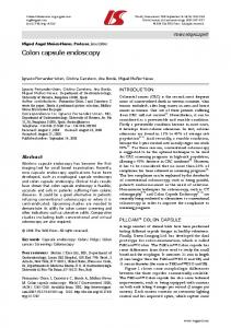
Figure 1 Macroscopic view of the duplicated colon and appendix vermiformis.
tubular str ucture with fecal material inside, and an appendix-like extra tubular structure with an obstructing fecalith in its lumen (Figure 1). This duplicated appendix was not perforated. Histologically, a colonic mucosa layer with accumulation of infiltrating lymphocyte nests, and a duplicated appendix was confirmed. Pathological examination revealed no evidence of malignancy (Figure 2).
DISCUSSION Gastrointestinal duplication, which may occur throughout the alimentary tract, is a rare congenital anomaly[1]. More than 80% of the cases present before the age of 2 years, as acute abdomen or bowel obstruction[1,2]. Most duplication is located in the ileum and presenting symptoms are bowel obstruction, intestinal bleeding and/or even perforation [3,4] . Apart from ileal duplication, colonic duplication is a rare abnormality. A thorough review of the international literature since 1950 has revealed 83 cases of colonic duplication to date, and only 4%-18% of all gastrointestinal duplications have been reported in the cecum[5-7]. Inclusion of the urogenital tract by an intestinal duplication has been reported in two cases[8,9]. Duplication of the tubular colon and vermiform appendix causing hydronephrotic atrophic kidney in adults has not previously been reported. Preoperative diagnosis of colon duplication is often difficult. Symptoms usually include abdominal pain, nausea and/or vomiting, palpable mass, weight loss, and bleeding, which often confuses colon duplication with other more common diagnoses. The pathogenesis of these lesions has not yet been well characterized. One of the most widely accepted theories is an abnormality of the embryonic gut that results in the formation of a diverticulum, a cyst, or twinning of a bowel segment[10]. Environmental factors such as trauma or hypoxia during early fetal development have also been suggested to play a role[11]. Although intestinal duplication is considered to be a benign lesion and mostly asymptomatic, they may result in significant morbidity and mortality if left untreated[12]. Indications for surgical intervention often arise in an acute setting, in the form of complications. Specifically, patients with previously undetected duplication may present with bowel obstruction or severe gastrointestinal hemorrhage (i.e., ulcerating www.wjgnet.com
January 28, 2008
Volume 14
Number 4
Figure 2 Histological appearance of the duplicated colon and appendix vermiformis.
gastric mucosa within a duplication cyst)[5]. If encountered incidentally, these lesions should be surgically addressed to avoid any future complication, including the possibility of malignant degeneration within the duplication cyst[5,12]. Duplication of the vermiform appendix is extremely rare. It is found in only 1/25 000 patients (0.004%) operated on for acute appendicitis [13] . The clinical presentation can vary depending on the location of the appendices in the colon. Cave and Wallbridge have classified the observed variants into several types: type A has incomplete duplication with both appendices having a common base; type B has complete duplication with the first appendix arising from its usual location at the confluence of the teniae coli, and the second appendix is located at various sites along the colon; and type C has complete duplication of the cecum, with each part having its own appendix[14,15]. Our case was diagnosed as type B. Although preoperative diagnosis has been made with the aid of radiological studies such as a barium enema, the majority of cases have been diagnosed at surgery or upon pathological examination. Barium enema studies are considered essential for the diagnosis of tubular colonic duplication, with opacification of two colons being the diagnostic sign. However, in certain cases, barium enema apparently does not show positive findings. A case has been reported in which a barium follow-through study established a complete diagnosis of duplication of the colon and terminal ileum. The findings were confirmed by laparotomy. Although the diagnosis of appendix duplication is a rarity, surgeons should be aware of the possibility, especially when clinical signs and symptoms point to appendicitis, although at laparotomy the appendix looks normal. Routine exploration for a second appendix is definitely not indicated because of its rarity and increased complication rate. Although duplication of the alimentary tract is rare, the possibility of congenital lesions of the alimentary tract should not be overlooked, even in adults presenting with vague or no gastrointestinal symptoms.
REFERENCES 1
Macpherson RI. Gastrointestinal tract duplications: clinical, pathologic, etiologic, and radiologic considerations. Radiographics 1993; 13: 1063-1080
Kabay S et al . Combined colon and appendix duplication 2 3 4 5 6 7
O’Neil J, Rowe M. Duplications of the gastrointestinal tract. Essentials of Pediatric Surgery. St. Louis: Mosby Yearbook, 1995: 520-525 Soares-Oliveira M, Carvalho JL, Campos M, Andrade M, Estevao-Costa J. Intestinal duplication. A diagnostic and therapeutic challenge. Acta Med Port 2002; 15: 365-368 Sakamoto K, Hasegawa S, Yamazaki Y, Makino T, Suda T, Imada T. Ileal duplication presenting as perforation: report of a case. Surg Today 2000; 30: 445-447 Holcomb GW 3rd, Gheissari A, O'Neill JA Jr, Shorter NA, Bishop HC. Surgical management of alimentary tract duplications. Ann Surg 1989; 209: 167-174 Puligandla PS, Nguyen LT, St-Vil D, Flageole H, Bensoussan AL, Nguyen VH, Laberge JM. Gastrointestinal duplications. J Pediatr Surg 2003; 38: 740-744 Fotiadis C, Genetzakis M, Papandreou I, Misiakos EP, Agapitos E, Zografos GC. Colonic duplication in adults: report of two cases presenting with rectal bleeding. World J Gastroenterol 2005; 11: 5072-5074
643 8
Decter RM, Kaplan KM, Eggli KD, Krummel TM. Colovesical fistula resulting from a perforated colonic duplication. Pediatrics 1998; 102: 654-656 9 Fechner G, Franke I, Willinek WA, Muller SC. Perforating colonic duplication as rare cause of renal abscess in children. Urology 2005; 66: 881 10 Hickey WF, Corson JM. Squamous cell carcinoma arising in a duplication of the colon: case report and literature review of squamous cell carcinoma of the colon and of malignancy complicating colonic duplication. Cancer 1981; 47: 602-609 11 Bishop HC, Koop CE. Surgical management of duplucations of the alimentary tract. Am J Surg 1964; 107: 434-442 12 Chen CC, Yeh DC, Wu CC, Li MC, Kwan PC. Huge cystic duplication of the ascending colon in adult. Zhonghua Yixue Zazhi (Taipei) 2001; 64: 174-178 13 Sobhian B, Mostegel M, Kunc C, Karner J. Appendix vermiformis duplex--a rare surprise. Wien Klin Wochenschr 2005; 117: 492-494 14 Cave AJ. Appendix Vermiformis Duplex. J Anat 1936; 70: 283-292 15 Wallbridge PH. Double appendix. Br J Surg 1962; 50: 346-347 S- Editor Liu Y L- Editor Kerr C
E- Editor Liu Y
www.wjgnet.com