sitive fluorescent microscopic method which allows measurement of DNA strand breaks in ... formamidopyrimidine-DNA glycosylase (FaPy)} or DNA inhibitors { ...
70
Gen. Physiol. Biophys. (1999), 18, Focus Issue, 70—74
Use of Single Cell Gel Electrophoresis (Comet Assay) Modifications for Analysis of D N A Damage E.
HORVÁTHOVA, D . S L A M E Ň O V Á A N D A.
Cancer Research Institute,
GÁBELOVÁ
Slovak Academy of Sciences, Bratislava, Slovakia
A b s t r a c t . Single cell gel electrophoresis (SCGE) or comet assay is a rapid and sen sitive fluorescent microscopic method which allows measurement of DNA s t r a n d breaks in individual cells. Modifications of S C G E conditions p e r m i t t e d t o de tect different types of D N A damage. In order to characterize DNA damage in duced by N-methyl-N'-nitro-N-nitrosoguanidine (MNNG) and hydrogen peroxide (H2O2) in Chinese hamster V79 cells, two approaches were used: (1) two p H values of unwinding and electrophoresis solutions (pH > 13.0 and pH = 12.1) t o specify t h e type of D N A lesions {the alkali-labile sites and true DNA singlestrand breaks (ssb)} and (2) DNA glycosylases {endonuclease III (EndoIII) a n d formamidopyrimidine-DNA glycosylase (FaPy)} or DNA inhibitors {hydroxyurea (HU) + l-(/3-D-arabinofuranosyl)cytosine (AraC)} t o characterize t h e types of DNA damage. Our results showed t h a t the lesions induced by H2O2 represented mainly the true DNA ssb, while MNNG formed predominantly alkali-labile sites, which were converted t o DNA ssb under strong alkaline conditions (pH > 13.0). T h e effects of DNA repair enzymes and DNA inhibitors were more significant un der lower pH (pH = 12.1) of unwinding and electrophoresis solution. Both, D N A glycosylases and DNA inhibitors increased t h e level of DNA ssb. K e y w o r d s : Hamster cells V79 — Comet assay — Repair enzymes — agent MNNG — Hydrogen peroxide H2O2
Alkylating
Introduction Single cell gel electrophoresis (SCGE) or comet assay is a rapid and sensitive fluo rescent microscopic m e t h o d which allows measurement of single-strand (ss) D N A breaks at low levels of damage in individual cells (Ostling and Johanson 1984; Singh et al. 1988; Collins et al. 1995). Ss DNA breaks result from a n u m b e r of different types of reactions in cells ( E a s t m a n and Barry 1992). However, s t a n d a r d methods for measuring ss breaks, including the comet assay, do not indicate t h e Correspondence to: Eva Horváthova, Cancer Research Institute, Slovak Academy of Sciences, Department of Mutagenesis and Carcinogenesis, Vlárska 7, 833 91 Bratislava, Slovakia. E-mail: exonhoreQsavba.sk
Modifications of Comet Assay
71
origin of these breaks. Generally, t h e monofunctional alkylating agent N-methyl-N'nitro-N-mtrosoguanidine (MNNG) causes instability of N-glycosyl bond of DNA, resulting in t h e appearance of alkali-labile sites and apurinic/apyrimidinic (AP) sites in DNA (Singer 1986) In contrast t o MNNG, hydrogen peroxide ( H 2 0 2 ) induces formation of highly reactive hydroxyl radicals ' O H , which attack DNA and create strand breaks, A P sites and base modifications in D N A (Halliwell and Aruoma 1991) We a t t e m p t e d t o increase t h e predictive value of t h e comet assay for characterisation of t h e n a t u r e and origin of ss DNA breaks induced by M N N G a n d H2O2 in Chinese hamster V79 cells by two steps. (1) parallel use of two p H values for unwinding and electrophoresis, namely pH = 12.1 a n d pH > 13.0, as generally alkali-labile sites are stable until pH is raised t o p H = 12 5 (Fortini et al 1996), (2) use of DNA glycosylases {endonuclease I I I (EndoIII), specific for oxidised pyrimidines and formamidopyrimidine-DNA glycosylase (FaPy), specific for damaged purines (Epe et al. 1993)} or DNA inhibitors {hydroxyurea (HU) + l-(/3-D-arabinofuranosyl)cytosine (AraC)} Materials and M e t h o d s Cell culture Quasidiploid Chinese hamster lung fibroblasts V79 were obtained from Prof A Abbondandolo, Laboratory of Mutagenesis, National Institute for Cancer Research, Genova, Italy Cells were grown at 37 °C in humidified atmosphere of 5% CO2 in Ea gle's MEM supplemented with 6% foetal calf serum and antibiotics (penicillin 100 U/ml, streptomycin and kanamycm 100 /xg/ml) Chemicals and treatment of cells N-methyl-N'-nitro-N-mtrosoguanidme (Aldnch, Ger many) stock solution in DMSO (60 mmol/1) was kept at -20 °C and diluted immediately before use Hydrogen peroxide (Lachema, Brno, Czech Republic) stock solution (10 mol/1) was kept at 4°C and diluted immediately before use m PBS buffer ( C a 2 + - and Mg 2+ -free) at 4°C Exponentially growing cells were treated either with MNNG (0 03, 0 06, 0 12 mmol/1 - 120 mm in coplete medium) or with H2O2 (25, 50, 100 /Limol/1 - 5 mm at 4°C in C a 2 + and Mg 2+ -free PBS buffer) Inhibitors, hydroxyurea (HU) and l-(/3-D-arabinofuranosyl)cytosine (AraC) (Sigma, St Louis, USA), were applied during 120 min treatment of cells 3 5 with MNNG at final concentrations of 2 x 10~ mol/1 and 2 x 10~ mol/1, respectively Single cell gel electrophoresis (comet assay) The procedure of Singh et al (1988) was used with minor modifications of Slameňová et al (1997) and Gábelová et al (1997) Control or treated V79 cells embedded m agarose were immersed in ice-cold lysis solution (2 5 mol/1 NaCl, 100 mmol/1 Na 2 EDTA, 10 mmol/1 Tris, pH 10 0, 1% Triton X-100) for 60 mm In experiments investigating the nature of induced damage, slides were after lysis washed 3 times for 5 mm in endonuclease buffer (40 mmol/1 HEPES-KOH, 0 1 mol/1 KC1, 0 5 mmol/1 EDTA, 0 2 mg/ml BSA, pH 8 0) and incubated at 37°C for 45 mm with endonuclease III (EndoIII), for 30 min with formamidopyrimidine-DNA glycosylase (FaPy) or with buffer alone EndoIII and FaPy were obtained as crude extracts from Dr A R Collins (Aberdeen) Then the slides were placed in electrophoresis boxes containing alkaline solutions pH > 13 0 (0 3 mol/1 NaOH, 1 mmol/1 Na 2 EDTA) or pH = 12 1 (0 03 mol/1 NaOH, 1 mmol/1 NažEDTA) for 40 mm at 4°C Electrophoresis was carried out for 30 mm at 4°C The slides were then neutralised with 0 4 mol/1 Tris-HCl, pH 7 5, for three 5-mm washing steps and stained with 20 fi\ ethidium bromide (10 /ig/ml) "Comets" were examined using a Zeiss epifluorescence microscope attached to an intensifying solid state
72
Horváthova et al
CCD camera and image analysis system (Komét 3 0, from Kinetic Imaging Ltd ) The "% of tail DNA" was used as the measure of DNA damage In the experiment 200 comets were scored per each sample The significance of differences between samples was assessed by Student's ť-test
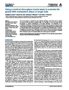
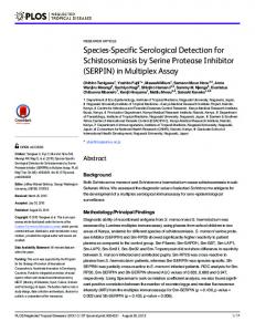
Results Discrimination between alkali-labile sites (pH > 13.0) and ss DNA breaks (pH = 12.1). T h e levels of induced ss DNA breaks in MNNG- and H 2 0 2 - t r e a t e d V79 cells are shown in Fig. 1. T h e levels of damage were measured immediately after t r e a t m e n t of cells with M N N G and H2O2 t o minimize removal of DNA lesions by cellular repair. Characterisation of MNNG-mduced damage by repair enzymes (EndoIII, FaPy) and inhibitors (HU + AraC) using two parallel pH values, pH > 13.0 and pH = 12.1, for unwinding and electrophoresis. T h e influence of DNA repair enzymes and DNA inhibitors on MNNG-treated V79 cells under standard S C G E alkaline conditions (pH > 13.0) is shown in t h e left p a r t of Fig. 2. T h e effects of D N A re pair enzymes and DNA inhibitors become evident under t h e decreased pH = 12.1 of unwinding and electrophoresis solution, which does not enable t h e detection of alkali-labile lesions in DNA (right p a r t of Fig. 2).
***
0
0.03
0.06
0.12
0
25
50
Concentrations of MNNG
Concentrations of H 2 0 2
(mmol/1)
(|imol/l)
100
Figure 1. Levels of induced ss DNA breaks in MNNG- (left) and m H 2 02-treated V79 cells (right) under standard SCGE alkaline conditions (• pH > 13 0) and under decreased o pH = 12 1 of unwinding and electrophoresis solution The results represent the mean of two independent experiments ± SD value Statistically significant differences *p < 0 05, **p < 0.01, ***p < 0 001 represent the difference between values measured at pH > 13 0 and 12.1
73
Modifications of Comet Assay
pH>13.0
C
0.06
pH=12.1
0.12
Concentrations of MNNG (mmol/1) • no enzyme BEndom HFaPy HINH1B
c
0.06
0.12
Concentrations of MNNG (mmol/1) • no enzyme B EndoIII • FaPy HINHIB
Figure 2. Influence of DNA repair enzymes {EndoIII and FaPy} and inhibitors {HU (2 x 10 - 3 mol/1) + AraC (2 x 10 - 5 mol/1)} on MNNG-treated V79 cells under standard SCGE alkaline conditions (pH > 13.0) (left) and under decreased pH = 12.1 of unwinding and electrophoresis solution (right). The results represent the mean of two independent experiments ± SD value. Statistically significant differences *p < 0.05; **p < 0.01; ***p < 0.001 represent the difference between samples without enzymes and samples with EndoIII, FaPy or DNA inhibitors. Discussion In previous papers both MNNG and H2O2 were shown to induce DNA ssb, as detected by several biochemical methods (Slameňová et al. 1997; Gábelová et al. 1997). To ascertain the ratio of alkali-labile sites and ss DNA breaks in MNNGand H20"2-treated V79 cells we assayed the samples at different pH of unwinding and electrophoresis solutions. The differences between the curves in the left part of Fig.l show that all detected breaks in MNNG-treated cells have the character of alkali-labile sites; at pH = 12.1 there was no significant increase in the number of ss breaks. Treatment with many alkylating agents, including N-methyl-N'-nitro-Nnitrosoguanidine (MNNG), leads to unstabilization of N-glycosyl bonds, opening of base rings, depurination/depyrimidination and the appearance of alkali-labile and apurinic/apyrimidinic (AP) sites by spontaneous hydrolysis or by the action of specific DNA glycosylases (Singer 1986). The removal of AP sites is accomplished by AP endonucleases which cleave DNA adjacent to AP sites and create ss DNA breaks. In samples treated with 50 or 100 /xmol/1 H2O2 the level of breaks induced at pH = 12.1 was significantly lower than that at pH > 13.0, yet the differences were small (Fig. 1 right part). We suggest that nearly all ss DNA breaks detected immedi-
74
Horváthova et al
ately after H202-treatment h a d t h e character of true DNA breaks and alkali-labile lesions represented only a minor type of lesions H2O2 crosses biological mem 1+ 2+ branes, penetrates to t h e nucleus and reacts with C u and F e ions t o form "OH radicals. Attack of "OH radicals on DNA leads to base modification, deoxyribose fragmentation, base loss a n d s t r a n d breaks (Halliwell a n d A r u o m a 1991). Most of these DNA lesions are repaired by direct rejoining or base excision repair. O u r d a t a suggest t h a t different types of lesions develop under different pH conditions. Fig. 2 suggests t h a t t h e effects of DNA repair enzymes a n d DNA inhibitors are more suitably examined under the lower pH (right p a r t of Fig. 2), which reduces detection of alkali-labile sites in DNA. EndoIII, F a P y and D N A inhibitors increased the level of ss DNA breaks. We assume t h a t this effect may have resulted from A P lyase activity of b o t h DNA repair enzymes. This hypothesis will be further examined by using an enzyme whose major physiological role is an A P endonuclease activity, 1. e. exonuclease I I I Applications of t h e modified comet assay with t h e inclusion of enzymes specific for certain kinds of lesion might be employed in genotoxicity testing and charac terisation of t h e damage caused by select carcinogens Acknowledgements. This study was supported by Inco Copernicus grant PL967095 References Collins A R , Ma Ai-guo, Duthie S J (1995) The kinetics of repair of oxidative DNA damage (strand breaks and oxidized pynmidine dimers) in human cells Mutat Res 336, 69-77 Eastman A , Barry A (1992) The origins of DNA breaks, A consequence of DNA damage, DNA repair, or apoptosis 7 Cancer Invest 10, 229-240 Epe B , Pflaum M , Haring M , Hegler J , Rudiger H (1993) Use of repair endonucleases to characterize DNA damage induced by reactive oxygen species in cellular and cell-free systems Toxicol Lett 67, 57-72 Fortmi P , Raspagho G , Falchi M , Doghotti E (1996) Analysis of DNA alkylation damage and repair in mammalian cells by the comet assay Mutagenesis 11, 169175 Gábelová A , Slameňová D , Ružeková Ľ , Farkašová T , Horváthova E (1997) Measure ment of DNA strand breakage and DNA repair induced with hydrogen peroxide using single cell gel electrophoresis, alkaline DNA unwinding and alkaline elution of DNA Neoplasma 44, 380-388 Halliwell B , Aruoma O I (1991) DNA damage by oxygen-derived species, Its mechanism and measurement m mammalian systems FEBS Lett 281, 9-19 Osthng O , Johanson J (1984) Microelectrophoretic studies of radiation-induced DNA damages m individual mammalian cells Biochem Biophys Res Commun 123, 291-298 Smger B (1986) O-Alkyl pynmidines in mutagenesis and carcinogenesis occurrence and significance Cancer Res 46, 4879-4885 Singh N P , McCoy M T , Tice R R , Schneider E L (1988) A simple technique for quantitation of low levels of DNA damage m individual cells Exp Cell Res 175, 184-191 Slameňová D , Gábelová A , Ružeková Ľ , Chalupa I , Horváthova E , Farkašová T , Bozsakyová E , Štetina R (1997) Detection of MNNG-induced DNA lesions in mammalian cells, Validation of comet assay against DNA unwinding technique, alkaline elution of DNA and chromosomal aberrations Mutat Res 383, 243-252