to show relatedness to VP12 of Colorado tick fever virus. (amino acid sequence identity 28%, calculated from alignment generated by ), while the ...
Journal of General Virology (2002), 83, 1941–1951. Printed in Great Britain ...................................................................................................................................................................................................................................................................................
Common evolutionary origin of aquareoviruses and orthoreoviruses revealed by genome characterization of Golden shiner reovirus, Grass carp reovirus, Striped bass reovirus and golden ide reovirus (genus Aquareovirus, family Reoviridae) Houssam Attoui,1 Qin Fang,2 Fauziah Mohd Jaafar,1 Jean-François Cantaloube,1 Philippe Biagini,1 Philippe de Micco1 and Xavier de Lamballerie1, 3 Unite! des Virus Emergents EA3292, Universite! de la Me! diterrane! e, Faculte! de Me! decine de Marseille, EFS Alpes-Me! diterrane! e, 13005 Marseille, France 2 Hydrobiology Institute, Wuhan Institute of Virology, CAS, Wuchang, 430071, Wuhan, Hubei, China 3 Maladies virales e! mergentes et syste' mes d’information UR 034, Institut de Recherche pour le De! veloppement, Marseille, France 1
Full-length and partial genome sequences of four members of the genus Aquareovirus, family Reoviridae (Golden shiner reovirus, Grass carp reovirus, Striped bass reovirus and golden ide reovirus) were characterized. Based on sequence comparison, the unclassified Grass carp reovirus was shown to be a member of the species Aquareovirus C. The status of golden ide reovirus, another unclassified aquareovirus, was also examined. Sequence analysis showed that it did not belong to the species Aquareovirus A or C, but assessment of its relationship to the species Aquareovirus B, D, E and F was hampered by the absence of genetic data from these species. In agreement with previous reports of ultrastructural resemblance between aquareoviruses and orthoreoviruses, genetic analysis revealed homology in the genes of the two groups. This homology concerned eight of the 11 segments of the aquareovirus genome (amino acid identity 17–42 %), and similar genetic organization was observed in two other segments. The conserved terminal sequences in the genomes of members of the two groups were also similar. These data are undoubtedly an indication of the common evolutionary origin of these viruses. This clear genetic relatedness between members of distinct genera is unique within the family Reoviridae. Such a genetic relationship is usually observed between members of a single genus. However, the current taxonomic classification of aquareoviruses and orthoreoviruses in two different genera is supported by a number of characteristics, including their distinct GMC contents, unequal numbers of genome segments, absence of an antigenic relationship, different cytopathic effects and specific econiches.
Introduction Viruses belonging to the family Reoviridae that infect aquatic animals are classified within the genus Aquareovirus. This genus was created by the International Committee on Author for correspondence : Houssam Attoui. Fax j33 4 91 32 44 95. e-mail virophdm!lac.gulliver.fr The accession numbers of sequences reported in this paper are AF450318–AF450324, AF403390–AF4034141 and AF418294–AF418304.
0001-8342 # 2002 SGM
Taxonomy of Viruses (ICTV) in 1991 (Francki et al., 1991). The type is Striped bass reovirus (SBRV, species Aquareovirus A), and five other species (Aquareovirus B to F) are recognized, along with a number of tentative species. The current classification has been established on the basis of three main criteria : RNA–RNA hybridization, electrophoretype analysis and antigenic properties (Mertens et al., 2000). Sequence data are recognized criteria for aquareovirus classification but, until very recently, analysis of the molecular relationships among aquareoviruses has been hampered by the paucity of the available genetic information. BJEB
H. Attoui and others
Aquareoviruses have been isolated from a wide variety of aquatic animals, including molluscs, finfish and crustaceans. In the past, they have been referred to as reovirus-like or rotavirus-like aquatic viruses. Like members of the genus Rotavirus, their genomes are composed of 11 segments of dsRNA. The genome is contained in a core surrounded by a double-layered icosahedral capsid that physically resembles capsids of mammalian orthoreoviruses (MRV), as demonstrated by cryoelectron microscopy (Shaw et al., 1996). Aquareoviruses grow in fish cell lines and produce large syncytia that represent the typical cytopathic effect of their replication. In fishes, their typical pathogenic effect is haemorrhage, which represents a serious threat to fish breeding. As an example, Grass carp reovirus (GCRV) provokes severe haemorrhage in fingerling and yearling grass carp, leading to 80 % mortality (Fang et al., 1989). Previously, partial genome sequences have been deposited in databases for Aquareovirus A, Aquareovirus B and GCRV. In this paper, we report the full-length genome sequences of Golden shiner reovirus (GSRV, Aquareovirus C) and GCRV (Aquareovirus C). We also have characterized genome segments 2, 3, 4, 8 and 10 of SBRV (Aquareovirus A) and segments 2 and 5 of golden ide reovirus (GIRV, unclassified). These molecular data provide the opportunity for the first robust analysis of the genetic relationships between aquareoviruses A, B and C and unclassified viruses. In addition, the analysis of complete genome sequences from three different aquareoviruses allowed the reassessment of the evolutionary relationship between viruses belonging to the genera Aquareovirus and Orthoreovirus.
aquareoviruses in its distinct cytopathic effect in cell culture (absence of syncytia and formation of focal aggregates of round cells) and by its sensitivity to chloroform. This virus was provided by Dr M. Neukirch. GIRV was grown in FHM cells at 20 mC. Other experimental conditions were as above. Striped bass reovirus (SBRV). SBRV is a member of the species Aquareovirus A. The virus was isolated in the USA from the salt-water striped bass fish (Morone saxatilis, family Centrarchidae) with haemorrhagic lesions of the skin (Subramanian et al., 1994). This virus was provided by Dr S. K. Samal as a suspension grown in a Chinook salmon embryo cell line (CHSE-214). The virus was studied without further propagation.
Preparation of virus dsRNA. Clarified supernatants from virusinfected cell cultures were subjected to ultracentrifugation. The pelleted viruses were resuspended in 200 µl EMEM and RNA was extracted with RNA-Now reagent (Biogentex) as described previously (Attoui et al., 2000). The dsRNA segments were separated on a 10 % polyacrylamide gel, excised and purified using the RNaid kit (Bio-101).
Cloning and sequence determination of genome segments of Aquareovirus C : GCRV and GSRV. Segments 1, 2 and 3 of GCRV873 were sequenced in a previous study (Fang et al., 2000 ; accession nos AF260511–AF260513). Cloning of segments 4–11 of GCRV-873 and segments 1–11 of GSRV was achieved using the single-primer amplification technique (Attoui et al., 2000), and this completed the genome characterization for both viruses.
Golden shiner reovirus (GSRV). GSRV is a member of the species Aquareo-
Partial sequence determination of genes 8 and 10 of GCRV : isolates GCRV-875, GCRV-876 and GCRV-991. dsRNAs from GCRV-875, GCRV-876 and GCRV-991 were heat-denatured at 99 mC for 1 min in the presence of 15 % DMSO and reverse-transcribed under standard conditions using Superscript-II (Invitrogen) and a random hexaprimer mixture. Primers were designed from the sequence of segments 8 and 10 of GCRV-873 and are reported in Table 1. PCR was performed under standard conditions using Taq polymerase (Invitrogen) at an annealing temperature of 55 mC. The PCR products were sequenced directly using the corresponding PCR primers, a D-Rhodamine DNA sequencing kit and an ABI Prism-377 sequence analyser (Perkin Elmer).
virus C. It has been isolated in the USA from the freshwater golden shiner fish [Notemigonus crysoleucas, family Cyprinidae (the minnow family)] showing petechial haemorrhage in skin (Plumb et al., 1979). The virus was purchased from the ATCC (reference VR-957). GSRV was propagated in fathead minnow (FHM) cells. Confluent monolayers of FHM were infected with 5 p.f.u. GSRV per cell in the presence of Eagle’s minimum essential medium with Hanks’ salts (EMEM) supplemented with 10 % foetal bovine serum. The cells were incubated for 3 days at 30 mC.
Sequence determination of GIRV (unclassified) and SBRV (Aquareovirus A). cDNAs of the genomes of SBRV and GIRV were synthesized by using either random hexaprimers or the single-primer amplification method. Segments 2 of SBRV and GIRV were amplified by PCR using primers designed from the most conserved nucleotide sequence alignments of the polymerases of orthoreoviruses and\or aquareoviruses. These primers are shown in Table 1. Segments 3, 4, 8 and 10 of SBRV and segment 5 of GIRV were gel-purified and cloned by using the single-primer amplification method.
Grass carp reovirus (GCRV). GCRV is now considered to belong to Aquareovirus C. Four isolates are now identified (Fang et al., 1989) as GCRV-873 (prototype strain), GCRV-875, GCRV-876 and GCRV-991. The virus was isolated from the freshwater grass carp fish (Ctenopharyngodon idellus, family Cyprinidae) in the People’s Republic of China (Chen & Jiang, 1984 ; Ke et al., 1990) and provokes severe haemorrhage in fingerling and yearling grass carp (Fang et al., 1989). GCRV was propagated in Ctenopharyngodon idellus kidney (CIK) cells. The cells were incubated for 3 days at 28 mC. Other experimental conditions were as above.
Sequences retrieved from databases. The nearly complete genome of Chum salmon reovirus (CSRV, Aquareovirus A) was retrieved from GenBank. Unpublished sequences of segments 1–11 of CSRV were deposited by S. Rao, G. R. Carner, W. Chen and J. R. Winton under accession numbers AF418294–AF418304. A partial sequence of genome segment 10 of Coho salmon reovirus CSR (Aquareovirus B) was retrieved from GenBank (accession number CSU90430).
Methods
The viruses and their propagation
Golden ide reovirus (GIRV). GIRV is an unclassified aquareovirus. The virus was isolated in Germany (Neukirch et al., 1999) from the freshwater golden ide fish (Leuciscus idus melanotus). It differs from other
BJEC
Sequence analysis methods. Sequence alignments were performed using (Thompson et al., 1994) and the local- program implemented in the DNATools package (version 5.01.661, S. W. Rasmussen). Phylogenetic analysis was carried out by the neighbourjoining method (Saitou & Nei, 1987) and the p-distance determination algorithm in the program (Kumar et al., 1993). Sequence relatedness
Taxonomy of aquareoviruses
Table 1. Primers used for the amplification of segments 2 of GIRV and SBRV and segments 8 and 10 of GCRV isolates Primer GSVseg2S SBRseg2rev ReAqSeg2S1 GIRVSeg2R ReAqSeg2S2 AquaSeg2R AquaIC8S AquaIC8R AquaIC10S AquaIC10R
Sequence (5h
3h)
CCAATACCTGTTAACTGATCTGATTAA TCCTTGACGATGAATTTGAGCGTGC CAAGCSATYATGAGRTCTCAATACGT GGATGGTCATATCCGAGAGGACACG GCCACCTCYACYGAGCAYACYGCTAATAA CATTCCTTGGTCWCCGGGGGGCATGTA GTTATTTTGTGATGGCACAGCGTCA GATGAAAGTCGTGAGGCAGCGGAGACG GCCCCCGATCATCACCACGATG GGGTGGGTAGGCCGGTGCTTA
Segment (origin) 2 (GSRV) 2 (SBRV) 2 (aquareovirusjorthoreovirus) 2 (GIRV) 2 (aquareovirusjorthoreovirus) 2 (aquareovirus) 8 (GCRV) 8 (GCRV) 10 (GCRV) 10 (GCRV)
Map position*
Orientation
689–719 Partial 1372–1397 Partial 2086–2114 2730–2704 1–25 1296–1270 12–33 879–859
Sense Antisense Sense Antisense Sense Antisense Sense Antisense Sense Antisense
* With respect to the GSRV sequence. is reported as percentage identity. Tree drawing was performed with the help of the program TreeView (Page, 1996). Comparisons of GSRV and GCRV sequence data with those available from nucleotide and protein databases were performed by using the NCBI program gapped (http :\\www3.ncbi.nlm.gov\blast). Hydropathy profiles were analysed by using amino acid sequence hydropathy values determined by the method of Kyte & Doolittle (1982) implemented in Microsoft Excel. Sequences aligned with were exported while all alignment-generated gaps were maintained (gap hydropathy valuel0). This permitted the comparison of positional hydropathy profiles for amino acid sequences of unequal lengths.
Results Sequence determination of Aquareovirus C viruses GCRV and GSRV
Sequences of genome segments 1–11 of GSRV were deposited in GenBank under accession numbers AF403398– AF403408. Sequences of segments 4–11 of GCRV were deposited under accession numbers AF403390–AF403397. The total RNA segment size, the longest ORF, the sizes of the 5h and 3h non-coding regions (NCR) and the size of the putative protein encoded were identified for each segment and are shown in Table 2. Partial sequence determination of genes 8 and 10 of GCRV isolates GCRV-875, GCRV-876 and GCRV-991
Partial sequences of segments 8 (1296 bp) and 10 (868 bp) of isolates GCRV-875, GCRV-876 and GCRV-991 were determined. The GenBank accession numbers are AF403412– AF403414 (segments 8) and AF403409–AF403411 (segments 10). Sequence determination of GIRV (unclassified) and SBRV (Aquareovirus A)
Primers ReAqSeg2S2\AquaSeg2R amplified a 645 bp sequence of segment 2 of SBRV. Sequencing of this product allowed the design of an SBRV sequence-specific reverse
primer designated SBRseg2rev. This primer, together with primer GSVseg2S, allowed PCR amplification of an overlapping sequence of 1491 bp. The final sequence obtained from SBRV segment 2 was 2026 bp long (accession no. AF450318). Primers ReAqSeg2S2\AquaSeg2R also amplified a 645 bp sequence of segment 2 of GIRV. Sequencing of this product allowed the design of a GIRV sequence-specific reverse primer designated GIRVseg2rev. This primer, together with primer ReAqSeg2S1, allowed PCR amplification of an overlapping sequence of 836 bp. The final sequence of GIRV segment 2 was 1360 bp long (accession no. AF450323). Segments 3, 4, 8 and 10 of SBRV and segment 5 of GIRV were separated and cloned by single-primer amplification. PCR amplification of cDNA from segment 3 using primer B (Attoui et al., 2000) has generated an amplicon that corresponded to a specific sequence of 1186 bp (accession no. AF450319). PCR from segment 4 resulted in a 1116 bp sequence (accession no. AF450320) located at the 3h terminus. Segments 8 and 10 were cloned as full-length sequences and were found to be respectively 1317 bp long (accession no. AF450321) and 987 bp long (accession no. AF450322). Segment 5 of GIRV was cloned as a full-length product and was found to be 2238 bp long (accession no. AF450324). Sequence analysis of Aquareovirus C viruses GCRV and GSRV Comparison of the genome electrophoretypes. The genomes of GCRV and GSRV showed identical electrophoretypes on a 1n2 % agarose gel. Slight variations were detected in the PAGE profiles. This could be explained by sequence variations, as observed with other viruses belonging to a single species within the family Reoviridae (Mertens et al., 2000). Comparison of GSRV with GCRV. The comparison of genome
segments 1–11 of GCRV and GSRV showed high degrees of identity (nucleotide sequences, 90n56–97n68 % ; amino acid sequences, 96–99n75 %). Cognate segments in the genomes of BJED
H. Attoui and others
Table 2. Properties of dsRNA segments of GSRV, GCRV and CSRV Putative encoded protein
5h NCR
3h NCR
Length (bp)
Length (aa)
Mass (Da)
Length (bp)
GSRV 1 2 3 4 5 6 7 8 9 10 11
3949 3877 3702 2320 2239 2039 1414 1297 1130 909 820
1299 1274 1214 742 728 648 146, 274* 412 352 276 244
141 266 141 585 132 058 79 463 80 249 68 557 15 705, 31 171 44 594 37 695 29 790 26 491
12 12 12 25 17 30 13 12 31 30 42 Consensus
5h GUUAUUU 5h GUUAUUU 5h GUUAUUU 5h GUUAUUG 5h GUUAUUU 5h GUUAUUU 5h GUUAUUU 5h GUUAUUU 5h GUUAUUU 5h GUUAUUU 5h GUUAUUG 5h GUUAUU\
UUCAUC 3h UUCAUC 3h UUCAUC 3h UUCAUC 3h AUCAUC 3h UUCAUC 3h UUCAUC 3h UUCAUC 3h AUCAUC 3h UUCAUC 3h UUCAUC 3h \UCAUC 3h
37 40 45 66 35 62 70 46 40 48 43
GCRV 1 2 3 4 5 6 7 8 9 10 11
3949 3877 3702 2320 2239 2039 1414 1296 1130 909 820
1299 1274 1214 742 728 648 146, 274* 412 352 276 244
141 406 141 536 132 104 79 642 80 243 68 600 15 706, 31 239 44 580 37 695 29 805 26 419
12 12 12 25 17 30 13 11 31 30 42 Consensus
5h GUUAUUU 5h GUUAUUU 5h GUUAUUU 5h GUUAUUG 5h GUUAUUU 5h GUUAUUU 5h GUUAUUU 5h GUUAUUU 5h GUUAUUU 5h GUUAUUU 5h GUUAUUG 5h GUUAUU\
UUCAUC 3h UUCAUC 3h UUCAUC 3h UUCAUC 3h AUCAUC 3h UUCAUC 3h UUCAUC 3h UUCAUC 3h AUCAUC 3h UUCAUC 3h UUCAUC 3h \UCAUC 3h
37 40 45 66 35 62 70 46 40 48 43
CSRV 1 2 3 4 5 6 7 8 9 10 11
3947 3867 3690 Partial 2242 2052 1395 1317 1118 985 781
1297 1240 1210 – 723 643 452 417 350 298 144, 154, 118†
140 930 137 579 131 949 – 80 151 68 878 49 842 45 335 38 057 32 410 15 117, 16 901, 13 041
13 115 18 21 53 5 12 25 27 24
5h GUUUUAU 5h GUUUUAU 5h GUUUUAU 5h GUUUUAU 5h GUUUUAU 5h GUUUUAU 5h GUUUUAU 5h GUUUUAG 5h GUUUUAG 5h GUUUUAG
AUCAUC 3h AUCAUC 3h UUCAUC 3h AUCAUC 3h UUCAUC 3h UUCAUC 3h UUCAUC 3h UUCAUC 3h UUCAUC 3h UUCAUC 3h UUCAUC 3h
5h GUUUUA\
\UCAUC 3h
Segment
Consensus
Terminal sequence
Terminal sequence
Length (bp)
40 29 39 117 49 67 31 51 28 61 52
* Segment is bicistronic. † Segment is tricistronic. , Not determined.
the two viruses had the same electrophoretic mobility. The detailed nucleotide and amino acid sequence identity values between cognate genes of these two viruses are shown in Table 3. Comparison of GSRV and GCRV with MRV. comparison of the genomes of GCRV and GSRV with a local Reoviridae BJEE
sequence database showed that they have remarkable similarity to the genome of MRV. Of the 11 segments of either GSRV or GCRV, eight segments showed significant identity (17–42 % : 42 % within the polymerase) to segments of MRV serotypes 1, 2 and 3 (Fig. 1). The correspondence was not always a function of the electrophoretic mobility, as shown in Fig. 1. Only segments 7, 10 and 11 did not show clear homology to genes
Taxonomy of aquareoviruses
Table 3. Comparison of segments 1–11 of the different aquareoviruses Values are percentage nucleotide (NUC) or amino acid (PROT) sequence identities. –, Sequence not available for GIRV or SBRV. GIRV Segment GSRV 1 2 3 4 5 6 7 8 9 10 11 GCRV 1 2 3 4 5 6 7 8 9 10 11 CSRV 1 2 3 4 5 6 7 8 9 10 11 SBRV 1 2
SBRV
CSRV
GCRV
NUC
PROT
NUC
PROT
NUC
PROT
NUC
PROT
– 71n7 – – 59n4 – – – – –
– 81n3 – – 56n6 – – – – – –
– 64n8 62n5 49n3 – – – 55n4 – 50n1 –
– 66n3 61n1 24n2 – – – 44n9 – 19n7 –
53n9 64n0 61n1 50n9 47n6 57n7 52n3 55n6 52n1 48n7 49n8
44n1 65n3 55n1 25n9 34n6 50n2 26 (NS31), 30 (NS16) 45n2 38n5 20n5 24 (ORF1), 25 (ORF2)
94n7 94n4 90n6 96n2 91n5 94n2 95n4 94n8 92n2 91n5 97n7
98n9 99n5 99n8 99n1 97n0 99n2 98 (NS31), 96(NS16) 98n8 100 96n4 98n8
– 71n7 – – 59n2 – – – – – –
– 81n3 – – 56n7 – – – – – –
– 65n3 62n5 49n8 – – – 54n7 – 48n9 –
– 66n5 61n1 24n5 – – – 45n2 – 19n7 –
54n0 63n9 62n5 50n9 47n5 56n9 52n2 55n2 52n8 48n6 50n5
44n0 65n5 55n1 26n2 34n4 50n2 26 (NS31), 30 (NS16) 44n9 38n5 20n5 24 (ORF1), 25 (ORF2)
– 62n0 – – 48n2 – – – – – –
– 64n8 – – 33n9 – – – – – –
– 78n7 78n0 70n8 – – – 77n9 – 74n1 –
– 95n9 84n4 71n2 – – – 86n8 – 80n9 –
– 63n0
– 65n0
of MRV. Such high values of amino acid sequence identity also help in the prediction of the function (and possibly the virus architecture) of the putative proteins of GSRV and GCRV (Fig. 1). ORF analysis has shown that segments 7 of GSRV and GCRV are bicistronic, encoding two proteins. The first protein (designated NS16, based on its theoretical molecular mass), encoded by the sequence between bases 14 and 454, was found to show relatedness to VP12 of Colorado tick fever virus (amino acid sequence identity 28 %, calculated from alignment
generated by ), while the second protein (NS31) showed no relatedness to any previously reported reovirus protein sequence. The orthoreovirus genome segment 7 (segment S1) is also bicistronic, encoding the outer capsid cell-attachment protein and a basic protein of unknown function. Based on their similar organization, it is possible that genome segments 7 of Aquareovirus C and MRV are equivalent. By analogy to Aquareovirus A and B, segment 10 of Aquareovirus C encodes outer capsid proteins. This is also true BJEF
H. Attoui and others Orthoreovirus (MRV-3) genes
Aquareovirus C (GSRV) genes
Aquareovirus A (CSRV) genes
Designation relative to SBRV
Putative function of GSRV/CSRV proteins compared to MRV
Structural
Guanylyl transferase (SP) (core protein)
Structural
RNA-dependent RNA polymerase (core protein)
Structural
Helicase, NTPase (SP) (core protein [T2 protein])
Non-structural
Non-structural protein (NSP)
Structural
NTPase (SP) (core protein)
Structural
Outer capsid protein (SP)
Non-structural
Cell-attachment protein (SP)
Structural
Core protein (SP)
Non-structural
Non-structural (NSP)
Outer capsid
Outer capsid protein (SP)
Non-structural
Fig. 1. Identities between Aquareovirus C (GSRV), Aquareovirus A (CSRV) and MRV serotype 3 (MRV-3). Bold double-headed arrows indicate homology between genes ; arrows carrying question marks indicate that assignment was based on aspects of organization. Percentage amino acid identities between homologous proteins of either GSRV or CSRV and MRV-3 are indicated by asterisks (*) ; (NV), no valid amino acid sequence identity ; ‡, genetic distances calculated from alignments generated with CLUSTAL W ; (NSx), theoretical size of the protein unknown because of the sequence is partial ; NSP, non-structural protein ; SP, structural protein. The putative functions of proteins from Aquareovirus A and Aquareovirus C were predicted by comparison to the already-established functions of MRV-3 proteins (Mertens et al., 2000). Designations relative to SBRV were taken from Subramanian et al. (1994).
for segment 10 of MRV. Analysis of the hydropathy plot of the proteins encoded by this segment of GSRV or GCRV and MRV showed similar hydropathy profiles in the aminoterminal part of the protein, with four domains in the order hydrophobic–hydrophilic–hydrophobic–hydrophilic (Fig. 2). Alignment of these protein sequences also showed numerous similar motifs (positions 21–27, GRLTLYT\GRVSIYS ; 45–55, CGRYTICAFCL\CGGAVVCMHCL ; 128–131, IVEL\LVEL ; 248–255, DDGHQARSA\DFGHFGLSH) with respect to the MRV sequence. Accordingly, it is likely that segments 10 of Aquareovirus C and MRV are equivalent. If this is true, segment 11 of Aquareovirus C would have no equivalent in the MRV genome. Analysis of the NCRs of GSRV and GCRV segments. Segments
1–11 of GCRV and GSRV were found to have conserved terminal sequences. All positive strands of each dsRNA segment had the motif 5h GUUAUUU\G 3h in common at the 5h end and the motif 5h A\UUCAUC 3h in common at the 3h end. The first and last nucleotides of each segment are inverted complements. In previous studies of reovirus genomes, comparable conserved motifs have been reported (Mertens et al., 2000). They are supposed to act as sorting signals, bringing a single copy of each genome segment into the nascent virus capsid (Anzola et al., 1987 ; Xu et al., 1989). Comparison of sequences of segments 8 and 10 among GCRV isolates. Sequence comparison of amplicons from different
virus isolates showed that, for segments 8 and 10, nucleotide and amino acid sequence identities were nearly 100 %. BJEG
Sequence analysis of Aquareovirus A viruses SBRV and CSRV Comparison of SBRV to CSRV. Sequences from segments 2, 3, 4, 8 and 10 of SBRV were characterized in this study. Sequence comparison of these segments to their cognates in CSRV revealed nucleotide sequence identities between 70n77 and 78n65 % and amino acid sequence identities between 71n2 and 95n87 % (the highest values being those of polymerase sequences ; Table 3). Comparison of Aquareovirus A with Aquareovirus C. The
nucleotide and amino acid sequence identities between SBRV or CSRV and GSRV or GCRV respectively ranged from 49n45 to 63n89 % and from 20n45 to 65n53 %. All segments of Aquareovirus C genomes have cognates in the genomes of Aquareovirus A viruses. The correspondence reflects the order of the electrophoretic mobility of the genomes perfectly, as shown in Fig. 1. Three obvious differences between Aquareovirus A and Aquareovirus C genomes were noticed, the first being the monocistronic character of segment 7 of CSRV, while those of GCRV and GSRV are bicistronic. Remarkably, both proteins NS16 and NS31 from Aquareovirus C (encoded by the two distinct ORFs of segment 7) showed similarity to the protein encoded by segment 7 of CSRV (NS49, encoded by a unique ORF) (sequence identities shown in Table 3). The second difference is the lack of similarity between the NS38 protein (segment 9) of CSRV and the σNS protein (segment 9) of
Taxonomy of aquareoviruses 0
30 60 90 120 150 180 210 240 270 300 330 360
3 1
A\UUCAUC 3h in common at the 3h end. Again, the first and last nucleotides of each segment are inverted complements. Comparison of terminal sequences of the NCRs of Aquareovirus A and Aquareovirus C. The conserved terminal sequences of
GSRV –1
Aquareovirus A are highly similar to those of Aquareovirus C, with one obvious difference. In the 5h motif, the adenine base is located at position 4 (5h GUUAUUU\G 3h) from the terminus for Aquareovirus C and at position 6 for Aquareovirus A (5h GUUUUAU\G 3h).
–3 3 1 SBRV –1 –3
Sequence analysis of GIRV segments 2 and 5
3
Comparison of GIRV with Aquareovirus A and Aquareovirus C.
1
The nucleotide and amino acid sequence identities between GIRV and Aquareovirus A viruses respectively ranged from 48n24 to 62n97 % and from 33n89 to 64n98 %. The nucleotide and amino acid sequence identities between GIRV and Aquareovirus C viruses respectively ranged from 59n24 to 71n69 % and from 56n73 to 81n34 %.
CSR –1 –3 3 1 MRV
–1 –3
Comparison of GIRV with MRV. comparison of GIRV 0
30 60 90 120 150 180 210 240 270 300 330 360
Amino acid position Fig. 2. Comparison of hydropathy profiles of aligned VP7 sequences (segment 10) of aquareoviruses (GSRV, SBRV, CSR) and σ3 sequence of MRV. The parts of the profiles represented by grey lines indicate the succession of the four domains : hydrophobic–hydrophilic–hydrophobic– hydrophilic.
MRV, while NS38 of GCRV and GSRV showed obvious similarity to this protein. The third difference is that segment 11 of CSRV is tricistronic (ORF1, nt 25–435 ; ORF2, nt 89– 553 ; ORF3, nt 375–731). Only the proteins encoded by ORF1 (NS13) and ORF3 (NS15) showed similarity to the NS26 protein (segment 11, monocistronic) of Aquareovirus C. Comparison of SBRV and CSRV with MRV. comparison of
the genomes of SBRV and CSRV with a local Reoviridae sequence database revealed similarity to the genome of MRV. Values of calculated amino acid sequence identity between CSRV or SBRV and MRV ranged between 18 and 40 %. Similarly to Aquareovirus C species, segments 1, 2, 3, 4, 5, 6 and 8 of CSRV exhibited similarity to segments 1, 2, 3, 4, 5, 6 and 8 in the order given in Fig. 1. The relationships between segments 7, 10 and 11 of CSRV and those of MRV were comparable to those between Aquareovirus C and MRV. As mentioned above, and in contrast to Aquareovirus C, the NS38 protein (segment 9) of CSRV did not exhibit similarity to the σNS protein (segment 9) of MRV. Analysis of NCRs of CSRV and SBRV segments. Segments 1–11 of CSRV and the full-length segments 8 and 10 of SBRV were found to have conserved terminal sequences. All positive strands of the sequenced dsRNA segments had the motif 5h GUUUUAU\G 3h in common at the 5h end and the motif 5h
genome with the local Reoviridae database showed similarity to the genome of MRV. Values of amino acid sequence identities between GIRV and MRV were 26 % in segment 5 and 41 % in segment 2. These values are comparable to those between Aquareovirus A or Aquareovirus C and MRV. Analysis of NCRs of GIRV segments : comparison with Aquareovirus A and Aquareovirus C. Only segment 5 of GIRV was
sequenced fully. The 5h- and 3h-terminal nucleotides of the positive strand of segment 5 were identical to those of Aquareovirus A. Global analysis of the sequenced genomes of aquareoviruses Sequence comparison of the putative RNA-dependent RNA polymerases of GCRV, GSRV, SBRV and GIRV and other members of the family Reoviridae. The amino acid sequences of the
polymerases of GCRV, GSRV, SBRV and GIRV were compared with the sequences of putative RNA-dependent RNA polymerases of representative viruses from nine genera of the family Reoviridae : Seadornavirus (12 segments), species Banna virus (isolate BAV-In6423 ; accession no. AF133430) and Kadipiro virus (isolate KDV-Ja7075 ; AF133429) ; Coltivirus (12 segments), species Colorado tick fever virus (isolate CTFVFl ; AF134529) ; Orthoreovirus (10 segments), species Mammalian orthoreovirus serotypes 1 (MRV-1 ; M24734), 2 (MRV2 ; M31057) and 3 (MRV-3 ; M31058) and Ndelle virus (NDEV ; AF368033) ; Orbivirus (10 segments), species African horse sickness virus serotype 9 (AHSV-9 ; U94887), Bluetongue virus serotypes 2 (BTV-2 ; L20508), 10 (BTV-10 ; X12819), 11 (BTV-11 ; L20445), 13 (BTV-13 ; L20446) and 17 (BTV-17 ; L20447) and Palyam virus isolate CHUV (Baa76549) ; Rotavirus (11 segments), species Rotavirus A (RV-A) strains BoRV-A\RF BJEH
H. Attoui and others
(b)
(a) 0·5
Frequency
0·4
0·3 30 % identity 0·2
0·1
33
66
99
Amino acid sequence identity (%) Fig. 3. (a) Frequency distribution histogram of amino acid sequence identities between polymerases of members of the Reoviridae. Sequence analysis included polymerase sequences of 34 viruses, including four isolates of MRV and five aquareoviruses. The vertical line at 30 % amino acid sequence identity indicates the limits of distinction between genera except for Rotavirus B, which is 22 % identical to other rotaviruses, and aquareoviruses, which show 40–42 % amino acid sequence identity to orthoreoviruses. (b) Radial neighbour-joining tree displaying relationships between the polymerase sequences of members of the Reoviridae. Bootstrap values of 500 replications are indicated at the nodes ($) within each genus.
(J04346), BoRV-A\UK (X55444), SiRV-A\SA11b (X16830), SiRV-A\SA11 (AF015955), PoRV-A\Go (M32805) and AvRV-A (Baa24146), Rotavirus B strain Hu\MuRV-B\IDIR (M97203) and Rotavirus C strain PoRV-C\Co (M74216) ; Fijivirus (10 segments), species Nilaparvata lugens reovirus strain NLRV-Iz (D49693) ; Phytoreovirus (12 segments), species Rice dwarf virus isolates RDV-Ch (U73201), RDV-H (D10222) and RDV-A (D90198) ; Oryzavirus (10 segments), species Rice ragged stunt virus strain RRSV-Th (U66714) ; and Cypovirus (10 segments), species Bombyx mori cytoplasmic polyhedrosis virus-1 strain Bm-1 CPV (AF323781). It was found that all members of a single genus exhibited amino acid sequence identities of over 30 % (Fig. 3a). The only exception was Rotavirus B, which was only 22 % identical to other rotaviruses. Between members of the genera Aquareovirus and Orthoreovirus, the amino acid sequence identity ranged from 40 to 42 %. This value is therefore comparable to the amino acid sequence identity observed between members of a single genus. The results of this analysis are illustrated by a radial neighbour-joining tree (Fig. 3b). Comparison of deduced amino acid sequences of segment 10 for isolates of viruses belonging to Aquareovirus A, Aquareovirus B and Aquareovirus C. In aquareovirus genomes, segment 10
encodes the major outer capsid protein VP7. VP7 sequences from different isolates of SBRV and CSRV (Aquareovirus A), BJEI
CSR (Aquareovirus B), GCRV and GSRV (Aquareovirus C) were compared. The results showed that amino acid sequence identities ranged from 19n7 to 100 % and that, within a species, amino acid sequence identities were greater than 80 %. The amino acid sequence identities were found to be 80n87 % among Aquareovirus A isolates, 95n77–99n62 % among Aquareovirus C isolates, 19n53–21n29 % between Aquareovirus A and Aquareovirus C isolates and 20n91–21n43 % between Aquareovirus B and Aquareovirus C isolates. Analysis of the GjC contents of the genomes of Aquareovirus A, Aquareovirus B, Aquareovirus C and GIRV. The GjC contents
of the genomes of GCRV and GSRV ranged between 53 and 60 mol % ; the highest value was calculated from segments 4 and 11. The GjC content of the genomes of GSRV and SBRV ranged between 53 and 57 mol % ; the highest value was calculated from segment 4. The GjC content of segment 2 of GIRV was 52 mol % and that of segment 5 was 54 mol %. The GjC content of segment 10 of CSR was found to be 56 mol %.
Discussion Aquareoviruses and orthoreoviruses share a number of common structural characteristics, in particular a similar ultrastructure in electron microscopy (Shaw et al., 1996).
Taxonomy of aquareoviruses
Besides having different numbers of genome segments, they are antigenically distinct and occupy distinct econiches. The first fish-virus isolates with polysegmented dsRNA genomes were obtained in the late 1970s and early 1980s and included GSRV, American oyster reovirus 13p2, CSRV and Channel catfish reovirus. These isolates were the first to be designated aquareoviruses (Winton et al., 1987). Numerous viruses belonging to the family Reoviridae have been isolated from aquatic animals since these initial isolations. The majority of these viruses were found to possess genomes of 11 dsRNA segments and, hence, to belong to the genus Aquareovirus. However, virus isolates such as the W and P2 viruses, isolated from Mediterranean crab, were found to possess genomes of 12 dsRNA segments and were hence excluded from classification within the genus Aquareovirus (Montanie et al., 1993). To date, aquareoviruses have been isolated from fish, molluscs and crustaceans (Mertens et al., 2000), while orthoreoviruses have been isolated from reptiles, birds and mammals. One of the first important findings of the current study pertains to the taxonomy of unclassified isolates. Firstly, the genome of GCRV was sequenced completely and genetic analyses have shown that it is almost identical to that of GSRV (Aquareovirus C) : (i) the genomes of the two viruses are of comparable sizes (GCRV, 23 695 bp ; GSRV, 23 696 bp) ; (ii) cognate segments of the viruses are of comparable sizes ; (iii) each genome segment is flanked by identical 5h and 3h conserved NCRs ; (iv) nucleotide and amino acid sequence identities between the two viruses are very high (respectively 90–97 % and 96–100 %) and the nucleotide sequence variation is mainly a function of the third codon position. These findings show clearly that GSRV and GCRV should be considered as isolates of the same species. Consequently, the hitherto unclassified GCRV belongs to the species Aquareovirus C. Secondly, segments 2 and 5 of the unclassified GIRV were sequenced. Assessment of the genetic relationship of GIRV to the species Aquareovirus B, D, E and F was impossible because of the absence of sequence data from these species, but sequence comparison permitted us to exclude GIRV from Aquareovirus A and Aquareovirus C. Besides the study of members of the genus Aquareovirus, the current study sheds new light on the relationship between orthoreoviruses and aquareoviruses. It is a general rule within the family Reoviridae, that the genetic relatedness of viruses belonging to different genera is very low. In the polymerase gene, the only one that allows sequence comparison between different genera, analysis of amino acid sequence identity frequency distribution (Fig. 3a) shows that viruses belonging to different genera have low amino acid sequence identity ( 20 %), denoting a very distant phylogenetic relationship. This is more evident in other genes for which comparison is practically impossible. The situation of orthoreoviruses and aquareoviruses is therefore a remarkable exception : (i) in the polymerase gene, the amino acid sequence identity between members of the two genera is up to 42 % (a value usually
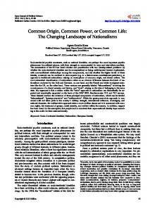
~510 My ago
Fig. 4. Simplified scheme of the evolution of the main hosts of orthoreoviruses and aquareoviruses. The separation between the hosts of aquareoviruses and orthoreoviruses about 510 My ago is shown by the diagonal line.
observed between members of a single genus) ; (ii) a clear genetic relationship can be observed between members of the two genera in seven other genes (see Fig. 1) where amino acid sequence identities range between 17 and 42 % ; (iii) the T2 protein (the innermost shell of the capsid) of members of the family Reoviridae is a protein that is structurally conserved between viruses belonging to different genera ; however, it is not possible to perform a significant sequence comparison. This protein is identified as the λ1 protein in the orthoreovirus genome. The equivalent aquareovirus protein, VP3, shows significantly high (37 %) amino acid sequence identity to λ1. This means that the genetic relationship between orthoreoviruses and aquareoviruses is comparable to that between viruses such as St Croix River virus and Bluetongue virus (belonging to the genus Orbivirus) or Kadipiro virus and Banna virus (both belonging to the genus Seadornavirus). Homology was also identified in the conservation of terminal sequences. For instance, the 5h and 3h conserved terminal sequences of GCRV segments are 5h GUUAUU 3h and 5h A\UUCAUC 3h, compared to 5h GCUAUU 3h and 5h A\UUCAUC 3h in genome segments 4, 7, 8 and 10 of MRV. Altogether, these data are undoubtedly an indication of the common evolutionary origin of these viruses. Fig. 4 shows a simplified scheme of the evolution of the main hosts of orthoreoviruses and aquareoviruses. The common ancestor of fish and the group ‘ reptilesjbirdsjmammals ’ existed around 510 million years (My) ago. It has been proposed that the molecular evolutionary rate of genomes of related dsRNA viruses is 10−) to 10−* mutations\nucleotide\year, which is equivalent to that of dsDNA (Attoui et al., 2002). If this is applied to the polymerases of orthoreoviruses and aquareoBJEJ
H. Attoui and others
viruses, it appears that divergence between these two groups occurred 49–520 My ago. Despite this imprecision in the evaluation of divergence, it cannot be excluded that orthoreoviruses appeared following the emergence of the evolutionary group that eventually gave rise to reptiles, birds and mammals. Finally, these results raise interesting questions concerning the taxonomic classification of orthoreoviruses and aquareoviruses in different genera on the one hand, and the relevance of quantitative taxonomy based on polymerase sequences in the Reoviridae on the other hand. Concerning the first question, it should be noted that the ICTV defines taxa as members of a polythetic class. Therefore, despite the unusual genetic relatedness between orthoreoviruses and aquareoviruses, there are strong arguments that justify their classification within two separate genera : (i) as reported above, the viruses originate from distinct econiches ; (ii) aquareoviruses have genomes composed of 11 segments and orthoreovirus genomes are composed of 10 segments of dsRNA ; (iii) the GjC content of orthoreoviruses ranges between 44 and 48 mol %, while that of aquareoviruses ranges between 52 and 60 mol % ; (iv) orthoreoviruses are nonsyncytializing viruses, in contrast to the majority of aquareoviruses ; and (v) there is no antigenic relationship between the two groups. Accordingly, the authors consider that the maintenance of the classification of these viruses in two different genera is justified and that orthoreoviruses and aquareoviruses constitute an interesting, but isolated, example of two genera that undoubtedly originate from a common evolutionary ancestor. Concerning quantitative taxonomy using polymerase sequences, analysis of amino acid sequence identity frequency distribution (Fig. 3a) shows that all members of a single genus have amino acid sequence identities of � 30 %. There is, however, one exception, Rotavirus B, which is only 22 % identical to other rotaviruses. Similarly, it can be observed that all viruses belonging to different genera have amino acid sequence identities of 30 %, the only exceptions being orthoreoviruses and aquareoviruses, as discussed above. Therefore, the only criterion that remains indisputable is the assignment of two viruses to different genera if their polymerase amino acid sequence identity is 20 %. Clearly, this is a modest contribution to phylogenetic classification. However, it is important to pursue the sequence characterization of representative members of the family Reoviridae to allow better definition of the quantitative basis of species delineation in the different genera and to try to improve the genetic criteria for the definition of genera. The authors wish to thank Dr S. K. Samal for providing SBRV and Dr M. Neukirch for providing GIRV. The authors also thank Dr S. Rao and colleagues for releasing the sequence of CSRV to the database. This work was supported by EU grant ‘ Reo ID ’ no. QLK2-2000-00143. The ‘ Unite! des Virus Emergents ’ is an associated research unit of the Institut de Recherche pour le De! veloppement (IRD). This study was supported in part by the IRD and the EFS Alpes-Me! diterrane! e. BJFA
References Anzola, J. V., Xu, Z. K., Asamizu, T. & Nuss, D. L. (1987). Segmentspecific inverted repeats found adjacent to conserved terminal sequences in wound tumor virus genome and defective interfering RNAs. Proceedings of the National Academy of Sciences, USA 84, 8301–8305. Attoui, H., Billoir, F., Cantaloube, J.-F., Biagini, P., de Micco, P. & de Lamballerie, X. (2000). Strategies for the sequence determination of
viral dsRNA genomes. Journal of Virological Methods 89, 147–158. Attoui, H., Mohd Jaafar, F., Biagini, P., Cantaloube, J.-F., de Micco, P., Murphy, F. A. & de Lamballerie, X. (2002). Genus Coltivirus (family
Reoviridae) : genomic and morphologic characterization of Old World and New World viruses. Archives of Virology 147, 533–561. Chen, B. S. & Jiang, Y. (1984). Morphological and physicochemical characterization of the hemorrhagic virus of grass carp. Kexue Tongbao 29, 832–835 (in Chinese). Fang, Q., Ke, L. H. & Cai, Y. Q. (1989). Growth characterization and high titre culture of GCHV. Virologica Sinica 3, 315–319. Fang, Q., Attoui, H., Cantaloube, J. F., Biagini, P., Zhu, Z., de Micco, P. & de Lamballerie, X. (2000). Sequence of genome segments 1, 2, and
3 of the grass carp reovirus (genus Aquareovirus, family Reoviridae). Biochemical and Biophysical Research Communications 274, 762–766. Francki, R. I. B., Fauquet, C. M., Knudson, D. L. & Brown, F. (1991).
Classification and Nomenclature of Viruses. Fifth Report of the International Committee on Taxonomy of Viruses. Wien : Springer-Verlag. Ke, L. H., Fang, Q. & Cai, Y. Q. (1990). Characteristics of a novel isolate of grass carp hemorrhage virus [J]. Acta Hydrobiologica Sinica 14, 153–159. Kumar, S., Tamura, K. & Nei, M. (1993). Molecular Evolutionary Genetics Analysis, version 1.01. Pennsylvania State University. Kyte, J. & Doolittle, R. F. (1982). A simple method for displaying the hydropathic character of a protein. Journal of Molecular Biology 157, 105–132. Mertens, P. P. C., Arella, M., Attoui, H., Belloncik, S., Bergoin, M., Boccardo, G., Booth, T. F., Chiu, W., Diprose, J. M., Duncan, R. and 34 others (2000). Family Reoviridae. In Virus Taxonomy. Seventh Report of the
International Committee for the Taxonomy of Viruses, pp. 395–480. Edited by M. H. V. van Regenmortel, C. M. Fauquet, D. H. L. Bishop, E. B. Carstens, M. K. Estes, S. M. Lemon, J. Maniloff, M. A. Mayo, D. J. McGeoch, C. R. Pringle & R. B. Wickner. San Diego : Academic Press. Montanie, H., Bossy, J.-P. & Bonami, J.-R. (1993). Morphological and genomic characterization of two reoviruses (P and W2) pathogenic for marine crustaceans ; do they constitute a novel genus of the Reoviridae family? Journal of General Virology 74, 1555–1561. Neukirch, M., Haas, L., Lehmann, H. & von Messeling, V. (1999).
Preliminary characterization of a reovirus isolated from golden ide Leuciscus idus melanotus. Diseases of Aquatic Organisms 35, 159–164. Page, R. D. (1996). TreeView : an application to display phylogenetic trees on personal computers. Computer Applications in the Biosciences 12, 357–358. Plumb, J. A., Bowser, P. R., Grizzle, J. M. & Mitchell, A. J. (1979). Fish viruses : a double stranded RNA icosahedral virus from a North American cyprinid. Journal of the Fisheries Research Board of Canada 36, 1390–1394. Saitou, N. & Nei, M. (1987). The neighbor-joining method : a new method for reconstructing phylogenetic trees. Molecular Biology and Evolution 4, 406–425. Shaw, A. L., Samal, S. K., Subramanian, K. & Prasad, B. V. V. (1996).
The structure of aquareovirus shows how the different geometries of the two layers of the capsid are reconciled to provide symmetrical interactions and stabilization. Structure 4, 957–967. Subramanian, K., McPhillips, T. H. & Samal, S. K. (1994). Charac-
Taxonomy of aquareoviruses terization of the polypeptides and determination of the genome coding assignments of an aquareovirus. Virology 205, 75–81. Thompson, J. D., Higgins, D. G. & Gibson, T. J. (1994). : improving the sensitivity of progressive multiple sequence alignment through sequence weighting, position-specific gap penalties and weight matrix choice. Nucleic Acids Research 22, 4673–4680.
properties of four members of a novel group of reoviruses isolated from aquatic animals. Journal of General Virology 68, 353–364. Xu, Z. K., Anzola, J. V., Nalin, C. M. & Nuss, D. L. (1989). The 3hterminal sequence of a wound tumor virus transcript can influence conformational and functional properties associated with the 5h-terminus. Virology 170, 511–522.
Winton, J. R., Lannan, C. N., Fryer, J. L., Hedrick, R. P., Meyers, T. R., Plumb, J. A. & Yamamoto, T. (1987). Morphological and biochemical
Received 16 January 2002 ; Accepted 24 March 2002
BJFB