In c.
PHARMACOLOGY - RESEARCH, SAFETY TESTING AND REGULATION
N
ov
a
Pu bl
is
he rs
COMMONLY USED DRUGS - USES, SIDE EFFECTS, BIOAVAILABILITY AND APPROACHES TO IMPROVE IT
In c.
PHARMACOLOGY - RESEARCH, SAFETY TESTING AND REGULATION Additional books in this series can be found on Nova‘s website under the Series tab.
N
ov
a
Pu bl
is
he rs
Additional e-books in this series can be found on Nova‘s website under the e-book tab.
In c.
PHARMACOLOGY - RESEARCH, SAFETY TESTING AND REGULATION
is
he rs
COMMONLY USED DRUGS - USES, SIDE EFFECTS, BIOAVAILABILITY AND APPROACHES TO IMPROVE IT
RAFIK KARAMAN
N
ov
a
Pu bl
EDITOR
New York
Copyright © 2015 by Nova Science Publishers, Inc.
For permission to use material from this book please contact us:
[email protected]
In c.
All rights reserved. No part of this book may be reproduced, stored in a retrieval system or transmitted in any form or by any means: electronic, electrostatic, magnetic, tape, mechanical photocopying, recording or otherwise without the written permission of the Publisher.
he rs
NOTICE TO THE READER The Publisher has taken reasonable care in the preparation of this book, but makes no expressed or implied warranty of any kind and assumes no responsibility for any errors or omissions. No liability is assumed for incidental or consequential damages in connection with or arising out of information contained in this book. The Publisher shall not be liable for any special, consequential, or exemplary damages resulting, in whole or in part, from the readers‘ use of, or reliance upon, this material. Any parts of this book based on government reports are so indicated and copyright is claimed for those parts to the extent applicable to compilations of such works.
is
Independent verification should be sought for any data, advice or recommendations contained in this book. In addition, no responsibility is assumed by the publisher for any injury and/or damage to persons or property arising from any methods, products, instructions, ideas or otherwise contained in this publication.
Pu bl
This publication is designed to provide accurate and authoritative information with regard to the subject matter covered herein. It is sold with the clear understanding that the Publisher is not engaged in rendering legal or any other professional services. If legal or any other expert assistance is required, the services of a competent person should be sought. FROM A DECLARATION OF PARTICIPANTS JOINTLY ADOPTED BY A COMMITTEE OF THE AMERICAN BAR ASSOCIATION AND A COMMITTEE OF PUBLISHERS. Additional color graphics may be available in the e-book version of this book.
a
Library of Congress Cataloging-in-Publication Data
N
ov
ISBN: 978-1-63463-828-9
Library of Congress Control Number: 2015931763
Published by Nova Science Publishers, Inc. † New York
In c.
he rs
Contents Preface
Drug Overview Hatem Amin Hejaz and Rafik Karaman
1
Chapter 2
Antibiotics Salma Jumaa and Rafik Karaman
41
Chapter 3
Pain Killers Samya Salah, Heba Awadallah and Rafik Karaman
75
Chapter 4
Lipid Lowering Medications - Uses, Side Effects, Pharmacokinetic Properties and Approaches to Improve Bioavailability 131 Wajd Amly and Rafik Karaman
Pu bl
is
Chapter 1
Antihyperglycemic Drugs Alaa Qtait and Rafik Karaman
173
Chapter 6
Anti-Hemorrhagic Agents Hiba Ghareeb and Rafik Karaman
201
Chapter 7
Osteoporosis Drugs Yahya khawaja and Rafik Karaman
219
Chapter 8
Fumaric Acid Esters as a Treatment for Psoriasis and Multiple Sclerosis Maryam Bader and Rafik Karaman
a
Chapter 5
249
About the Author
263
Index
265
ov
N
vii
a
ov
N is
Pu bl he rs
In c.
In c.
he rs
Preface
N
ov
a
Pu bl
is
Pharmacology is the science of medicine used in animals and humans. Many prescribed drugs are dispensed by pharmacists on a daily basis, and this is considered one of the most dangerous and important activities practiced by pharmacists. Comprehensive knowledge and critical understanding of the pharmacology principles which include the pharmacokinetics and pharmacodynamics of drugs is the solid basis for a safe and effective therapeutic practice. The focus of this book is on providing comprehensive, authoritative, and readable chapters on classes of commonly used drugs for medical and pharmacy students, nurses, junior doctors, pharmacists and other allied professionals in the health sciences. This book is a collaborative effort by the editor and some of his colleagues and graduate students as coauthors. The book is mainly devoted on describing the pharmacology, pharmacokinetics, uses, side effects and ways to improve the bioavailability for some of the most commonly used drugs. The first chapter introduces a comprehensive overview on the definition of drugs, approaches of drugs classification and some important aspects of drugs design and development, drug effectiveness and safety, and drug errors. The second chapter describes the three antibiotic classes, their mechanism of action, clinical uses, side effects, and their resistance by different bacteria. These described antibiotics include vancomycin, penicillins, cephalosporins, carbapenems tetracyclines, aminoglycosides, macrolides, chloramphenicol, fluoroquinolones and sulfonamides. The third chapter discusses the current used medicines for treating pain. These medications include the non-steroidal anti-inflammatories (NSAIDs), acetaminophen and opiates and their combination. The fourth chapter describes a number of drugs used to lower lipid levels in the blood such as statins, their adverse effects and methods to improve their bioavailability. The fifth chapter is devoted to the description of the pathogenesis and types of diabetes and the current used drugs to treat this disease. In addition, a detailed description of the pharmacokinetic properties, mechanism of action, side effects of known anti-hyperglycemic agents is presented. The sixth chapter describes the two different classes of anti-hemorrhagic agents (hemostatic agents): the first class includes systemic drugs such as tranexamic acid, ωaminocaproic acid, anti-inhibitor coagulant complex-heat treated, anti-hemophilic factor, factor IX, carbazochrome, fibrinogen concentrate, oprelvekin and phylloquinone, and the
viii
Rafik Karaman
N
ov
a
is
Pu bl
Rafik Karaman, Ph.D. Editor
he rs
In c.
second class of hemostatic agent is the local acting agents such as cellulose, collagen, gelatin, thrombin and thrombin combination products. The seventh chapter is devoted to osteoporosis (a progressive bone disease) and the various therapies available for treating this disease such as bisphosphonates, raloxifene, calcitonin, teriparatide and denosumab. The last chapter is devoted to the description of two different autoimmune diseases; multiple sclerosis (MS) and psoriasis. Up to date there is no known cure for MS and psoriasis. The available treatments approved by the FDA are mainly considered symptom relieving drugs and their major therapeutic action is only to slow the disease progression, and they lack the ability to eliminate the disease completely. A great devotion in this chapter was given to discuss several aspects regarding fumarate acid esters including; pharmacokinetics, mode of action, recommendations and specifically their potential as an effective treatment for psoriasis and MS. I want to express my sincere gratitude to all coauthors for their great efforts and cooperation. It has been a pleasure for me to be in the center of composing this nice piece of pharmacology book. I hope that this book will provide a succinct review of pharmacology with over 750 references. It is especially helpful to undergraduate and graduate students preparing for variety types of examinations.
ISBN: 978-1-63463-828-9 © 2015 Nova Science Publishers, Inc.
In c.
In: Commonly Used Drugs Editor: Rafik Karaman
Chapter 1
he rs
Drug Overview
Hatem Amin Hejaz1 and Rafik Karaman1,2 1
Pharmaceutical Sciences Department, Faculty of Pharmacy Al-Quds University, Jerusalem, Palestine 2 Department of Science, University of Basilicata, Potenza, Italy
is
Abstract
Pu bl
A drug is a chemical substance with known biological effects on humans or other animals. In the pharmacology field, a drug is defined as a chemical substance used in the treatment, cure, prevention, or diagnosis of disease or used to otherwise enhance physical or mental well-being. Drugs usually affect either normal or abnormal physiological processes. Drugs may be used for a limited duration, or on a regular basis for chronic disorders. The way drugs are classified or grouped are confusing. Therefore, a new approach of drugs classification is presented in this chapter along with general information on drugs which includes definition, drugs and diseases types, drugs administration, drugs interactions and drug names. In addition, the chapter describes some important aspects of drugs design and development, drug effectiveness and safety, and drug errors.
N
ov
a
Keywords: Drugs, Disease, Drug classification, Drug administration, OTC drugs, Prescription drugs, Drug interactions, Drug safety
ADHD ADME AIDS API BCS BP
Abbreviations Attention Deficit Hyperactivity Disorder Absorption, Distribution, Metabolism and Excretion Acquired Immune Deficiency Syndrome Active Pharmaceutical Ingredients Biopharmaceutical Classification System Blood Pressure
2
Hatem Amin Hejaz and Rafik Karaman
is
he rs
In c.
Behind-the-Counter Central Nervous System Coenzyme A Catechol O-Methyltransfarase Cyclooxygenase-1 Cyclooxygenase-2 Dichlorodiphenyltrichloroethane Disease- Modifying Antirheumatic Drugs Deoxyribonucleic Acid Dipeptidyl Peptidase-IV Food and Drug Administration Gastroesophageal Reflux Disease Gastrointestinal Tract Hydrochloric Acid 3-Hydroxy-3-Methylglutaryl-Coenzyme A Intravenous Injection Intramuscular Injection International Normalized Ratio Institutional Research Board Kilocalorie Lysergic Acid Diethylamide Monoamine Oxidases Monoamino Oxidase Inhibitors Nonsteroidal Anti-inflammatory Drugs Organic Medicinal Agents Over-the-Counter Prescription Only Medicines Research and Development Deoxyribonucleic acid Medical Prescriptions S-Adenosylmethionine Structure Activity Relationships Subcutaneous Serotonin Nonselective Reuptake Inhibitor Selective Serotonin Reuptake Inhibitors Triiodothyronine Thyroxine Tricyclic Antidepressant Tumor Necrosis Factor
N
ov
a
Pu bl
BTC CNS CoA COMT Cox-1 COX-2 DDT DMARDs DNA DPP-IV FDA GERD GIT HCl HMG-CoA I.V IM INR IRB Kcal LSD MAO MAOIs NSAIDs OMAs OTC POM R&D RNA Rx SAM SAR Sc SNRI SSRIs T3 T4 TCA TNF
Drugs Definition
A drug is a chemical substance that has known biological effects on humans or other animals, used in the treatment, cure, mitigation, prevention, or diagnosis of disease or used to
3
Drug Overview
he rs
Disease Classification
In c.
enhance physical or mental well-being. Drugs may be used for a limited duration, or on a regular basis for chronic disorders. Drugs are generally taken to cure and/or relieve any symptoms of an illness or medical condition, or may be used as prophylactic medicines. A drug usually interacts with either normal or abnormal physiological process in a biological system, and produces a desired and positive biological action. If the drug‘s effect helps the body, the drug is called a medicine, whereas, if its effect causes harm to the body, the drug is classified as a poison [1-3]. The drugs can treat different types of diseases such as infectious diseases, non-infectious diseases, and non-diseases (alleviation of pain, prevention of pregnancy and anesthesia).
a
Pu bl
is
There are three various ways of expressing human ill-health: disease, illness and sickness. Disease is abnormal pathophysiological conditions affects either part or all or the body organisms and associated with a group of signs, symptoms and laboratory findings linked by a common pathophysiologic sequence. Disease may be caused due to external sources, such as infectious diseases such as bacteria (pneumonia, salmonella), viruses (common cold, AIDS), fungi (thrush, athletes foot) and parasites (malaria) or it may be caused due to internal dysfunctions, such as autoimmune diseases or non-infectious diseases such as disorders of the human body caused by genetic malfunction, environmental factors, stress, old age etc. (e.g., diabetes, heart disease, cancer, hemophilia, asthma, mental illness, stomach ulcers, arthritis). Diseases are most likely affect people physically, and/or emotionally. Diseases could be acute (short e.g., common cold, respiratory infections) or chronic (lasts for a long time, above six months e.g., diabetes, asthma, arthritis, cancer). Illness is a condition of being unhealthy in the body or mind or it is the subjective state of the individual who feels aware of not being well. The ill individual may or may not be suffering from disease (illness can include lethargy, depression, anorexia, sleepiness, hyperalgesia and inability to concentrate). Sickness is the social role assumed by an individual suffering from an illness. Other terms for ill-health are syndrome and conditions; when the signs and symptoms have not yet clearly been placed in a common pathophysiologic sequence the disease is referred to as a syndrome. Diseases of a chronic nature are sometimes called conditions, especially if they are present since birth [4-5]. Thus; a disease is a condition of impaired health resulting from a disturbance in the structure or function of the body.
N
ov
Diseases may be classified into the following major categories:
Infectious diseases: also known as transmissible or communicable diseases. They are caused by microorganisms, viruses, rickettsia, bacteria, fungi, protozoa and worms. They are contagious illnesses that can be transmitted between people or animals in a variety of ways. All communicable diseases are transmitted via some form of infectious pathogen. A communicable disease that passes through sexual contact is
4
Hatem Amin Hejaz and Rafik Karaman
In c.
Pu bl
he rs
is
called a sexually transmitted disease. Antibiotics, antivirals, antifungals, antiprotozoal and anthelminthic agents are the drugs used to treat infectious diseases. Non-communicable diseases: non-infectious and non-transmissible. They are not mainly caused by an acute infection and result in long-term health consequences and often create a need for long-term treatment and care. They are chronic and considered as the number one cause of death and disability in the world. These include chronic lung disease, autoimmune disease, heart disease, stroke, cancers, asthma, diabetes, chronic kidney disease, osteoporosis, Alzheimer's disease, injuries and mental health disorders and more. Allergic diseases: are caused by antigens and foreign substances. Metabolic disorders: are caused by defects in the body's ability to carry out normal reactions, these may be hereditary, deficiency, and congenital defects. Cancer: is a group of diseases involving abnormal cell growth with the potential to invade or spread to other parts of the body. The majority of cancers are due to environmental factors and the remaining are due to inherited genetics. Toxic diseases: are caused by the consumption of substances, which are harmful to the human body (caused by poisons). Psychosomatic and mental diseases: psychosomatic disorders are diseases which involve both mind (psyche) and body (soma). These may include affective emotional instability, behavioral dysregulation, and/or cognitive dysfunction or impairment. These include major depression, generalized anxiety disorder, schizophrenia, and attention deficit hyperactivity disorder (ADHD). These diseases affected the ability of a person to work or study and harm his life including interpersonal relationships. Miscellaneous diseases: Among this class are foodborne illness or food poisoning, airborne diseases, lifestyle diseases (any disease that appears to increase in frequency as countries become more industrialized and people live longer, especially if the risk factors include behavioral choices like a sedentary lifestyle or a diet high in unhealthful foods such as refined carbohydrates, trans fats, or alcoholic beverages) and organic diseases (caused by a physical or physiological change to some tissue or organ of the body, excluding infections and mental disorders).
a
The general classification of diseases which is most widely used, is that based on pathogenesis or disease mechanisms. Most diseases can be assigned in the following classification:
N
ov
(i) Congenital diseases: also referred as birth defects; the conditions existing at birth and often before birth, involve defects in or damage to a developing fetus. They may be genetic (inherited or sporadic mutations, inheritance of abnormal genes from the parents) and non-genetic (environmental or accidental). The causes also include fetal alcohol exposure, toxic substances (drugs and/or environmental toxin during pregnancy), infections, lack of nutrients (folic acid), radiation and physical restraint [5]. Congenital anomalies and preterm birth are important causes of childhood death, chronic illness, and disability in many countries.
5
Drug Overview
Drugs Classification
In c.
(ii) Acquired diseases: the medical condition which develops after birth. For example, inflammatory, hemodynamic, growth disorders, injury and disordered repair, disordered immunity, metabolic and degenerative disorders [5].
There are different ways to group or classify drugs; therefore, different classification systems for therapeutic agents exist. A drug may be classified by its chemical structure; by the way it is used to treat a particular condition, by its source or by its mechanism of action. Each drug can be classified into one or more drug classes [3, 6].
he rs
Drug can be classified by the following ways:
N
ov
a
Pu bl
is
1. By its pharmacological effect; drugs are classified by their biological effect they have, e.g., anti-inflammatory, analgesics, antiviral, anticancer, antianxiety, antidepressants, antipsychotics, antihypertensive, antibacterial agents, antiarrhythmic, diuretics and others [7-9]. 2. By its chemical structure; drugs are grouped together by their chemical structures based on a common skeleton they have. Some examples include sulfonamides, sulfonylurea, tricyclic antidepressants, -lactams (penicillins), barbiturates, opiates, steroids, catechol amines, aminoglycosides and others. All drugs of a certain chemical group have the same uses, or act on the same site of action [9]. 3. By its target system; drugs in this class are classified according to a certain target or target organ system in the body where they affect. Examples for such class include drugs acting on the cardiovascular system, drugs acting on the nervous system, drugs acting on the gastrointestinal system and drugs acting on the musculoskeletal system [9]. 4. By its site of action; drugs are classified according to the drug targets (receptors, enzymes, cell lipids, pieces of DNA or RNA and carbohydrates) where they interact. Most of drugs interact with enzymes or receptors to give their biological action and others interact with other drug targets such as, drugs inhibit the enzyme acetylcholinesterases. This classification is specific as most of drugs targets have been identified. The drugs in this group have a common mechanism of action [9]. 5. By its mechanism of action; there are a difference between actions of drugs and their effects. Drugs are classified by a specific biochemical interaction through which a drug substance produces its pharmacological effect. Drugs are divided into six classes according to their biochemical mechanism of action: (i) signal-transduction systems, (ii) other components of plasmatic membranes, (iii) intracellularly, (iv) a gene therapy, (v) extracellularly and (vi) invasive agents [9-11]. A mechanism of action usually includes the specific molecular targets to which the drug binds or interacts. For example; the mechanism of action of non-steroidal anti-informatory (NSAIDs) drugs is by inhibiting cyclooxygenase enzyme and thus stopping the production of prostaglandins and thromboxane and as a result reducing the pain and inflammation. One major problem of pharmacology is that there is no such a drug
6
Hatem Amin Hejaz and Rafik Karaman
he rs
In c.
which produces only a single effect. The primary effect is the desired therapeutic effect. Secondary effects are all other effects beside the desired effect which may be either beneficial or harmful. Drugs are chosen to exploit differences between normal metabolic processes and any abnormalities which may be present. Since the differences may not be great, drugs may be nonspecific in action and alter normal functions as well as the undesirable ones which lead to unwanted side effects. The mechanisms of action of some drugs are still unknown. Many drugs have multiple mechanisms of action thus; it is sometimes difficult to agree on how to classify a particular drug. 6. By its physicochemical properties; this classification is also know the biopharmaceutical classification system (BCS). It is the measures of permeability, solubility and dissolution of the drugs. The system is designed mainly for oral drug delivery as most of the drugs are administered orally. BCS is a tool in a drug product development used by the industry. The primary purpose of the BCS is to help in qualifying drug products for a waiver of in vivo bioequivalence studies. The aim of the BCS is the measurement of the permeability and solubility of the drug in vitro and thus prediction of its performance in vivo. The information will help in the drug‘s formulation. The BCS places a given active pharmaceutical ingredients (API) in one of four categories depending on its permeability and solubility [12]:
N
ov
a
Pu bl
is
Class I: high permeability, high solubility; a drug substance is considered ―highly soluble‖ when the highest dose strength is soluble in less than 250 ml water over a pH range of 1–7.5 and 37 °C. A drug substance is considered ―highly permeable‖ when the extent of the absorption (parent drug plus metabolites) is more than 90% of the administered dose. Those compounds are well absorbed and their absorption rate is usually higher than their excretion. Examples include metoprolol, amiloride, chloroquine, cyclophosphamide, diazepam, digoxin, doxycycline, fluconazole and etc. Class II: high Permeability, low Solubility; drug‘s absorption in this class is generally slower than those in Class I. The dissolution-rate of these drugs is limited. However, specific techniques may be considered to enhance their dissolution rate. The bioavailability of those products is limited. Examples for this class include glibenclamide, bicalutamide, ezetimibe, carbamazepine, dapsone, ibuprofen, nifedipine, sulfamethoxazole, trimethoprim and etc. Class III: Low Permeability, High Solubility; drugs in this class are quite soluble and generally have rapid dissolution rates; however, their absorption is limited due to their low permeation. The formulation method to be used should change the permeability or gastrointestinal duration time and thus enhance intestinal absorption. Examples include cimetidine, abacavir, acetaminophen, acyclovir, allopurinol, atenolol, captopril, codeine phosphate, metformin, promethazine, sodium cloxacillin, ibuprofen and etc. Class IV: Low Permeability, Low Solubility; drugs in this class have significant problems due to their low solubility and low permeability. Those compounds have a poor bioavailability. Usually they are not well absorbed over the intestinal mucosa and a high variability is expected. Techniques can be considered regarding selection of excipients designed to enhance their dissolution rates and absorption. Examples include hydrochlorothiazide, furosemide, ritonavir, acetazolamide and etc.
7
Drug Overview 7. By its source or origin; drugs can be classified according to their origin:
N
ov
a
Pu bl
is
he rs
In c.
(i) Natural compounds: materials obtained from either plant or animal, such as vitamins, hormones, amino acids, antibiotics, alkaloids and glycoside. Natural products (secondary metabolites) have been the most successful source of potential drug leads. (ii) Synthesis compounds: they are chemically produced in a laboratory, either pure synthesis or synthesis of organic compounds whose structures are closely related to those of naturally occurring compounds. (iii) Semi-synthesis compounds: some compounds either cannot be purely synthesized or cannot be isolated from natural sources in low cost. Therefore, the natural intermediate of such drugs could be used for the synthesis of the desired product such as semisynthetic penicillins [13]. 8. By its activity: drug activity can be classified as structurally non-specific drugs or structurally specific drugs. The actions of structurally non-specific drugs result from accumulation of a drug in some vital part of a cell with lipid characteristics such as general anesthetics, hypnotics, some bactericidal and insecticides. The structurally non-specific drug depends on physical properties like solubility, partition coefficients and vapor pressure and not on the presence or absence of some chemical groups [14]. Structurally specific drug is dependent upon the interaction of the drug with a cellular receptor. It is dependent upon factors such as the presence or absence of certain functional groups, intramolecular distance, and shape of the molecules. The drug activity is not easily correlated with any physical property and small changes in the structure often lead to changes in activity. 9. By its route of administration: route of administration is the path by which a drug or other substance is taken into the body. Route of administration are generally classified by the location at which the drug is applied or where the target of action is. Each route of administration has specific purposes, advantages, and disadvantages [15-17]. Drugs are introduced into the body by several routes: (a) Gastrointestinal/enteral: administration through the gastrointestinal tract (GIT) is termed enteral or enteric administration (meaning 'through the intestines'). Usually includes oral and rectal administration. Sublingual (under the tongue) and buccal are sometime classified as enteral. Enteral administration can be used for systemic administration such as tablets and capsules, as well as local (topical), such as enema. Many drugs can be administered orally as liquids, capsules, tablets, or chewable tablets. The oral route is the most often used and most convenient because it is the safest and least expensive. Some drugs are placed under the tongue (taken sublingually) or between the gums and teeth (buccal). Nitroglycerin (used to treat angina) is given sublingually, has a rapid absorption and an immediate effect. Many drugs that are administered orally can also be administered rectally as a suppository. A suppository is prescribed for people who cannot swallow (pediatrics, elderly). Other routes of administration are used when the oral route cannot be used. For example, when a person cannot take anything by mouth or a drug must be administered rapidly or in a precise or very high dose, or a drug is poorly or erratically absorbed from the digestive tract. b) Central nervous system: this includes epidural (injection or infusion into the epidural space), intracerebral (into the cerebrum) direct injection into the brain (e.g., treatment of brain malignancies and
8
Hatem Amin Hejaz and Rafik Karaman
In c.
intracerebroventricular (into the cerebral ventricles) and (c) Other locations: this includes; intravenous, intramuscular, intravaginal, intrauterine, epicutaneous or topical (application to the skin). Topical: local effect, substance is applied directly where its action is desired to a specific location. Examples, epicutaneous, inhalational, enema, ophthalmic drugs/ eye drops (onto the conjunctiva), and otic drugs (ear drops). Enteral: administration involves any part of the GIT. The desired effect is systemic; drug is given via the digestive tract. The drug may be introduced orally by mouth or by gastric feeding tube, duodenal feeding tube, or gastrostomy. Parenteral: desired effect is systemic; substance is given by routes other than the digestive tract. Examples: intravenous, intra-arterial, intra-muscular, intracerebral, intracerebroventricular and subcutaneous (hypodermoclysis).
is
he rs
10. By its safety during pregnancy: many drugs are used in pregnancies. The most commonly used drugs include antiemetics, antacids, antihistamines, analgesics, antimicrobials, diuretics, hypnotics, tranquilizers, and social and illicit drugs. Medications taken by the pregnant woman can cross the placenta and enter the developing baby's bloodstream. A medicine's effect on the unborn baby depends on the medication and the trimester in which the medicine is taken. Drugs that cross the placenta may have a direct toxic effect or a teratogenic effect. The food and drug administration (FDA) classifies drugs into 5 categories (A, B, C, D and X, Table 1) based on the potential for producing birth defects or safety for use during pregnancy. Drugs that fall into either class A or B are considered safe and are routinely used [1819].
N
ov
a
Pu bl
Category A: these drugs have been tested and found to be safe during pregnancy. Controlled studies in women fail to demonstrate a risk to the fetus in the first trimester (and there is no evidence of a risk in later trimesters), and the possibility of fetal harm appears remote. Category A includes drugs such as folic acid, vitamin B6, and some thyroid medicines in prescribed doses. Category B: these drugs are frequently used during pregnancy and do not appear to cause major birth defects or other problems. Animal-reproduction studies have not demonstrated a fetal risk but there are no controlled studies in pregnant women, and animal-reproduction studies have shown an adverse effect that was not confirmed in controlled studies in women in the first trimester (and there is no evidence of a risk in later trimesters). Category B includes some antibiotics, acetaminophen, aspartame, famotidine, prednisone, insulin, and ibuprofen. Pregnant women should not take ibuprofen during the last three months of pregnancy. Category C: no studies in animals have revealed adverse effects on the fetus (teratogenic or embryocidal or other) and there are no controlled studies in women, or studies in women and animals are not available. It is wise to avoid taking the medications during pregnancy. Drugs should be given only if the potential benefit justifies the potential risk to the fetus. Always the pregnant woman should consult the doctor about taking any medications, whether prescription or over-the-counter. Category D: there is a positive evidence of human fetal risk, but the benefits from use in pregnant women may be acceptable despite the risk (e.g., if the drug is needed in a life-
9
Drug Overview
Table 1. Classification of some drugs [19]
Pharmaceutical agent Tetracycline Triamcinolone (skin) Chloramphenicol Sulfonamide Misoprostol Finasteride Methimazole Valproic Acid Metronidazole Warfarine Lithium Alcohol Theophylline
Category D C X X X X X X X X X X C
he rs
Category B D B B B C X X B D D C X
is
Pharmaceutical agent Acetaminophen/Paracetamol Acetylsalicylic acid/Aspirin Amoxicillin Amoxicillin with clavulanic acid Cefotaxime Diclofenac Isotretinoin Leflunomide Loperamide Paroxetine Phenytoin Rifampicin Thalidomide
In c.
threatening situation or for a serious disease for which safer drugs cannot be used or are ineffective). Category X: studies in animals or humans have demonstrated fetal abnormalities, or there is evidence of fetal risk based on human experience or both, and the risk of the use of the drug in pregnant women clearly outweighs any possible benefit. The drug is contraindicated in women who are or may become pregnant.
Pu bl
As mentioned previously, drugs can be classified according to various criteria including chemical structure or pharmacological action or origin. Since there is uncertainty in the relationship between chemical structure and pharmacological activity, it would be unwisely to classify all drugs on the basis of their chemical structures or origin. Therefore, it is advantageous to classify them according to their medicinal use. Using this classification we may divide drugs into the following main groups [20]:
a
Chemotherapeutic agents: those drugs which are used to fight pathogenic, used to cure infectious diseases and cancer such as sulphonamides, antibiotics, antimalarial agents, antivirals, anticancer agents and etc. Pharmacodynamic agents: drugs that act on the various physiological functions of the body, used in non-infectious diseases such as cholinergic, adrenergic, hallucinogenic and sedatives. Miscellaneous agents: such as narcotic analgesics and local anesthetics.
N
ov
Drugs are classified into different groups according to their chemical characteristics, structure and how they are used to treat specific disease. There are about eighty such broad categories of drugs under therapeutic classification, they are: [7-8, 13, 21-24].
10
Hatem Amin Hejaz and Rafik Karaman
N
ov
a
Pu bl
is
he rs
In c.
Analgesics: agents that relieve pain without causing loss of consciousness. Examples include acetaminophen, ibuprofen and aspirin. Anesthetics: produce lack of feeling either local or general depending upon the type and way of administration. Examples include lidocaine and procaine. Antacids: neutralize acid such as magnesium hydroxide, aluminum hydroxide and calcium carbonate. Anthelmintics: used to treat worm infections such as albendazole, mebendazole, triclabendazole, levamisole, aminoacetonitrile, flubendazole and tiabendazole. Antianemics: prevent or cure anemia or increase the number of red blood cells or the amount of hemoglobin in the blood, deficiencies of which characterize the disorder known as anemia. Examples of this class are ferrous sulfate, folic acid and vitamin B12. Antianginals: prevent or relieve angina attacks, used in the treatment of angina pectoris, a symptom of ischemic heart disease. Examples include nitroglycerin, isosorbide dinitrate, isosorbide mononitrate), acebutolol, oxprenolol, verapamil, amlodipine, nifedipine and Diltiazem. Antianxiety agents: relieve anxiety and muscle tension such as chlordiazepoxide, valium, alprazolam, clonazepam, oxazepam, clorazepate, rromazepam and afobazole. Antiarrhythmics: control cardiac arrhythmias such as lidocaine, propranolol, quinidine, procainamide, phenytoin, atenolol, amiodarone and verapamil. Antibiotics: destroy or inhibit growth of microorganisms; agents target the bacterial cell wall e.g., penicillins and cephalosporins or cell membrane e.g., polymyxins, those targets protein synthesis e.g., macrolides, lincosamides, tetracyclines, aminoglycosides, those interferes with essential bacterial enzymes e.g., rifamycins, lipiarmycins, quinolones, and sulfonamides. Others antibiotics include cyclic lipopeptides (such as daptomycin), glycylcyclines (such as tigycycline), oxazolidinones (such as linezolid) and lipiarmycins (such as fidaxomicin). Anticholinergics: block parasympathetic nerve impulses; decrease oral and respiratory secretions such as atropine, trihexyphenidyl, benzatropine, ipratropium bromide and bupropion. Anticoagulants: prevent or delay blood clotting such as heparin, warfarin, dabigatran, rivaroxaban and apixaban. Anticonvulsants: prevent or relieve convulsions/seizures such as phenytoin, ethosuximide, carbamazepine, valproic acid, eslicarbazepine acetate, pregabalin, vigabatrin, felbamate and stiripentol. Antidepressants: prevent or relieve symptoms of depression; tricyclic antidepressant such as imipramine, monoamino oxidase inhibitors (MAOIs) such as phenelzine and isocarboxazid, and selective serotonin re-uptake inhibitors (SSRIs) such as fluoxetine and fluvoxamine. Antidiabetics: used to manage diabetes. Antidiarrheal agents: prevent or relieve diarrhea; inhibit peristalsis and reduce fecal volume. Examples include methylcellulose, bismuth subsalicylate, electrolyte and bulking agents such as fibers. Antidotes: counteract poisons and their effects. Examples include naloxone: antidote of opioids, flumazenil: antidote of benzodiazepines, N-Acetyl cysteine: antidote of paracetamol, deferoxamine: antidote of iron, ethanol or fomepizole: antidote of methanol and ethylene glycol.
11
Drug Overview
N
ov
a
Pu bl
is
he rs
In c.
Antidysrhythmics: control and prevent cardiac dysrhythmias Antiemetics: prevent or relieve nausea and vomiting. Examples include trimethobenzamide, dimenhydrinate, metoclopramide, promethazine and dronabinol. Antiflatulents: relieve gas and bloating in GI tract. Examples include simethicone, lactase, beano, marmite, epazote and asafetida. Antifungals: kill or inhibit growth of fungi. Examples include amphotericin B, candicidin, natamycin, nystatin, econazole, ketoconazole, tioconazole, fluconazole, terbinafine, benzoic acid, flucytosine and griseofulvin. Antigout agents: inhibit production of uric acid. Examples include colchicine, allopurinol, benzbromarone, febuxostat, sulfinpyrazone and Probenecid. Antihistamines: act to prevent the action of histamine (allergies). Examples include fexofenadine, diphenhydramine, brompheniramine, cyproheptadine, loratadine and bromodiphenhydramine. Antihyperlipidemic agents: used to lower abnormally high blood levels of fatty substances (lipids). Examples include atorvastatin, gemfibrozil, lovastatin, niacin and simvastatin. Antihypertensive agents: prevent or control high blood pressure. Examples include clonidine, methyldopa, metoprolol, enalapril and nifedipine. Anti-impotence agents: used to treat erectile dysfunction. Examples for such class include sildenafil; Viagra, tadalafil; Cialis, vardenafil, prostaglandin E1, papaverine and phentolamine. Anti-infective agents: kill and inhibit growth of bacteria. Anti-inflammatory agents: prevent inflammation. Examples include ibuprofen, naproxen and aspirin. Antimanic agents: used for treatment of manic episode of manic-depressive and bipolar disorder. Examples for such class include lithium, haloperidol, clonazepam and lorazepam. Antimigraine agents: cause vasoconstriction in large intracranial arteries. Examples include 2-bromo-lysergic acid, amidrine, cafergot, ergotamine, iprazochrome, lasmiditan, methysergide, migraleve, oxetorone, telcagepant, treximet and antimigraine. Antineoplastic agents: prevent the replication of neoplastic cells; used to treat tumors. Examples include busulfan and cyclophosphamide. AntiParkinsonian agents: used for palliative relief of major symptoms of Parkinson disease. Examples include L-dopa, ropinirole, amantadine, deprenyl and apomorphine. Antiprotozoal agents: destroy protozoa. Examples include metronidazole, furazolidone, eflornithine, melarsoprol, ornidazole, paromomycin, pentamidine, pyrimethamine and tinidazole. Antipsychotic agents: used to treat psychotic disorders. Examples include amisulpride, aripiprazole, clozapine, olanzapine, quetiapine, risperidone, sertindole, chlorpromazine, flupentixol, haloperidol, levomepromazine, pericyazine, perphenazine, pimozide, sulpiride, trifluoperazine and zuclopenthixol. Antipyretics: reduce fever. Among the examples included in this class are paracetamol and aspirin. Antiretroviral agents: used to manage HIV infections. This class includes maraviroc, enfuvirtide, zidovudine, abacavir, lamivudine, emtricitabine, ritonavir, elvitegravir and nevirapine.
12
Hatem Amin Hejaz and Rafik Karaman
N
ov
a
Pu bl
is
he rs
In c.
Antispasmodics: control hyper motility in IBS. Examples include anisotropine, atropine, cindinium, hyoscyamine, dicycloverine, mebeverine, donnatal, metaxalone, methocarbamol, chlorzoxazone, dantrolene and baclofen. Antituberculosis agents: used in treatment of tuberculosis; inhibit growth of mycobacteria. Among examples included in this class are isoniazid, ethambutol, rifabutin, rifapentine, pyrazinamide, rifampin, streptomycin, kanamycin, ciprofloxacin and thioridazine. Antitumor necrosis factor agents: slow and halt the destruction of joints by disrupting the activity of tumor necrosis factor (TNF). Examples include etanercept, infliximab, adalimumab, golimumab and certolizumab pegol. Antitussive agents: prevent or relieve cough. This class includes codeine, dextromethorphan, guaifenesin and benzonatate. Antiulcer agents: used in treatment of active duodenal ulcer and for pathological hyper secretory; controls stomach acid. Examples include cimetidine, ranitidine, nizatidine and famotidine. Antivirals: combat a specific viral disease such as herpes. Examples include acyclovir, zanamivir, penciclovir, oseltamivir, Zidovudine and famciclovir. Bone resorption inhibitors: treat and prevent osteoporosis. Examples include alendronate, risedronate, ibandronate, zoledronic acid, raloxifene, denosumab and teriparatide. Bronchodilators: dilate the bronchi. This class includes albuterol, isoproterenol, salbutamol, salmeterol, formoterol, ipratropium, theophylline and tiotropium. Cardiac glycosides: exert a positive inotropic effect on the heart; increase strength and force of contractions and slow heart rate. Examples include digitalis preps; digoxin and digitoxin. Contraceptives: device, method or agent that prevents conception. Examples include intrauterine device, progestogens and estrogens. Corticosteroids: suppress inflammation and modify normal immune response. This class includes cortisol, prednisone, methylprednisolone, triamcinolone acetonide, fluocinonide, betamethasone, dexamethasone and hydrocortisone 17-butyrate. COX-2 Inhibitors: inhibit cyclooxygenase (COX-2) enzyme found in joints and other areas affected by inflammation. Examples include rofecoxib and celecoxib. Decongestants: reduce nasal congestion and/or swelling; produce vasoconstriction. Examples for such class are pseudoephedrine, phenylephrine, naphazoline and xylometazoline. Disease-modifying antirheumatic drugs (DMARDs): may influence the course of disease progression of rheumatoid arthritis. Examples include leflunomide, penicillamine, cyclophosphamide, methotrexate and auranofin. Diuretics: increase the excretion of urine. Examples include loop diuretics such as furosemide, thiazides diuretics such as hydrochlorothiazide, carbonic anhydrase inhibitors such as acetazolamide and methazolamide, potassium-sparing diuretics such as spironolactone and amiloride, calcium-sparing diuretics such as thiazide (chlorothiazide and hydrochlorothiazide), Osmotic diuretic such as mannitol and low ceiling diuretic such as bendroflumethiazide and hydrochlorothiazide. Electrolytes: treat or prevent electrolyte depletion. They are used to replace fluids and minerals such as sodium and potassium lost due to diarrhea and vomiting. They help preventing or treating the loss of too much body water (dehydration).
13
Drug Overview
N
ov
a
Pu bl
is
he rs
In c.
Emetic agents: used to induce vomiting. Examples include apomorphine, ipecac syrup, hydrogen peroxide and xylazine. Expectorants: facilitate the removal of secretion from bronco-pulmonary mucous membrane. Examples include guaifenesin, carbocysteine, potassium iodide and potassium guaiacol sulfonate. Gastric acid-pump inhibitors: suppress gastric acid secretions; also used for gastroesophageal reflux disease (GERD). Examples include pantoprazole, omeprazole, lansoprazole and rabeprazole. Hemostatic agents: control or stop bleeding. Examples for such class include vitamin K, aminocaproic acid, chitosan, fibrinogen and aluminum sulfate. Hormone replacement (HRT) agents: treat vasomotor symptoms of menopause. Examples for such agents are Estrogen and progestin derivatives. Hypnotics: produce sleep or hypnosis; depress CNS. Examples include chloral hydrate, ethchlorvynol, secobarbital, phenobarbital, methaqualone, alprazolam, lorazepam, diazepam, clonazepam and zopiclone. Hypoglycemics: lower blood glucose level. Examples include Insulin and oral hypoglycemic agents such as Sulfonylurea e.g., chlorpropamide, tolbutamide, glipizide, glibenclamide, glimepiride, α-Glucose inhibitors e.g., acarbose, meglitinides e.g., repaglinide, mitiglinide and nateglinide, thiazolidinediones (Glitazones) e.g., rosiglitazone, pioglitazone, lobeglitazone, troglitazone, biguanides e.g., metformin; glucophage®, phenformin, buformin, dipeptidyl peptidase-IV (DPP-IV) inhibitors e.g., sitagliptin, vildagliptin, saxagliptin, linagliptin, anagliptin, teneligliptin, alogliptin, gemigliptin, dutogliptin, berberine and lupeol. Immunologic agents: induce immunity and prevent infectious diseases; stimulate body to produce antibodies. Examples for such class are Varicella vaccine, MMR vaccine, DPT vaccine, Hepatitis B vaccine. Immunosuppressants: treat and prevent rejection of transplanted organs. Examples include glucocorticoids, cytostasis, cyclophosphamide, methotrexate, azathioprine, fluorouracil, antibodies, interferons, infliximab, etanercept, adalimumab, mycophenolic acid, fingolimod, ciclosporin, tacrolimus and sirolimus. Laxatives: loosen and promote normal bowel elimination; relieve constipation. Examples for such class include bisacodyl, sennosides, magnesium hydroxide; milk of magnesia®, psyllium, methyl cellulose, polycarbophil calcium, glycerine, sorbitol, lactulose, magnesium sulfate, castor oil, lubiprostone, cisapride and tegaserod. Leukotriene receptor antagonist blockers: used for treatment and management of asthma. Examples include zafirlukast, zileutin and montelukast. Lipid-lowering agents: this class includes cholesterol-lowering drugs, statins such as atorvastatin, fluvastatin, lovastatin, pitavastatin, pravastatin, rosuvastatin and simvastatin, niacin, bile-acid resins, fibric acid derivatives (fibrates), Cholesterol absorption inhibitors (ezetimibe) and omega-3- fatty acids. Mucolytic agents: break chemical bonds in mucus, lowering the viscosity. Examples include acetyl cysteine, erdosteine, bromheksin, carbocysteine, guaifenesin and iodinated glycerol. Muscle relaxants: produce relaxation of skeletal muscle. Examples include diazepam, metaxalone, orphenadrine, chlorzoxazone and methocarbamol, Neuroleptic: modifies psychotic behavior (Risperdal, Zyprexa, Stelazine)
14
Hatem Amin Hejaz and Rafik Karaman
Pu bl
is
he rs
In c.
Ophthalmic anti-infective agents: treat eye infections. Examples include levofloxacin, tobramycin, ciprofloxacin, azithromycin, gatifloxacin, natamycin, moxifloxacin, idoxuridine, chloramphenicol and gentamicin. Otic preparations: treat ear conditions. Examples include antibiotics, hydrogen peroxide, isopropyl alcohol, glycerin, boric acid, hydrocortisone, ethyl alcohol, acetic acid, aminoglycosides antibiotics, aluminum acetate, triamcinolone and dexamethasone. Platelet inhibitors: inhibit the ability of platelets to adhere to each other; similar to coagulant. Examples include aspirin, triflusal, clopidogrel, prasugrel, ticagrelor, ticlopidine, vorapaxar, abciximab, tirofiban and dipyridamole. Selective serotonin reuptake inhibitors (SSRIs): selectively inhibit serotonin reuptake and result in potentiation of serotonergic neurotransmissions. Examples include fluvoxamine, paroxetine, fluoxetine; Prozac and sertraline. Serotonin nonselective reuptake inhibitors (SNRIs): inhibit the reuptake of both serotonin and norepinephrine. Examples for such class are venlafaxine, desvenlafaxine, duloxetine, milnacipran, levomilnacipran and sibutramine. Smoking deterrents: used to manage nicotine withdrawal. Examples include bupropion, topiramate, varenicline, nicotine and clonidine. Thrombolytic agents: dissolve an existing thrombus (clot) when administered soon after their occurrence. Examples include streptokinase, anistreplase, alteplase and urokinase. Thyroid hormone agents: increase basic metabolic rate (Triiodothyronine; T3, Thyroxine; T4) Vasodilators: produce relaxation of blood vessels; lowers blood pressure. Examples include Iisosorbide dinitrate and nitroglycerin. Vasopressors: produce contraction of muscles of capillaries and arteries; elevate blood pressure; used to treat allergic reactions (Norepinephrine, Metaraminol) Weight control agents: used to manage obesity. Examples include a combination of phentermine and topiramate, orlistat, lorcaserin, sibutramine, rimonabant, amphetamine, Phenylpropanolamine, metformin and pramlintide.
Drug Types
a
There are four types of drugs: 1-medication, 2-recreational, 3-spiritual and 4-nootropic [26-35].
N
ov
Medication Drugs: A medication or medicine is a drug taken to cure and/or improve any symptoms of an illness or medical condition, or may be used as preventive medicine that has future benefits but does not treat any existing or pre-existing diseases or symptoms. Medications are classified in various ways. One of the key divisions is between traditional small molecule drugs, usually derived from chemical synthesis, and biopharmaceuticals which include recombinant proteins, vaccines, and blood products used therapeutically, gene therapy and cell therapy. Dispensing of medication is often regulated by governments into three categories:
15
Drug Overview
In c.
Over-the-counter (OTC): medications which are available in pharmacies and supermarkets without special restrictions and without a need of prescription. The list of OTC medicines varies from country to country. Behind-the-counter (BTC): drugs that are dispensed by a pharmacist without a need for a doctor's prescription and are available in pharmacies. They are called pharmacy medicines in the United Kingdom. These medicines are only sold in registered pharmacies, by or under the supervision of a pharmacist. These medications are designated by the letter P on the label. The number of medicines available without a prescription varies from country to country. Prescription only medicines (POM): these medications must be prescribed by a licensed medical professional, usually a physician and are available in pharmacies or clinics.
is
he rs
Medications are typically produced by pharmaceutical companies and are often patented to give the developer exclusive rights to produce them, but they can also be derived from naturally occurring substance in plants called herbal medicine. Those that are not patented (or their patent has expired) are called generic drugs. Generic drugs can be produced by other companies without restrictions or licenses from the patent holder. A pharmaceutical drug (medicine or medication and officially medicinal product) is any chemical substance formulated or compounded as a single active ingredient or in combination of other pharmacologically active substance, it may be in a separate but packed in a single unit pack as combination product intended for internal, or external or for use in the medical diagnosis, cure, treatment, or prevention of diseases [36-44].
a
Pu bl
Examples of Important OTC Drugs: in the United States, the FDA classified whether medicines are prescription or nonprescription based on the safety and effectiveness of the drug if used under a physician care or not. The term prescription (Rx) refers to medicines that are safe and effective when used under a doctor's care. Nonprescription or OTC drugs are medicines that are safe and effective for use without a doctor's prescription. The FDA also has the authority to decide when a prescription drug is safe enough to be sold directly to consumers over the counter. The regulatory process known as Rx-to-OTC switch is allowing Americans to take a more active role in their health care. As a result of this process, more than 700 products are sold as OTC. There are more than 80 classes of OTC drugs, ranging from allergy medicines to pain relievers to weight loss products [45-47]. OTC medicines vary from country to country.
N
ov
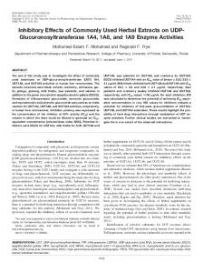
OTC Drugs for Aches, Pains, and Headaches: OTC pain medicines can help with headache, arthritis pain, sprains, and other minor joint and muscle problems. Acetaminophen and nonsteroidal anti-inflammatory drugs (NSAIDs) such as aspirin, ibuprofen and naproxen (Figure 1) are used as OTC drugs for pain relief. Acetaminophen (Tylenol) and ibuprofen (Advil, Motrin) used to reduce fever in children and adults.
16
Hatem Amin Hejaz and Rafik Karaman O CH3
O
OH O
CH3
In c.
HN
O
OH
Acetaminophen
Aspirin
O
CH3 OH
CH3
he rs
O
H3C
H3C
CH3
HO
Naproxen
O
Ibuprofen
Figure 1. Chemical structures of acetaminophen, aspirin, naproxen and ibuprofen.
Pu bl
is
OTC Drugs for Cold, Sore Throat and Cough: Cold medicines can treat symptoms not the specific cold viruses. Treatment the cold symptoms make the patient feels better, they don't cure the cold, but they can bring relief, lighter symptoms, or maybe even shorten the cold. Using zinc supplements within 24 hours of the start of a cold may reduce the symptoms and duration of the cold. Cough medicines include guaifenesin (Figure 2) which helps break up mucus, menthol throat lozenges which soothe "tickle" in the throat. Liquid cough medicines with dextromethorphan (Figure 2) suppress the cough (Benylin, Delsym, Robitussin DM, and Simply Cough, Vicks, and store brands).
O
CH3
H3C
OH
a
O
OH
ov
Guaifenesin
N
O
H
N
CH3
Dextromethorphan OH NH CH3
CH3
Pseudophedrine
Figure 2. Chemical structures of guaifenesin, dextromethorphan and pseudoephedrine.
17
Drug Overview
he rs
In c.
Decongestants help clear a runny nose and relieve postnasal drip. Examples of oral decongestants include pseudoephedrine (Figure 2) (Contact Non-Drowsy, Sudafed, and store brands), phenylephrine (Sudafed PE and store brands). Examples of decongestant nasal sprays are oxymetazoline (Afrin, Neo-Synephrine Nighttime, Sinex Spray), phenylephrine (Neo-Synephrine, Sinex Capsules) [45-47]. OTC Drugs for Allergy: In general, there is no cure for allergies, but there are several types of medications available, both OTC and prescription. These medications help ease and treat annoying symptoms like congestion and runny nose. These allergy drugs include antihistamines, decongestants, combination drugs, corticosteroids, and others. For allergies antihistamine pills and liquids work well for treating allergy symptoms. Among the Antihistamines that may cause sleepiness are diphenhydramine (Benadryl), chlorpheniramine (Chlor-Trimetron) (Figure 3), brompheniramine (Dimetapp), or clemastine (Tavist). Examples of antihistamines that cause little or no sleepiness are loratadine (Alavert, Claritin, Dimetapp ND), fexofenadine (Allegra), cetirizine (Zyrtec) (Figure 3). O
O
CH3 H3C
OH
N
N
is
N
Pu bl
N
Cl
Chlorpheniramine
Cl
Cetirizine
Figure 3. Chemical structures of chlorpheniramine and cetirizine.
N
ov
a
OTC Drugs for Diarrhea, Nausea and Vomiting: Diarrhea, vomiting and nausea are common complaints that can result from diseases such as gastrointestinal and inflammatory bowel diseases, conditions such as food allergies, and from usage of antibiotics or other drugs. The fluid and electrolytes lost during diarrhea and vomiting need to be replaced promptly. Anti-diarrhea medicines such as loperamide (Imodium) are effective for terminating diarrhea. Medicines that contain bismuth may be taken for mild diarrhea (Kaopectate, Pepto-Bismol). Rehydration fluids may be used for moderate and severe diarrhea (Enfalyte or Pedialyte). For mild nausea and vomiting, liquids and pills for stomach upset may help (Emetrol; Pepto-Bismol). Rehydration fluids may be used to replace fluids from vomiting (Enfalyte or Pedialyte). Medicines for motion sickness such as dimenhydrinate (Dramamine), meclizine (Bonine, Antivert, Postafen, and Sea Legs) can be effective in nausea and vomiting conditions [45-47].
18
Hatem Amin Hejaz and Rafik Karaman
O
H3C H3C
he rs
In c.
OTC Drugs for Skin Rashes and Itching: Oral antihistamines may help in itching and allergy conditions. Hydrocortisone cream may help with mild rashes (Cortaid, Cortizone 10). Anti-fungal creams and ointments may help with diaper rashes and rashes caused by yeast (nystatin, miconazole, clotrimazole, and ketoconazole) [45-47]. Top Twenty OTC Medications Sold in US: The following are the top twenty OTC drugs sold in the united states: Advil (ibuprofen), Aleve (naproxen), Cēpacol® antibacterialmulti-protection mouthwash (cetylpyridinium chloride), Children's Dimetapp Cold and Cough (brompheniramine maleate, dextromethorphan HBr, and phenylephrine HCl), Claritin (loratadine), Colace (docusate sodium), Cortaid Maximum Strength (hydrocortisone acetate), Dulcolax (docusate sodium or bisacodyl), Excedrin Extra Strength (acetaminophen, aspirin, caffeine), Gaviscon (aluminum hydroxide, magnesium carbonate), Lotrimin® AF Antifungal (miconazole nitrate, Figure 4), Maalox Antacid (aluminum hydroxide, magnesium carbonate), Midol (ibuprofen), Motrin IB (ibuprofen), Orajel Maximum Strength (benzocaine Figure 4), Pepto-Bismol (bismuth subsalicylate), Rolaids® Multi-Symptom (calcium carbonate, dimethicone or simethicone), Tagamet HB (cimetidine), Tylenol® (acetaminophen) and Zantac75® (ranitidine hydrochloride, Figure 4) [47].
+
O
N
N
S
O
NH
Pu bl
is
NH
H2N
O
CH3
-
CH3
Ranitidine
Cl
Cl N
N
O
Benzocaine
O Cl
Cl
Miconazole
N
ov
a
Figure 4. Chemical structures of ranitidine, benzocaine and miconazole.
Drug Administration
Drugs, both medicinal and recreational, can be administered by a number of ways. Many drugs can be administered in a variety of ways rather than just one as the following [48]. Bolus: is the administration of a medication, drug or other compound that is given to raise its concentration in blood to an effective level. The administration can be given intravenously, by intramuscular, intrathecal or subcutaneous injection. An intravenous injection can be more difficult to administer than a subcutaneous or intramuscular injection because inserting a needle or catheter into a vein may be difficult, especially if the person is obese. The
19
Drug Overview
N
ov
a
Pu bl
is
he rs
In c.
subcutaneous route is used for many protein drugs because such drugs would be destroyed in the digestive tract if they are taken orally. Inhaled: Inhaled medications can be absorbed quickly, and act both locally and systemically. They are breathed into the lungs as an aerosol or dry powder. This includes smoking a substance. Usually, this method of administration is used to administer drugs that act specifically on the lungs, such as aerosolized antiasthmatic drugs in metered-dose containers (called inhalers) and gases used for general anesthesia. Injected: They can be as a solution, suspension or emulsion. They are given either intramuscular, or intravenous, or intraperitoneal or intraosseous. Intravenous administration is the best way to deliver a precise dose quickly and in a well-controlled manner throughout the body. The intramuscular route is preferred over the subcutaneous route when larger volumes of a drug are needed. Insufflation: snorted into the nose. Drugs administered by this route generally work quickly. Some of them irritate the nasal passages. Orally: The oral route is the most common route of drug administration. It may be in the form of a liquid or solid (tablets, capsules, syrup, emulsions or powders) that is absorbed through the intestines. The advantages of this route are its convenience, cheapest, easy to use, safe and acceptable. While the disadvantages of this route include less amount of a drug reaches the target tissue, some of the drug is destroyed by gastric juices, the drug‘s absorption is slow, gastric irritation may be caused by the drug, objectionable in taste and discoloration of teeth. Rectally: Drugs in solid forms such as suppositories or in liquid forms such as enema are given by this route and absorbed by the rectum or colon. This route is mostly used in old patients and pediatrics that have difficulties in swallowing. Drugs may have local or systemic actions after absorption. The advantages of this route are preferred in unconscious or uncooperative patients, avoiding nausea or vomiting can be achieved using this route and drugs are not destroyed by enzymes (avoid hepatic first pass metabolism, drugs given by rectal route have 50% first pass metabolism). While the disadvantages are patients dislike suppositories or may be not acceptable by the patients. Locally acting drugs include glycerin and bisacodyl suppository and systemic acting drugs include indomethacin (antiinflammatory) and aminophylline (bronchodilator). Sublingually: drugs are diffused into the blood through tissues under the tongue. Topically: usually as a cream or ointment. A drug administered in this manner may be given to act locally or systemically. Vaginally: Some drugs may be administered vaginally to women as a solution, tablet, cream, gel, suppository or ring. The drug is slowly absorbed through the vaginal wall. Suppositories are primarily used to treat vaginal infections. This route is often used to give estrogen to women during menopause to relieve vaginal symptoms such as dryness, soreness and redness. Recreational Drugs: Recreational drugs are chemical substances that affect the central nervous system (CNS) such as opioids or hallucinogens. They may be used for effects on perception, consciousness, personality and behavior. They are taken for enjoyment, or leisure purposes, rather than for medical reasons. Alcohol, nicotine, and caffeine are the most widely consumed psychotropic drugs worldwide. Some drugs can cause addiction and habituation and all of these drugs have side effects [26-35]. The use of these drugs is incredibly common around the world and it very often leads to disaster and crime.
20
Hatem Amin Hejaz and Rafik Karaman
CH3 O
N
CH3
O
H
CH3
HN
Lysergic acid diethylamide
OH
he rs
HN N
In c.
Spiritual Drugs (Entheogen): Spiritual drugs are chemical substances, typically of plant origin or may be synthesized, that are ingested to produce unordinary state of consciousness for religious or spiritual purposes. These drugs can cause severe damage to mental or physical health. There are a number of drugs that are said to induce spiritual experiences including lysergic acid diethylamide (LSD) (Figure 5), peyote, ayahuasca, psilocybin (magic mushrooms), ecstasy, marijuana, mescaline and etc. [26-35].
Cl
S N O CH 3
O
Tianeptine
is
CH3
NH2
Amphetamine
Pu bl
Figure 5. Chemical structures of lysergic acid diethylamide, tianeptine and amphetamine.
N
ov
a
Nootropic Drugs: Nootropic drugs also referred to as smart drugs, memory enhancers, neuro enhancers, cognitive enhancers, and intelligence enhancers, are drugs, supplements, nutraceutical, and functional foods that improve one or more aspects of mental function, such as working memory, motivation, and attention. These drugs are used primarily to treat people with cognitive or motor function difficulties attributable to such disorders as Alzheimer's disease, Parkinson's disease, Huntington's disease and ADHD. Examples for such drugs include stimulants (amphetamine, Figure 5), methylphenidate, eugeroics (armodafinil and modafinil), xanthines and nicotine. Certain stimulants will enhance cognition in the general population, but only when used at low (therapeutic) concentrations. Relatively high doses of stimulants will result in cognitive deficits. Other examples are phosphatidylserine, tianeptine (Figure 5), valproate and isoflavones [26-35].
Drug Interactions
A drug interaction occurs when a drug affects the activity of another drug when both are administered together. Drug interactions can result in unwanted side effects, reduce the effectiveness of a medicine or possibly increase the action of a particular medicine or produce a new effect that neither produces on its own. Drug interactions are usually divided into four
21
Drug Overview
N
ov
a
Pu bl
is
he rs
In c.
groups: antagonism (drug's reduces or blocks the effect of another), synergism (drug's effect is increased), potentiation (drug A boosts the effects of drug B), and interaction with metabolism. There are different risk factors of drug interactions: age (elderly or young), multiple disease, multiple drug therapy, renal and liver impairment, narrow therapeutic index (e.g., Insulin, Lithium, Digoxin, Warfarin, Phenytoin, Theophylline, Phenobarbitone) and enzyme inhibitors or inducers. The outcomes of drug interactions might be loss of therapeutic effect, toxicity, unexpected increase in pharmacological activity, beneficial effects e.g., additive and potentiation (intended) or antagonism (unintended) and chemical or physical interaction (e.g., I.V incompatibility in fluid or syringes mixture). Drug interactions may result from pharmacokinetic interactions (absorption, distribution, metabolism, and excretion) or from interactions at drug receptors. These interactions may occur out of accidental misuse or due to lack of knowledge about the active ingredients involved in the relevant substances. Interactions may also exist between drugs and foods (drug-food interactions), as well as drugs and medicinal plants or herbs (drug-plant interactions). Drug-food interactions can happen with both prescription and OTC medicines. Not all medicines are affected by food, but many medicines can be affected by what we eat and when we eat. An example of a drugfood interaction; patients who take monoamine oxidase inhibitors (antidepressants) should not take food containing tyramine (found in cheese) because hypertensive crisis may occur [49-53]. The pharmacological interactions of drugs are important to know and understand. For example; if a person is taking two drugs and one increases the effect of the other it is possible that an overdose may occur (toxicity occurs). The interaction of the two drugs may also increase the risk of side effects. On the other hand, if the action of a drug is reduced it may reduce or diminish any therapeutic effect because of under dosage. Nevertheless, sometimes the interactions may improve the therapeutic effect. Examples of this include the use of codeine with paracetamol to increase the analgesic effect of the medication. The combination of clavulanic acid with amoxicillin is another example to overcome the bacterial resistance to antibiotics. The interactions that are important are those that have negative effects on patients. The risk that a pharmacological interaction will cause is increased as a function of the number of drugs administered to a patient at the same time. As mentioned before, it is possible that an interaction will occur between a drug and another substance present in the body (such as food or alcohol). In certain specific situations a drug may even react with itself (hypersensitivity). In other situations, the interaction does not involve any effect on the drug. In certain cases, the presence of a drug in an individual's blood may affect certain types of laboratory analysis (analytical interference). It is also possible for interactions to occur outside the organism before the administration of a drug. This can occur when two drugs are mixed together. Some classic examples of this type of interaction include thiopentone and suxamethonium (Figure 6) which should not be placed in the same syringe and the same is also true for benzylpenicillin and heparin.
22
Hatem Amin Hejaz and Rafik Karaman CH3 Et
HN S
CH3 N H
O
Thiopentone
CH3
O H3C H3C
+
N
+
O
CH3
O O
Suxamethonium
Figure 6. Chemical structures of thiopentone and suxamethonium.
CH3
In c.
O
N
CH3
he rs
Drug interactions may be due to various processes such as alterations in the pharmacokinetics of the drug (alterations absorption, distribution, metabolism, and excretion (ADME)) or/and interactions in the pharmacodynamic properties of the drug (coadministration of a receptor antagonist and an agonist for the same receptor). Pharmacodynamics is related to the pharmacological activity of the interacting drugs e.g., synergism, antagonism, altered cellular transport, effect on the receptor site [49-53].
is
Types of Drug Interactions
Pu bl
As mentioned before, drug interaction is the modification of the effect of one drug (the object drug) by the prior concomitant administration of another (precipitant drug). There are different types of drug interaction: (i) pharmacokinetic interactions, (ii) pharmacodynamic interactions, (iii) drug-nutrient interactions, (iv) drug-disease interactions and (v) pharmaceutical interactions. The outcomes of drug interactions are loss of therapeutic effect, toxicity, unexpected increase in pharmacological activity, beneficial effects such as additive and potentiation (intended) or antagonism (unintended) and chemical or physical interaction such as I.V incompatibility in fluid or syringes mixture. There are essentially two types of drug interactions: pharmacodynamic and pharmacokinetic.
a
Pharmacokinetic Interactions
N
ov
Pharmacokinetic interactions involve the effect of a drug on another and the drugs absorption, distribution, metabolism and excretion are the important factors playing roles in such interactions. Pharmacokinetic interactions are those in which one drug results in an alteration (increase or decrease) of the concentration of another drug in the system. Different parameters can be affected by pharmacokinetic interactions, including a drug‘s bioavailability, volume of distribution, peak level, clearance and half-life. Such changes can lead to changes in drug plasma concentrations and ultimately increase the risk of side effects or diminish the efficacy of one or more drugs. Pharmacokinetic interactions are more complicated and difficult to predict because the interacting drugs often have unrelated actions.
23
Drug Overview
N
ov
a
Pu bl
is
he rs
In c.
The paragraphs below are a brief summary and some examples of the four pharmacokinetic processes that may be involved individually or collectively in various drug interactions. Absorption: Drug absorption refers to the route or method by which the drug reaches the blood supply (the movement of drugs into the body depends on how the drug is administered). There are three areas in which interactions might occur at the level of drug absorption. One drug may affect the rate and/or extent of absorption of other drugs if it alters GI motility, gastric pH or chemically binds with other drugs to form insoluble, nonabsorbable complexes. Alteration of GIT absorption can occur as a result of either (1) alteration of pH (such as antacids and H2 antagonists decrease the pH and thus decrease the absorption of Ketoconazole, (2) alteration bacterial flora (e.g., In 10% of patients receive digoxin about 40% or more of the administered dose is metabolized by the intestinal flora). If antibiotics are administered with digoxin, a toxicity might occur as digoxin concentration increases because antibiotics kill a large number of the normal flora of the intestine, (3) formation of drug chelates or complexes (e.g., tetracycline interacts with iron preparations, milk products and antacids. Aluminum or magnesium hydroxides decrease the absorption of tetracycline by 85% due to chelation, (4) drug induced mucosal damage (e.g., antineoplastic agents such as cyclophosphamide, vincristine, procarbazine which inhibit the absorption of several drugs such as digoxin) and (5) altered GIT motility (e.g., metoclopramide (antiemetic), increases the toxicity of cyclosporine because it increases its absorption due to the increase of stomach empting time). Displaced protein binding (distribution): Distribution is the movement of the drugs around the body. Once the drug is absorbed, it is rapidly distributed around the blood supply, and then slowly distributed to the various tissues and organs. The rate and extent of the drug distribution depends on the blood flow, tissue size, and affinity of the drug to plasma protein (albumin) and tissue components, and permeability of tissue membranes. The most likely bound drugs are capable to displace others. The free drug is increased by displacement by another drug with higher affinity. For example, drugs that are highly bound to plasma protein such as phenytoin, tolbutamide and warfarin (Figure 7) can displace other agents with lower affinity to plasma protein such as aspirin, sulfonamides and phenylbutazone. Warfarin and methotrexate bound to albumin and plasma protein in the blood and they will be unavailable to interact with their targets. When another drug is taken with these medications which have the ability to compete for plasma protein binding (e.g., sulphonamide), a certain percentage of previously bounded drug (warfarin or methotrexate) is released, thus increasing the free form of the drug and consequently its effect. Altered metabolism: the effect of one drug on the metabolism of the other is well documented. The liver is the major site of drug metabolism but metabolism can occur in other organs such as WBC, skin, lung, and GIT. Cytochrome P450 family is the major metabolizing enzyme in phase I (oxidation process). Therefore, the effect of drugs on the rate of metabolism of others can involve the followings: (a) Enzyme induction; a drug may induce the enzyme that is responsible for the metabolism of another drug or even itself such as in the case of carbamazepine (antiepileptic drug, Figure 8) which increases its own metabolism. Phenytoin increases hepatic metabolism of theophylline (Figure 8), leading to a decrease in the latter‘s concentrations level and
24
Hatem Amin Hejaz and Rafik Karaman
reduces its therapeutic action. Enzyme induction involves protein synthesis. Therefore, it needs time up to 3 weeks to reach a maximal effect.
NH
S O
O
OH
O H3C
In c.
CH3
CH3
NH O
O
Warfarin
he rs
Tolbutamide
O
Figure 7. Chemical structures of tolbutamide and warfarin.
O
H N
O H2N
N H
Phenytoin
Pu bl
Carbamazepine
O
H3C
is
N
O
O
N
N
H N N
CH3
Theophylline
Figure 8. Chemical structures of carbamazepine, pheytoin and theophylline.
N
ov
a
(b) Enzyme inhibition; it is a decrease of the rate of metabolism of a drug by another one. This will lead to an increase of the concentration of the target drug and consequently to an increase of its toxicity. Inhibition of the enzyme may be due to the competition on its binding sites. When an enzyme inducer such as carbamazepine is administered with an inhibitor such as verapamil (Figure 9), the effect of the inhibitor will be predominant. Erythromycin inhibits the metabolism of astemazole and terfenadine, thus increases their serum concentrations and leads to an increase of the life threatening cardio-toxicity. Another example is omeprazole (Figure 9) which inhibits the oxidative metabolism of diazepam (Figure 9). Excretion: Renal excretion (active tubular secretion); it occurs in the proximal tubules, a portion of renal tubules. The drug combines with a specific protein to pass through the proximal tubules. When a drug has a competitive reactivity to the protein that is responsible for the active transport of another drug this will reduce a drug excretion increasing its concentration and hence its toxicity. For example, probenecid (Figure 10) decreases tubular secretion of methotrexate (Figure 10). Passive tubular reabsorption; excretion and reabsorption of drugs occur in the tubules by passive diffusion which is regulated by concentration and lipid pharmacodynamics interactions solubility. Ionized drugs are reabsorbed in a lower extent than non-ionized ones. Sodium bicarbonate increases lithium clearance and decreases its action. Antacids increase salicylates clearance and decrease their action [54-58].
25
Drug Overview
O
CH3
CH3
N
O
H3C
O
Cl
CH3 CH3
N
N
CH3
CH3
Verapamil
Diazepam H3C
O
O
CH3 CH3
N
he rs
H3C
O
N
O
In c.
H3C
S N H
N
O
Omeprazole
Figure 9. Chemical structures verapamil, diazepam and omeprazole.
N
S
is
O
H3C
O
Pu bl
O
CH3
OH
Probenecid
HO
O
O
a
NH2
N
N
ov
H2N
N
N
N
OH
NH
N CH3
Figure 10. Chemical structures of probenecid and methotrexate.
O
Methotrexate
26
Hatem Amin Hejaz and Rafik Karaman
Pharmacodynamic Interactions
In c.
Pharmacodynamic interactions are alteration of the dug action without a change in its serum concentration by pharmacokinetic factors. The Pharmacodynamics interactions types are:
S O H2N
N N H
O
N
CH3
H2N
Pu bl
Sulfamethoxazole
O
N
is
O
he rs
A synergism (1+1=3): When the therapeutic or toxic effects of two drugs are greater than the effect of an individual drug, it is called synergism. Drug synergism is of two types: additive effect and potentiation. Propranolol + verapamil is synergistic or additive effect Additive effect (1+1=2): when the net effect of two drugs used together is equal to the sum of the individual drug effects, the drugs are said to have an additive effect. For example, the combination of a thiazide diuretic and a beta adrenergic blocking drug used for the treatment of hypertension. Potentiation (1+0=2): when the net effect of two drugs used together is greater than the sum of the individual drug effects, the drugs are said to have potentiation effect. For example, the combination of sulfamethoxazole (Figure 11) and trimethoprim (Figure 11) used as antimicrobial agents.
NH2
O
O
CH3 CH3
CH3
Trimethoprim
Figure 11. Chemical structures of sulfamethoxazole and trimethoprim.
N
ov
a
Antagonism (1+1=0 or 0.5): the effect of one drug can be reduced or abolished by the presence of another drug and this effect is termed drug antagonism. Drug antagonism is of three types- chemical, physiological and pharmacological. Physiological and pharmacological antagonisms involve the interaction of an agonist with an antagonist. Chemical antagonism; when a drug antagonizes the effect of another drug by simple chemical reaction without activating a receptor. For example, antacid neutralizes the gastric acid. Physiological antagonism; when the physiological effect of a drug is antagonized by another drug by acting on two different types of receptors. For example, acetylcholine causes contraction of an intestinal smooth muscle by acting on muscarinic choline-receptors. Whereas this action of acetylcholine is antagonized (that is relaxation of the intestinal smooth muscle) by adrenaline. Pharmacological antagonism; when a drug antagonizes the effect of another drug by acting on the same receptor it is called pharmacological antagonism. Pharmacological antagonism is of two types: competitive and noncompetitive. Competitive antagonism; competitive antagonism is reversible. The inhibitory effect of an antagonist is overcome by using large amount of agonist. Here, both, the agonist and antagonist compete for the same receptor and are able to displace each other at the receptor site. For example, acetylcholine causes contraction of intestinal smooth muscle. Atropine
27
Drug Overview
H3C
N
H
H
HO
CH3
O
O
In c.
(Figure 12) blocks the effect of acetylcholine (Figure 12). When a low concentration of an agonist is plotted against the responses, a sigmoid shaped curve will be obtained. The maximum response of the agonist is obtained in the presence of competitive antagonist. The dose-response curve remains parallel but is shifted to the right.
+
H3C
Atropine
O
CH3
CH3
he rs
O
N
Acetylcholine
Figure 12. Chemical structures of atropine and acetylcholine.
Noncompetitive antagonism; the maximum response of an agonist in the presence of antagonist is reduced. The inhibitory effect of a drug is not overcome by using large amount of agonist [48, 54, 57, 59-63].
is
Drug-Nutrient Interactions
N
ov
a
Pu bl
Nutrition can affect the body's response to drugs; conversely, drugs can affect the body's nutrition. Food, beverages, and dietary supplements alter the effects of drugs the person takes. Foods can enhance, delay, or decrease drug absorption. The presence of food in the digestive tract may reduce the absorption of a drug. Often, such interactions can be avoided by taking the drug 1 hour before or 2 hours after eating. Dietary supplements are regulated as foods, not as drugs, so they are not tested comprehensively. However, they may interact with prescription or OTC drugs. People who take dietary supplements should inform their doctors and pharmacists, so that interactions can be avoided. For instance, taking alcohol with metronidazole can cause flushing, headache, palpitations, and nausea and vomiting. Drugs such as terfenadine, cyclosporine and felodipine interact with grapefruit juice. Other food, such as orange juice, coffee, or mineral water, may reduce the absorption and effectiveness of these drugs. Anticoagulants interact with foods containing high concentrations of vitamin K (such as broccoli, brussels sprouts, spinach, and kale). Such foods may reduce the effectiveness of anticoagulants such as warfarin and increase the risk of clotting. Intake of such foods should be limited, and the amount consumed daily should remain constant. Digoxin interacts with oat meals, the fiber in oat meal and other cereals, when consumed in large amounts, can interfere with the absorption of digoxin. Tetracycline interacts with calcium or foods containing calcium, such as milk and other dairy products. These foods can reduce the absorption of tetracycline which should be taken 1 hour before or 2 hours after meals [64-67].
28
Hatem Amin Hejaz and Rafik Karaman
Drug-Disease Interactions
he rs
Pharmaceutical Interactions
In c.
Sometimes, drugs that are helpful in one disease are harmful in another. Drug-disease interactions can occur in any age group but are common among old people, who tend to have more diseases. For example, some beta-blockers taken for heart disease or high blood pressure can worsen asthma. This interaction makes it hard for people with diabetes to tell when their blood sugar is too low. Some drugs taken to treat a cold may worsen glaucoma. People should tell their doctor all of the diseases they have before the doctor prescribes a new drug [68-72].
a
Pu bl
is
Pharmaceutical interactions occur when two drugs react chemically or physically during administration or absorption so that the amount of drug available for absorption (of one or both drugs) is altered. Pharmaceutical interactions also occur as a result of mixing two or more antagonistic substances and an undesirable product is formed which may affect the safety, efficacy and appearance of pharmaceutical preparation. The types of interactions are: (i) physical interaction, examples immiscibility, insolubility, precipitation and liquefaction, (ii) chemical interaction; (1) tolerated: the chemical interaction can be minimized by changing the order of mixing or mixing the solutions in dilute forms but no alteration is made in the formulation, (2) adjusted: the chemical interaction can be prevented by an addition or substitution of one of the reacting ingredients of a prescription with another of equal therapeutic value [73-76]. Serious loss of potency can occur from incompatibility between an infusion fluid and a drug that is added to it. Similarly, an addition of more than one drug to the same infusion fluid may result in interactions which cause loss of activity. The immediate effect of soluble insulin is reduced if it is drawn up with potassium zinc insulin in the same syringe or drip [73-76]. Examples of pharmaceutical interactions are mixture of thiopentone and suxamethonium (precipitation), mixture of diazepam and infusion fluid (precipitation), mixture of phenytoin and infusion fluid (precipitation), mixture of heparin and hydrocortisone (inactivation of heparin), mixture of kanamycin and hydrocortisone (inactivation of kanamycin) and mixture of carbenicillin and gentamicin (inactivation of gentamicin). Table 2 illustrates some examples of drug interactions.
N
ov
Table 2. Examples of some important drug interactions
Interaction SSRIs with selegiline or nonselective monoamine oxidase inhibitor SSRIs with tramadol
Effect Hypertensive crisis
Time to Effect Soon after initiation
Recommendations Avoid.
Seizures; serotonin syndrome
Any time
Monitor the patient for signs and symptoms of serotonin syndrome
29
Drug Overview Time to Effect Any time Possibly after initial dose
Warfarin effects increased TCA level increased
Any time
Warfarin with ciprofloxacin, clarithromycin, erythromycin, metronidazole Warfarin with acetaminophen
Warfarin effects increased
within 1 week
Bleeding & INR increased
Any time
Warfarin with aspirin
Bleeding & INR increased
Any time
Warfarin with NSAIDs
Bleeding & INR increased
Any time
Fluoroquinolones with sucralfate Carbamazepine with cimetidine, erythromycin, clarithromycin or fluconazole Phenytoin with cimetidine, erythromycin, clarithromycin or fluconazole Phenobarbital with cimetidine, erythromycin, clarithromycin or fluconazole Phenytoin with rifampin
Fluoroquinolones absorption decreased Carbamazepine levels Increased
Any time
a
Phenobarbital with rifampin
N
ov
Carbamazepine with rifampin Lithium with NSAID or diuretic Oral contraceptive with rifampin
Monitor for anticholinergic excess and consider lower dosage of TCA Use another antibiotic
Reduce the dose of acetaminophen and monitor INR. Reduce the dose of aspirin to 100/ day and monitor INR. Avoid combination or use a cyclooxygenase-2 inhibitor instead of Cox-1 inhibitor and monitor INR. Space administration by 2 -4 hr. Monitor carbamazepine levels
he rs
Any time
within 1 week
Pu bl
SSRIs with tricyclic antidepressant (TCA)
Recommendations Avoid Avoid. monitor the patient for signs and symptoms of serotonin syndrome Monitor INR.
In c.
Effect Serotonin syndrome Serotonin syndrome
is
Interaction SSRIs with St. John's wort SSRIs with naratriptan, rizatriptan, sumatriptan or zolmitriptan Lovastatin with warfarin
Phenytoin levels increased
within 1 week
Monitor phenytoin levels
Phenobarbital levels increased
within 1 week
Monitor phenobarbital levels
Phenytoin levels decreased Phenobarbital levels decreased Carbamazepine levels decreased Lithium levels increased
within 1 week
Monitor phenytoin levels.
within 1 week
Effectiveness of contraception decreased
Any time
Monitor phenobarbital levels. Monitor carbamazepine levels. Decrease lithium dosage by 50% and monitor lithium levels Avoid combination or use contraceptive pill with a higher estrogen or use different method of contraception
within 1 week Any time
30
Hatem Amin Hejaz and Rafik Karaman Table 2. (Continued) Time to Effect Any time
Recommendations Avoid combination or use another contraceptive
Any time
Cisapride with erythromycin, clarithromycin, fluconazole, itraconazole, ketoconazole, nefazodone Cisapride with class IA or class III antiarrhythmic agents, tricyclic antidepressants or phenothiazine Sildenafil (Viagra) with nitrates
Cisapride metabolism inhibited
within 1 week
Avoid combination or use contraceptive pill with a higher estrogen or use different method of contraception Avoid. Or use metoclopramide
Sildenafil with cimetidine, erythromycin, itraconazole or ketoconazole HMG-CoA reductase inhibitor with niacin, gemfibrozil, erythromycin or itraconazole
Sildenafil levels increased
he rs
Prolongation of QT interval along with arrhythmias
Any time
Avoid. Or use metoclopramide.
Acute hypotension
Directly after taking sildenafil Any time
Absolute contraindication
Rhabdomyolysis
Avoid combination
Any time
Pu bl
Oral contraceptive with troglitazone
In c.
Effect Effectiveness of contraception decreased Effectiveness of contraception decreased
is
Interaction Oral contraceptive with antibiotics
Initiate sildenafil at a 25-mg dose
Prescription or Nonprescription Drugs
N
ov
a
According to laws, drugs are divided into two groups: prescription drugs and nonprescription drugs (OTC). Prescription drugs are used only under medical supervision and dispensed only with a prescription from a licensed medical professional (physician, dentist or veterinarian). Nonprescription drugs are used without medical supervision (such as aspirin) and are sold as OTC. In the United States, the FDA is the government agency that decides which drugs require a prescription and which don't require prescription [77-80]. Dietary supplements do not require the FDA approval before marketing; thus their safety and efficacy standards are different and not the same as prescription or OTC drugs because these products are not classified as drugs. Dietary supplements include medicinal herbs and nutraceutical which given as supplement to the diet. These products may contain vitamins, amino acids, minerals, and herbs or other plant-derived material. They can act in the same way as a drug does in the body and may cause health problems if not used correctly or if taken in large amounts. These products may not claim to treat specific medical conditions because they do not meet the FDA standards for safety and efficacy [77-80].
31
Drug Overview
Drug Names
Pu bl
is
he rs
In c.
Drugs have three or more names including a chemical name, brand or trade name (proprietary, given by the pharmaceutical company it is developed by), and generic or common name (nonproprietary or official, scientific name, named for the active ingredient of the medicine). The chemical name is assigned according to rules of nomenclature of chemical compounds. The brand name is always capitalized and is selected by the manufacturer. The generic name refers to a common established name irrespective of its manufacturer. For example, sildenafil is the generic name of a medicine used to treat erectile dysfunction. However, the company that makes sildenafil, Pfizer, sells it under the brand name Viagra. In most cases, a drug bearing a generic name is equivalent to the same drug with a brand name. Some knowledge of drug names can help in understanding drug product labels. The names of medicines can often be confusing, as the same medicine can sometimes be called differently. The chemical name describes the atomic or molecular structure of the drug. The chemical name is usually too complex and cumbersome for general use. So an official body assigns a generic name to a drug. The generic names for drugs of a particular type (class) usually have the same ending. For example, the names of all beta-blockers, which are used to treat such disorders as high blood pressure, end in "lol" such as metoprolol and propranolol. The trade name is chosen by the pharmaceutical company that manufactures or distributes the drug. Patented drugs are usually sold under a trade name. Generic versions of trade-name drugs manufactured after expiration of the pharmaceutical company's patent may be sold under the generic name (for example, ibuprofen) or under the manufacturer's own trade name (for example, Advil) [81-84].
Drug Development
N
ov
a
Before a drug is tested in human, the drug should be tested in a laboratory and animals to determine how the drug works and to evaluate its safety in humans. Drug Development is the process of bringing a new drug to the market after identification of the lead compound through a drug discovery process. Drug development includes pre-clinical research where the experiments are conducted in microorganisms/animals and clinical trials where studies are carried out on humans. The drug development process may include obtaining an approval from regulatory bodies to market the drug. The drug discovery process begins by focusing on specific diseases and patient needs. Researchers search for biological targets within the body that play a role in a given disease. Targets can be part of the body (such as a protein, receptor, or gene) or foreign (such as a virus or bacteria). Researchers identify, design, and synthesize promising molecules, screening perhaps tens of thousands of compounds, to assess their effect on the relevant biological targets. Molecules that have the desired effect on the target and meet other design criteria become ―lead‖ molecules that go on to the next phase of development. The structure of the lead compound is usually modified many times to optimize its specificity and selectivity to target site, its potency, efficacy, effect, safety, stability, absorption, pharmacokinetics and pharmacodynamics properties. The structure modification of the lead compound is known as structure activity relationship (SAR). In general, SAR also
32
Hatem Amin Hejaz and Rafik Karaman
N
ov
a
Pu bl
is
he rs
In c.
overcome the problems associated with certain drugs such as physicochemical properties (solubility, acidity/basicity, reactivity, binding affinity), pharmacokinetics (absorption, distribution, metabolism, excretion), pharmacodynamics (onset of action, duration of action), pharmaceutics (formulation, stability) and pharmacological (effect, side effects, mode of action) in order to have optimum drugs that are suitable for clinical uses. These factors involve what the body does to the drug and what the drug does to the body. Ideally, when a drug is highly selective for its target site it has minimal or no side effects. The drug should also be highly potent and effective, thus low doses are used in order to minimize the side effects of the drug. The drug should be effective when taken by mouth, absorbed well from the digestive tract, and reasonably stable in body tissues and fluids so that, ideally, one dose a day is adequate. During a drug development, standard or average doses are determined. However, people respond to drugs differently. Many factors must be considered when doctors determine the dose for a particular person, including age, weight, genetic make-up, and the presence of other disorders, affect drug response. Development of new drugs is a complex and costly process. It takes an average of 10 years and about $950 million to get a new drug from the laboratory to the market. Only one in 1000 compounds which begin laboratory testing will make it to human testing [85-94]. Early development: After a drug that may be useful in treating a disorder is identified or designed, it will undergo extensive study on animals. Early development gathers information about how the drug works, it‘s effectively, and what toxic effects it produces, including possible effects on reproductive capacity and the health. However, little will be known about the safety, toxicity, pharmacokinetics and metabolism of the drug in humans. The pre-clinical study is the function of drug development to assess all of these parameters prior to human clinical trials. Many drugs are rejected at this stage because they are shown to be too toxic or not effective. If a drug seems promising after early development, a program describing the clinical study must be approved by an appropriate institutional research board (IRB) and an investigational new drug application is filed with the FDA. If the FDA approves the application, the drug is allowed to be tested in people (a phase called clinical studies) [85-94]. Clinical studies: clinical studies involve three or four steps. These studies occur in several phases and only in volunteers who have given their full consent. Phase I trials determine safety and dosing usually in healthy volunteers (20 to 80). The investigation in Phase I is to evaluate the safety and tolerability of the drug as well as its pharmacokinetic and pharmacodynamic properties. In phase I, different amounts of the drug are given to a small number of healthy, young, usually male people to determine the dose at which toxicity first appears. Phase II trials are used to get an initial reading of efficacy and further explore safety in small numbers of sick patients (100 to 500 patient volunteers). In phase-II, safety and tolerability, pharmacokinetic and pharmacodynamic properties, efficiency and dosage to effect relationship are investigated. Phase II evaluates what effect the drug has on the target disorder and what the right dose might be. Different amounts of the drug are given to up to 100 people who have the target disorder to see whether there is any benefit. Just because a drug is effective in animals in early development does not mean it is effective in people. Phase III trials are large, pivotal trials to determine safety and efficacy in sufficiently large number of patients (up to 1000 or more patient volunteers). In this phase, the drug is
33
Drug Overview
N
ov
a
Pu bl
is
he rs
In c.
tested in a much larger (often hundreds to thousands, 300-30,000) group of people who have the target disorder to monitor reaction to long term drug use. These people are selected to be as similar as possible to the people who might use the drug in their life. The drug's effectiveness is studied further, and any new side effects are noted. Phase III tests usually compare the new drug to standard drug or to placebo, or both in multicenter and multinational trials. In addition to determining a drug's effectiveness, studies in people focus on the type and frequency of side effects and on factors that make people susceptible to these effects (such as age, sex, other disorders, and the use of other drugs). The overall aim of phase III is risk-benefit evaluation. The outcome of phase III studies is crucial for the decision taking of the regulatory authorities. Approval: After extensive study and testing, a candidate drug is submitted for regulatory approval. The decision of the regulatory body determines whether or not the drug can be marketed to patients for approved uses. The review process usually takes 6 -24 months or longer. If studies indicate that the drug is effective and safe, a new drug application is filed with the FDA, which reviews all the information and decides whether the drug is sufficiently effective and safe to be marketed. If the FDA approves, the drug becomes available for use. The whole process usually takes about 10 years. On average, only about 5 out of 4,000 drugs studied in the laboratory are studied in people, and only about 1 out of 5 drugs studied in people is approved and prescribed. Overall drug development process on average to bring one drug to market takes 12-15 years. Pre-clinical testing takes 1-3 years, on average 18 months. Clinical research (phase I-III) takes 2-10 years, on average 5 years. The drug development phase is significantly more expensive in terms of time and money than either lead discovery or drug design and many drugs will fail during the wayside. On average, for every 10000 synthesized structures during a drug design, 500 will reach animal testing, 10 will reach phase I clinical trials and only 1 will reach the market place. Phase IV (post marketing): individuals may respond quite differently to the same medication. Thus, after a new drug is approved, the manufacturers continue monitor the use of the drug and promptly report any additional, previously undetected side effects to the FDA. Usually approved medications have been subjected to extensive testing for safety and efficacy. But even after medications are approved for general use, companies continue to collect product-safety information and monitor the safety profile of all products. In fact, the monitoring increases over time, through the collection of information from ongoing clinical studies, spontaneous adverse-event reports voluntarily reported directly from healthcare providers, and patients using the medicine. The FDA may withdraw approval if new evidence indicates that a drug may cause severe side effects [85-94]. Placebos: Dosage form is made to look exactly like a real drug but do not contain an active ingredient (inactive), usually contains sugar or starch used in research studies. Despite there being no active ingredients, some people who take a placebo feel better. Some others develop side effects. This phenomenon, called the placebo effect. The placebo effect is mainly on symptoms rather than the actual disease. When a new drug is being developed, investigators conduct studies to compare the effect of the drug with that of a placebo because any drug can have a placebo effect, unrelated to its action. The true drug effect must be distinguished from a placebo effect. Typically, half the study's participants are given the drug, and half are given an identical-looking placebo. Ideally, neither the participants nor the investigators know who received the drug and who received the placebo.
34
Hatem Amin Hejaz and Rafik Karaman
In c.
When the study is completed, all changes observed in participants taking the active drug are compared with those in participants taking the placebo. The drug must perform significantly better than the placebo to justify its use [95-96].
Drug Effectiveness and Safety
Pu bl
is
he rs
The main goals of drug development are effectiveness and safety, however, there are three main issues involved in the drug development process: (i) the drug has to be tested to ensure that it is not only safe and effective, but can be administered in a suitable fashion. This step involves preclinical and clinical trials to test toxicity, drug metabolism, stability, formulation, and pharmacological aspects, (ii) there are the various patenting and legal issues and (iii) the drug has to be synthesized in ever-increasing quantities for testing and manufacturing. All drugs can harm as well as help, safety is relative, and this depends upon the dose or quantity of the drug. The difference between the usual effective dose and the dose that causes severe or life-threatening side effects is called the margin of safety. A wide margin of safety is desirable, but when treating a dangerous condition or when there is no other alternatives, a narrow margin of safety must be accepted. The most useful drugs are effective and, for the most part, safe. Penicillin is such a drug. Except for people who are allergic to it, penicillin is nontoxic, even in large doses. On the other hand, barbiturates, which were once commonly used as sleep aids, can interfere with breathing, lower blood pressure, and cause death if taken in excess. Sometimes it is difficult to design effective drugs with a wide margin of safety and few side effects. Consequently, some drugs must be used even though they have a very narrow margin of safety. For example, warfarin, which is taken to prevent blood clotting, can cause bleeding, but it is used when the need is so great that the risk must be tolerated. Clozapine is another example. This drug often helps people with schizophrenia when all other drugs have proved ineffective. But clozapine has a serious side effect: it can decrease the production of white blood cells, which are needed to protect against infection. Because of this risk, people who take clozapine must have their blood tested frequently as long as they take the drug [9799].
a
Drug Errors
N
ov
Drug errors are mistakes made by doctors, health care practitioners, pharmacists, and patients when drugs are prescribed, given, taken, or stored. Drug errors can make people ill or allow diseases to worsen or even losses their life. Drug mistakes may be caused by the following:
People become confused and take drugs incorrectly. Doctors choose the wrong drug or write a prescription for the wrong dose. Pharmacists incorrectly read the prescription and give the wrong drug or dose. Health professionals incorrectly read the label of the drug container and give the wrong drug or dose.
35
Drug Overview
The pharmacist or person incorrectly stores the drug, weakening the drug's strength. People use an expired drug.
References
[2] [3] [4] [5]
is
[6]
Pu bl
[7] [8]
Drug. Dictionary.com Unabridged (v 1.1), Random House, Inc., via dictionary.com. Retrieved on 20 September 2007. Drug. The American Heritage Science Dictionary, Houghton Mifflin Company, via dictionary.com. Retrieved on 20 September 2007. Definition and classification of Drug or Pharmaceutical Regulatory aspects of drug approval Accessed 30 Dec 2013. Dorland's Medical Dictionary McWhinney IR. (2007) Health and disease: problems of definition. Canadian Medical Association Journal, 136 (8): 815. Simon Cross. (2013) Principles of Disease Classification. Underwood's Pathology: a Clinical Approach, 6th Edition, Chapter2 Pharmacological classification of drugs with doses and preparations fourth edition, KD Tripathi, 2006, 3ed edition. Finkel, Richard; Cubeddu, Luigi; Clark, Michelle. (2009) Lippencott's Illustrated Reviews: Pharmacology 4th Edition. Lippencott Williams & Wilkins, pp. 1–4. Patrick (1995). An introduction to medicinal chemistry, Graham Chapter-1. Kulinskiĭ LS1, Kolesnichenko LS. (1998) Classification of drugs by biochemical mechanism of action, Vopr Med Khim. Mar-Apr;44 (2):179-84. Imming Pl, Buss T, Dailey LA, Meyer A, Morck H, Ramadan M, Rogosch T. (2004) A classification of drug substances according to their mechanism of action; Pharmazie, 59 (8):579-89. Amidon GL, Lennernäs H, Shah VP et al. (1995) A theoretical basis for a biopharmaceutic drug classification: The correlation of in vitro drug product dissolution and in vivo bioavailability. Pharm. Res; 12 (3): 413-420. http://www.epgonline.org database of prescription pharmaceutical products including drug classifications Medicinal chemistry, by Ashutosh Kar, (2007) Physical-chemical factors and biological activity, structurally specific drugs and structurally nonspecific drugs, chapter 2, page 23. Lees P, Cunningham FM, Elliott J. (2004) Principles of pharmacodynamics and their applications in veterinary pharmacology. J. Vet. Pharmacol. Ther., 27 (6): 397– 414.doi:10.1111/j.1365-2885.2004.00620.x. PMID 15601436
he rs
[1]
[9] [10] [11]
a
[12]
ov
[13]
N
In c.
People should be sure they understand how and when to take a drug when they pick up a prescription. People should keep a written list of all their current drugs and dosages and bring the list to every health care appointment or emergency department visit. If there is any doubt as to which drugs are being used, people are instructed to bring all their drugs to their health care appointments for review [100-103].
[14]
[15]
36
Hatem Amin Hejaz and Rafik Karaman
N
ov
a
Pu bl
is
he rs
In c.
[16] De Boer AG, Moolenaar F, de Leede LG, Breimer DD. (1982) Rectal drug administration: clinical pharmacokinetic considerations. Clin. Pharmacokinetics, 7 (4):285-311. [17] Routes for Drug Administration. Emergency Treatment Guidelines Appendix. Manitoba Health. (2003). Retrieved April 2, 2013. [18] Classifying drugs in pregnancy, volume 37, No. 2: April 2014, Australian Prescriber. [19] Appendix 4: Pregnancy. British National Formulary. 55 edition. March 2008. [20] Hong-Fang Ji, Xue-Juan Li, and Hong-Yu Zhang. (2009) Natural products and drug discovery. Can thousands of years of ancient medical knowledge lead us to new and powerful drug combinations in the fight against cancer and dementia? EMBO Rep.; 10 (3): 194–200. [21] Tariq Ahmad Pharmacological classification of drugs first edition [22] Drug List by Therapeutic Class, Blue Cross and Blue Shield of Texas. 2009 Preferred Drug Guide. Page 1-27. http://www.bcbstx.com/pdf/drug_list_category.pdf [23] KD Tripathi (2010) Pharmacological Classification of Drugs, 4th Edition, [24] James Ritter, Lionel Lewis, Timothy Mant, Albert Ferro. (2008) A Textbook of Clinical Pharmacology and Therapeutics. 5th edition, [25] Drugs from A to Z: A Dictionary, Richard R. Lingeman, 1974 [26] Marc-Antoine Crocq. (2003) Alcohol, nicotine, caffeine, and mental disorders., Dialogues in Clinical Neuroscience Dialogues Clin. Neurosci.; 5(2): 175–185. [27] Charles E. Faupel; Alan M. Horowitz; Greg S. Weaver, Jay Corzine. (2013) The Sociology of American Drug Use. McGraw Hill. p. 366. [28] Patt Denning, Jeannie Little, Adina Glickman. Over the Influence: The Harm Reduction Guide for Managing Drugs and Alcohol Paperback – November 15, 2003 [29] Aarti Sharma, Abraham Jacob, Manas Tandon, and Dushyant Kumar. (2010) Orphan drug: Development trends and strategies. Journal of Pharmacy and Bioallied Sciences. J. Pharm. Bioallied Sci., 2(4): 290–299. [30] Federal Food, Drug, and Cosmetic Act (FD&C Act). [31] Rafael Sampaio Octaviano de SouzaI; Ulysses Paulino de AlbuquerqueI; Júlio Marcelino MonteiroI; Elba Lúcia Cavalcanti de Amorim. a review of its traditional use, phytochemistry and pharmacology... Brazilian Archives of Biology and Technology. Braz. arch. biol. technol. vol.51 no.5 Curitiba Sept./Oct. 2008 [32] Godlaski, Theodore M. (2001) The God within. Substance Use and Misuse., 46 (10): 1217–1222. [33] Carl A. P. Ruck; Jeremy Bigwood; Danny Staples; Jonathan Ott; R. Gordon Wasson. (1979) Entheogens. Journal of Psychedelic Drugs, 11 (1–2): 145–146. [34] Cristina Lanni1, Silvia C. Lenzken1, Alessia Pascale, Igor Del Vecchio, Marco Racchi, Francesca Pistoia, Stefano Govoni. (2008) Cognition enhancers between treating and doping the mind. Pharmacological Research, 57 (3): 196–213. [35] Giurgea C. (1972) Pharmacology of integrative activity of the brain. Attempt at nootropic concept in psychopharmacology. Actual Pharmacol., 25: 115–56. [36] Monetary and Economic Department, Statistical release OTC derivatives statistics at end June 2013, Bank for International Settlements (BIS), retrieved 12 April 2014. http://www.bis.org/publ/otc_hy1311.pdf
37
Drug Overview
N
ov
a
Pu bl
is
he rs
In c.
[37] Market Review of OTC Derivative Bilateral Collateralization Practices, International Swaps and Derivatives Association (ISDA), 1 March 2010, retrieved 12 April 2014. http://www.isda.org/c_and_a/pdf/Collateral-Market-Review.pdf [38] L. Douglas Ried, Sally A. Huston, Suzan N. Kucukarslan, Elliott M. Sogol, Kenneth W. Schafermeyer, and Sujit S. Sansgiry. (2011) Risks, benefits, and issues in creating a behind-the-counter category of medications. J. Am. Pharm. Assoc.; 51:26–39 [39] Frederick H. Branding, R.Ph., J.D.Rahul Narula. Reed Smith. A Middle Class in the United States? Behind the Counter (BTC) Drugs. Page 1-35 http://www.nabp.net /events/assets/Branding12508NABP.pdf [40] Information and Resources FDA Weighs Behind-the-Counter Drugs. Behind-theCounter Drugs Would Be Sold by Pharmacists Without Prescriptions. By Todd Zwillich WebMD Health News. http://www.webmd.com/news/20071114/fda-weighsbehind-counter-drugs?page=2&rdspk=active [41] Alexander GC, O'Connor AB, Stafford RS. (2011) Enhancing prescription drug innovation and adoption. Annals of Internal Medicine. 154 (12): 833–837. [42] Jeffrey K. Aronson. (2009) From prescription-only to over-the-counter medicines (‗PoM to P‘): time for an intermediate category. British Medical Bulletin; 90:63-9. [43] Patients and prescribing: rights and responsibilities. British Medical Association. 23 March 2006. Retrieved 18 April 2010. http://bma.org.uk/ap.nsf/Content/ InfoOnPrescrib0904~Pats&Prescrib [44] Information and Guidance on Prescribing In General Practice. General Practitioners Committee Guidance for GPs. British Medical Association http://www.pennine-gptraining.co.uk/prescribing-in-general-practice-gpc-2004.pdf [45] Over-the-counter medicines. MedlinePlus. A service of the U.S. National Library of Medicine National Institutes of Health. http://www.nlm.nih.gov/medlineplus/ency /article/002208.htm [46] Over-the-Counter Drug Listing, Conexis. 2003, page 1-3. https://www.countyofsb.org /hr/pdf/benefits/04FlexOTCDrugListing.pdf [47] Over the Counter Medications. Drugs. Com. http://www.drugs.com/otc/ [48] Lees P, Cunningham FM, Elliott J. (2004) Principles of pharmacodynamics and their applications in veterinary pharmacology". J. Vet. Pharmacol. Ther., 27 (6): 397–414 [49] Merle L, Laroche M-L, Dantoine T, Charmes J-P. (2005). Predicting and Preventing Adverse Drug Reactions in the Very Old". Drugs and Aging, 22 (5): 375–392. [50] Elizabeth Lipp. (2008) Tackling Drug-Interaction Issues Early On Researchers Explore a Number of Strategies to Better Predict Drug Responses in the Clinic. Genetic Engineering & Biotechnology News, Jun 15, (Vol. 28, No. 12). [51] Louis Merle, Marie-Laure Laroche, Thierry Dantoine, Jean-Pierre Charmes. (2005) Predicting and Preventing Adverse Drug Reactions in the Very Old. Drugs & Aging., Volume 22, Issue 5, pp 375-392 [52] Bailey DG, Malcolm J, Arnold O, Spence JD. (1998) Grapefruit juice-drug interactions. Br. J. Clin. Pharmacol., 46 (2): 101–10. [53] Haider SI, Johnell K, Thorslund M, Fastbom J. (2007) Trends in polypharmacy and potential drug-drug interactions across educational groups in elderly patients in Sweden for the period 1992–2002. International Journal of Clinical Pharmacology and Therapeutics, 45(12): 643–53.
38
Hatem Amin Hejaz and Rafik Karaman
N
ov
a
Pu bl
is
he rs
In c.
[54] American Nurse Today. Adverse Drug Reactions in the Elderly. Shari J. Lynn. (2012) Am Nurs Today; 7 (1) [55] Kriger's Textbook of Pharmacology. (2012) By Kriger Research Center. 5th edition. Page 2-16. [56] Derelanko MJ, Hollinger MA. (1995) Metabolism and toxicokinetics of xenobiotics. In:Derelanko MJ, Hullinger MA, editors. Handbook of Toxicology. 1st edn. New York: CRC Press. p. 539-79. [57] Quinn DI Wodak A Day RO (1997). "Pharmacokinetic and Pharmacodynamic Principles of Illicit Drug Use and Treatment of Illicit Drug Users". Clinical Pharmacokinetics, 33 (5): 344–400. [58] Brandon EFA, Raap CD, Meijerman I, et al. (2003) An update on in vitro test methods in human hepatic drug biotransformation research: pros and cons. Toxicol. Appl. Pharmacol.;189:233-46. [59] Markus Hinder. (2011) Pharmacodynamic Drug–Drug Interactions. Drug Discovery and Evaluation: Methods in Clinical Pharmacology., pp 367-376 [60] Jump up Carruthers SG. (1980) Duration of drug action. Am. Fam. Physician., 21 (2): 119–26. [61] Barbara J Pleuvry. (2005) Pharmacodynamic and pharmacokinetic drug interactions. Anaesthesia & Intensive Care Medicine., Volume 6, Issue 4, Pages 129–33. [62] Isabelle Ragueneau, Jean-Marie Poirier, Nathalie Radembino, Anne Buu Sao, Christian Funck-Brentano, and Patrice Jaillon. (1999) Pharmacokinetic and pharmacodynamic drug interactions between digoxin and macrogol 4000, a laxative polymer, in healthy volunteers. Br. J. Clin. Pharmacol.; 48(3): 453–56. [63] Yu-Mei Tan, Harvey Clewell, Jerry Campbell, and Melvin Andersen. (2011) Evaluating Pharmacokinetic and Pharmacodynamic Interactions with Computational Models in Supporting Cumulative Risk Assessment. Int. J. Environ. Res. Public Health.; 8(5): 1613–30. [64] Brown, R. O. (2005) Drug-nutrient interactions. Nutrition support for the critically ill patient: a guide to practice. pp. 341-55. [65] J. A. Thomas. (1995) Drug–Nutrient Interactions.. Nutrition Reviews, Volume 53, Issue 10, pages 271–82. [66] Srinivas Guptha Gunturu, T. S. Dharmarajan, Drug–Nutrient Interactions. Geriatric Gastroenterology 2012, pp 89-98. [67] Trovato A1, Nuhlicek DN, Midtling JE. (1991) Drug-nutrient interactions. Am. Fam. Physician.; 44 (5):1651-8. [68] Richard M Goldberg, John Mabee, Linda Chan, Sandra Wong. (1996) Drug-drug and drug-disease interactions in the ED: Analysis of a high-risk population.. The American Journal of Emergency Medicine, Volume 14, Issue 5, , Pages 447–50. [69] Catherine I. Lindblad1, Joseph T. Hanlon, Cynthia R. Gross, Richard J. Sloane, Carl F. Pieper, Emily R. Hajjar, Christine M. Ruby, Kenneth E. Schmader, (2006) Multidisciplinary Consensus Clinically important drug-disease interactions and their prevalence in older adults.. Clinical Therapeutics, Volume 28, Issue 8, Pages 1133–43. [70] Svetlana Vladislavovna Doubova, Hortensia Reyes-Morales, Laura del Pilar TorresArreola and Magdalena Suárez-Ortega. (2007) Potential drug-drug and drug-disease interactions in prescriptions for ambulatory patients over 50 years of age in family medicine clinics in Mexico City.. BMC Health Services Research, 7:147.
39
Drug Overview
N
ov
a
Pu bl
is
he rs
In c.
[71] Kurnik, Daniel; Loebstein, Ronen; Farfel, Zvi; Ezra, David; Halkin, Hillel; Olchovsky, David. Complex Drug-Drug-Disease (2004) Interactions Between Amiodarone, Warfarin, and the Thyroid Gland. Medicine: Volume 83 - Issue 2 - pp 107-13. [72] D. Craig Brater, (1986) Drug-drug and drug-disease interactions with nonsteroidal antiinflammatory drugs.. The American Journal of Medicine, Volume 80, Issue 1, Supplement 1, 17 January, Pages 62–77. [73] Nita K. Pandit and Robert Soltis. (2012) Introduction to the Pharmaceutical Sciences: An Integrated Approach. 2nd edition published by Wolters Kluwer. Drug–Drug Interactions, chapter 16. [74] S-M Huang, R Temple, D C Throckmorton and L J Lesko. (2007) Drug Interaction Studies: Study Design, Data Analysis, and Implications for Dosing and Labeling. Clinical Pharmacology & Therapeutics, 81, 298–304. [75] Chi-Yuan Wu, Leslie Z. Benet. (2005) Predicting Drug Disposition via Application of BCS: Transport/Absorption/ Elimination Interplay and Development of a Biopharmaceutics Drug Disposition Classification System. Pharmaceutical Research, Volume 22, Issue 1, pp 11-23. [76] Matthijs L. Becker, Marjon Kallewaard, Peter W.J. Caspers, Tom Schalekamp, Bruno H.C. Stricker. (2005) Potential Determinants of Drug-Drug Interaction Associated Dispensing in Community Pharmacies. Drug Safety, Volume 28, Issue 5, pp 371-78. [77] G G Fillenbaum, J T Hanlon, E H Corder, T Ziqubu-Page, W E Wall, Jr, and D Brock. (1993) Prescription and nonprescription drug use among black and white communityresiding elderly. American Journal of Public Health: Vol. 83, No. 11, pp. 1577-82. [78] Gerda G. Fillenbaum, Ronnie D. Horner, Joseph T. Hanlon, Lawrence R. Landerman, Deborah V. Dawson, Harvey J. Cohen. (1996) Factors predicting change in prescription and nonprescription drug use in a community-residing black and white elderly population.Journal of Clinical Epidemiology, Volume 49, Issue 5, Pages 587– 93. [79] Dima M. Qato; G. Caleb Alexander; Rena M. Conti; Michael Johnson; Phil Schumm; Stacy Tessler Lindau. Use of Prescription and Over-the-counter Medications and Dietary Supplements Among Older Adults in the United States. JAMA. 2008;300(24):2867-78. [80] Tim R. (2006) Covington. Nonprescription Drug Therapy: Issues and Opportunities. Am. J. Pharm. Educ.; 70(6): 137. [81] James M. Hoffman, Susan M. Proulx. (2003) Medication Errors Caused by Confusion of Drug Names.Drug Safety, Volume 26, Issue 7, pp 445-52. [82] Bruce L Lamberta, b, Ken-Yu Changa, Swu-Jane Lina. (2001) Effect of orthographic and phonological similarity on false recognition of drug names. Social Science & Medicine, Volume 52, Issue 12, Pages 1843–57. [83] Grzegorz Kondrak, Bonnie Dorr. (2004) Identification of confusable drug names: a new approach and evaluation methodology., Published in: Proceeding COLING '04 Proceedings of the 20th international conference on Computational Linguistics. Article No. 952. [84] Drugs.com. International Drug Names. http://www.drugs.com/international/ [85] Michael Williams, Jeffrey B. Malick. (1987) Drug Discovery and Development. Drug Discovery and Development, pp 3-29.
40
Hatem Amin Hejaz and Rafik Karaman
N
ov
a
Pu bl
is
he rs
In c.
[86] Joseph A DiMasia, Ronald W Hansenb, Henry G Grabowskic. (2003) The price of innovation: new estimates of drug development costs. Journal of Health Economics, Volume 22, Issue 2, Pages 151–85. [87] Jiunn H. LinFNa and Anthony Y. H. Lu. (1997) Role of Pharmacokinetics and Metabolism in Drug Discovery and Development. The American Society for Pharmacology and Experimental Therapeutics, Vol. 49, No. 4, 403-49. [88] Lewis B. Sheiner. (1997) Learning versus confirming in clinical drug development. Clinical Pharmacology & Therapeutics, 61, 275–91. [89] Collins JM, Grieshaber CK, Chabner BA. (1990) Pharmacologically Guided Phase I Clinical Trials Based Upon Preclinical Drug Development. J. Natl. Cancer Inst., Volume 82, Issue 16, 1321-26. [90] Van Spall HG, Toren A, Kiss A, Fowler RA. (2007) Eligibility criteria of randomized controlled trials published in high-impact general medical journals: a systematic sampling review. JAMA, 297 (11): 1233–40. [91] DeMets, D., Friedman, L., and Furberg, C. (2010.) Fundamentals of Clinical Trials. Springer 4th Edition. [92] Hughes B. (2010) Drug Approvals. Nature Reviews Drug Discovery, 9, 89-92. [93] Holland, John . (2013) Fixing a broken drug development process. Journal of Commercial Biotechnology, vol.19 (1) [94] Robey, R. R (2004). A five-phase model for clinical-outcome research. Journal of Communication Disorders, 37, 401–11. [95] Moerman DE, Jonas WB (2002). Deconstructing the placebo effect and finding the meaning response. Ann. Intern. Med., 136 (6): 471–6. [96] L. B. Sheiner1 and J.-L. Steimer (2000). Pharmacokinetic/Pharmacodynamic Modeling in Drug Development, Annual Review of Pharmacology and Toxicology, Vol. 40: 6795. [97] Susan Thaul (2012). How FDA Approves Drugs and Regulates Their Safety and Effectiveness. Congressional Research Service. http://fas.org/sgp/crs/misc/R41983.pdf [98] Robert L North (1973). Drug Effectiveness and Safety. Ann. Intern. Med.; 79(3):45556. [99] Jack V. Tu, James Bowen, Maria Chiu, Dennis T. Ko, Peter C. Austin, Yaohua He, Robert Hopkins, Jean-Eric Tarride, Gord Blackhouse, Charles Lazzam, Eric A. Cohen, and Ron Goeree (2007). Effectiveness and Safety of Drug-Eluting Stents in Ontario. N Engl. J. Med.; 357:1393-1402. [100] Jill Gladstone BA MSc RGN (1995). Drug administration errors: a study into the factors underlying the occurrence and reporting of drug errors in a district general hospital. Journal of Advanced Nursing, Volume 22, Issue 4, pages 628–37. [101] Timothy S. Lesar; Ben M. Lomaestro; Henry Pohl (1997). Medication-Prescribing Errors in a Teaching Hospital A 9-Year Experience, Arch. Intern. Med.; 157(14):156976. [102] L. A. Bordun and W. Butt. (1992) Drug errors in intensive care. Journal of Paediatrics and Child Health, Volume 28, Issue 4, pages 309–311. [103] Rosemary M Preston. (2004) Drug errors and patient safety: the need for a change in practice. Br. J. Nurs. 11; 13(2):72-8.
ISBN: 978-1-63463-828-9 © 2015 Nova Science Publishers, Inc.
In c.
In: Commonly Used Drugs Editor: Rafik Karaman
Chapter 2
he rs
Antibiotics Salma Jumaa1 and Rafik Karaman1,2 1
is
Pharmaceutical Sciences Department, Faculty of Pharmacy Al-Quds University, Jerusalem, Palestine 2 Department of Science, University of Basilicata, Potenza, Italy
Abstract
Pu bl
Antibiotics are the most active chemotherapeutics among drugs; they exert their therapeutic effect by antagonizing the growth of bacteria. Since 1910 many antibiotics have been developed with different mechanisms of action including: (1) inhibition of bacteria‘s cell wall synthesis; this class of antibiotics includes vancomycin and β-lactam antibiotics such as penicillins, cephalosporins and carbapenems, (2) inhibition of protein synthesis including tetracyclines, aminoglycosides, macrolides and chloramphenicol and (3) DNA synthesis inhibitors such as fluoroquinolones and sulfonamides that inhibit folic acid synthesis. In this chapter we describe the three antibiotic classes, their mechanism of action, clinical uses, side effects, and their resistance by different bacteria.
N
ov
a
Keywords: antibiotics, cell wall synthesis inhibitors, penicillins, β-lactam antibiotics, mechanism of action, cephalosporins, side effects, protein synthesis inhibitors, protein synthesis inhibitors, bacterial resistance
PBP DHP1 I.V UTI G.I
Abbreviations Penicillin binding proteins Human renal dehydrogenase 1 Intravenous Urinary tract infection Gastrointestinal
42
Salma Jumaa and Rafik Karaman Adenosine monophosphate Sexually transmitted diseases
History
In c.
AMP STD
Pu bl
is
he rs
Infections were the major cause of death during the nineteenth century. The introduction of antibiotics not only helped in the treatment of infections but also have a major role in decreasing mortality and morbidity. In 1910 Paul Ehrlich developed the first antimicrobial salvarsan for the treatment of syphilis, a disease that was almost incurable back then [1]. In 1932 prontosil, a sulfonamide antibiotic was discovered and since it was cheap, many companies were encouraged to mass produce many derivatives of prontosil [2]. During the second half of the nineteenth century and before the important discovery of Fleming many researchers recorded observations regarding the antibacterial properties of penicillium fungi [2]. In 1929, Alexander Fleming introduced "penicillin" as a compound with antibacterial properties, when he observed that a bacterial growth was terminated by a mold, however, because prontosil was available there was not much interest in penicillin. Till 1941, the purity of extracted penicillin was only 0.3 to 7%, which was not sufficient to be clinically used. In 1945, Dorothy (Crowfoot), Hodgkin and Barbara Low used x-ray crystallography to determine the chemical structure of penicillin and in 1950 penicillin was chemically synthesized [3]. The isolation of 6-aminopenicillanic acid (Figure 1) in 1958 led to the semisynthesis of new penicillins such as ampicillin, methicillin and carbenicillin [3]. Few years later, ticarcillin (1971) and piperacillin (1977) were synthesized and in 1989 the combination of piperacillin- tazobactam was introduced and was widely used because of its high activity against gram positive bacteria [4].
O
N
ov
a
H2N
N
H S
Figure 1. Chemical structure of 6-aminopenicillanic acid.
OH
O
43
Antibiotics
In c.
Selman Waksman, a soil microbiologist, and coworkers isolated the first aminoglycoside streptomycin which was effective against tuberculosis, the main cause of death in the 19th century [5]. In 1953, Newton and Abraham discovered cephalosporin C; its antibacterial activity was low but was not susceptible to hydrolysis by penicillinase. Cephalothin, the first cephalosporin available for clinical use, was introduced in 1964, but it was only available for parenteral use [4]. Lilly Research Laboratories developed cephalexin, which was effective upon oral administration and total synthesis of cephalosporin C was accomplished in 1970. Beecham laboratories discovered β-lactamases inhibitors and Merck developed thienamycin antibiotic which was followed by the discovery of sulfazecin, the monocyclic β-lactam antibiotic, by Takeda Chemical Industries [3].
he rs
Cell Wall Synthesis Inhibitors
Penicillin binding proteins (PBP) are bacterial proteins that bind covalently to penicillins and other β-lactam antibiotics. These antibiotics bind and acylate the binding site of PBP leading to inhibition of cell wall synthesis [6].
Β - Lactam Antibiotics
Pu bl
is
β-lactam antibiotics have wide spectrum of activity and low toxicity because the drug targets bacterial cell wall that has no analogues in higher organisms.
Mechanism of Action
N
ov
a
The inhibition of cell wall synthesis leads to loss of osmotic support and eventually cell lysis. The last step in cell wall synthesis is the cross linking of peptidoglycans between the carboxyl of D-alanine in one peptidoglycan chain and an amino group in the next chain, this reaction is catalyzed by transpeptidase. The cross linking of the adjacent glycan chains causes the rigidity of the cell wall. Binding of penicillin to the transpeptidase enzyme forms an acyl enzyme complex via the penicillin β-lactam ring cleavage, which leads to transpeptidase enzyme inactivation and eventually cell lysis [7]. Few novel antibacterial drugs were synthesized in the past few years, some of these drugs are inhibitors of peptidoglycan synthesis, such as MurA inhibitors; MurA is an enzyme that catalyses the first step in peptidoglycan synthesis, the natural fosfomycin is an irreversible inhibitor of this enzyme. Inhibitors of MurB, MurC, MurD and MurE were developed as well [8].
Penicillins
Penicillins are the oldest available antibiotics, their bacterial activity is due to the inhibition of cell wall synthesis, despite the development of resistance penicillins remain the
44
Salma Jumaa and Rafik Karaman
In c.
most useful antibiotics today. Natural penicillins have narrow spectrum of activity while the newer penicillins have wide spectrum, they are effective against many gram negative bacteria such as H influenza and E coli. Penicillins are effective in the treatment of nose, throat, lower respiratory tract and genitourinary tract soft tissue infections [9]. Penicillin G (Figure 2) is natural penicillin with narrow spectrum of activity, short half life of 30-60 minutes and easily inactivated by gastric secretions, which makes it mostly used in parenteral administration. Bezathine penicillin is a slow release form of penicillin G and is used for the treatment of early and late stage syphilis as I.M injection [9]. Penicillin V (Figure 3) is natural penicillin that is used orally because of its stability in the gastric secretions. Synthetic penicillins were developed because of the emergence of resistance due to βlactamases.
H N
he rs
H S
O
N
is
O
Pu bl
O
OH
Penicillin G
Figure 2. Chemical structure of penicillin G.
H N
S
N
O O
N
ov
a
H
O
HO O
Penicillin V
Figure 3. Chemical structure of penicillin V.
45
Antibiotics
H
H N
NH2
is
S
he rs
In c.
Ampicillin (Figure 4) was the first penicillin in this category, developed by the addition of an amino group to benzylpenicillin. Ampicillin is available as oral and parenteral dosage forms and is administered every 6 hours. Amoxicillin (Figure 5) replaced ampicillin because of better absorption when administered orally, bioavailability of 95%, dosing every 8 hours and less G.I side effects. Both ampicillin and amoxicillin are used as prophylaxis before genitourinary and gastrointestinal procedures and prophylaxis against bacterial endocarditis. Further, both penicillins have broader spectrum than the natural penicillins [9]. In the case of chronic bronchitis, H. influenza infections and H. influenza meningitis amoxicillin is the drug of choice [10].
O
N
Pu bl
O
HO
Ampicillin
O
Figure 4. Chemical structure of ampicillin.
N
ov
a
H2N
O
O
HN
O N OH
H HO
Amoxicillin Figure 5. Chemical structure of amoxicillin.
S
46
N
ov
a
Pu bl
is
he rs
Figure 6. Chemical structure of β-lactamase inhibitors.
In c.
Salma Jumaa and Rafik Karaman
Figure 7. Chemical structures of ticarcillin, carbenicillin, azlocillin and piperacillin.
47
Antibiotics
In c.
The development of bacterial β-lactamases caused inactivation and resistance to β-lactam antibiotics. Clavulanic acid, sulbactam and tazobactam (Figure 6) are suicidal inhibitors that bind irreversibly to β-lactamase enzymes. Amoxicillin-clavulanic acid was the first β-lactamβ-lactamase combination introduced into clinical practice [11]. This combination is the only orally available and most widely used in skin and intra-abdominal infections. Ampicillinsulbactam and piperacillin-tazobactam are available only for intravenous administration [9]. Carboxypenicillins are extended spectrum penicillins such as ticarcillin, carbenicillin, azlocillin and piperacillin (Figure 7), they are used for the treatment of complicated infections [9].
he rs
Side Effects of Penicillins
Penicillins are the safest among all antibiotics; they are classified by the FDA as pregnancy category B and are safe for use in breastfeeding women. The most common side effect and allergic symptom is skin rash. In the case of ampicillin G.I side effects are the most common. Candidiasis is common in the use of broad spectrum penicillins. Allergy and hypersensitivity are also common side effects which may cause anaphylaxis [9].
is
Cephalosporins
N
ov
a
Pu bl
Cephalosporins are a large group of β-lactam antibiotics with broad spectrum of activity, compared to older antibiotics cephalosporins have good pharmacokinetic profile and low drug toxicity. Based on their spectrum of activity they are classified into four generations [12]. Semisynthetic cephalosporins are produced by modifications on cephalosporin C molecule (Figure 8). Substitutions on C7 result in providing compounds with more stability against β-lactamases, which caused increase in activity and broader spectrum such as cefuroxime, cefotaxime, ceftriaxone and ceftazidime. Substitutions on C3 yield compounds with longer half life such as in the case of ceftriaxone and ceftazidime [12]. Cephalosporins like other β-lactam drugs bind to the PBP by covalent bond and inactivate transpeptidase enzymes, leading to inhibition of the cell wall synthesis [12].
NH2
OH O
O N
O
HO
O
O
N H
cephalosporin C
Figure 8. Chemical structure of cephalosporin C.
H
O S
48
Salma Jumaa and Rafik Karaman
First Generation Cephalosporins
In c.
The first generation cephalosporins were introduced to clinical practice in 1960. Antibiotics of this generation are most active against aerobic gram positive cocci [12]. They include cephalothin (Figure 9), which has a short half life of 0.67 hour [12] and cephaloridine (Figure 10) that causes nephrotoxicity. Both have a broad spectrum of activity but they are susceptible to β-lactamases and ineffective against gram negative bacteria [13]. This generation also includes cephalexin and cefaclor (Figure 11) that are administered orally three to four times daily, they are absorbed in the brush border membrane of the small intestine via a dipeptide transporter, these drugs are best to be administered on empty stomach [12].
he rs
Second Generation Cephalosporins The second generation cephalosporins are more stable against β-lactamases but not effective against some gram negative bacilli [13]. Cefoxitin and cefotetan (Figure 12) are more active against anaerobic bacteria [12].
HO
O
N
is
S
O
O
H
O
S
Pu bl
N H
O
Cephalothin
Figure 9. Chemical structure of cephalothin.
-
O
O
a ov
N
O
S
N+
N
O N H
H
S
Cephaloridine Figure 10. Chemical structure of cephaloridine.
49
H N
H
S
NH2 O
N O O
H N
H
S
O
OH
O
Cephalexin
NH2
O
N
Cl
In c.
Antibiotics
OH
he rs
Cephaclor
Figure 11. Chemical structures of cephalexin and cephaclor.
is
S
H HN
S
Pu bl
O
O
H2N
N
O
N
O
HO
O
O
Cephoxitin
N
N
ov
a
N
N
S
O S H
-
Na+
O
N O
O O
O-
NH2
S H N
Na+
S
O
O
Cephotetan
Figure 12. Chemical structures of cephoxitin and cephotetan second generation cephalosporins.
50
Salma Jumaa and Rafik Karaman
Third Generation Cephalosporins
NH2 S
In c.
The third generation cephalosporins such as cefotaxime (Figure 13), ceftazidime, cefdinir, cefixime and ceftriaxone are more stable against many β-lactamases and has a broader specrum of activity [14]. Ceftazidime (Figure 14) is highly effective against aerobic gram negative bacteria and most active against pseudomonas aeruginosa [12]. The half life of ceftazidime is 1.9 hours, it is excreted unchanged in the urine [13].
he rs
N
O
O
H N
H
S
N
O
N
is
O
O
OH
Pu bl
O
Cefotaxime
Figure 13. Chemical structure of cefotaxime.
NH2
S
O
a ov
N
HO
N
S N+
H
H N
N O
N O -O
O
Figure 14. Chemical structure of cephtazidime.
Cephtazidime
O
51
Antibiotics
In c.
Cefdinir (Figure 15) was approved by the FDA in 1997. Diarrhea is the main side effect of cefdinir, it is mainly excreted by kidneys and has a half life of approximately 1.5 hours [15]. Cefixime (Figure 16) can be administered once daily because it has a half life of three to four hours, which is the longest half life of the orally administered cephalosporins [12]. Ceftriaxone (Figure 17) is administered parenterally and has the longest half life of all βlactam drugs, it is administered once daily [12].
NH2 S
he rs
N
OH
H N
H
S
N
N
O
Cefdinir
OH
Pu bl
O
is
O
Figure 15. Chemical structure of cefdinir.
NH2
O
N
ov
a
S
S
H
H N
O HO
O
Cefixime Figure 16. Chemical structure of cefixime.
O N
O
N
OH
N
52
Salma Jumaa and Rafik Karaman
Fourth Generation Cephalosporins
H N
N S
N
O
he rs
O
In c.
The fourth generation cephalosporins were developed by the addition of a quaternary ammonium group at the C3 position, which makes zwitterionic compounds with the ability to penetrate gram negative bacterial outer membrane [12]. These compounds are parenterally administered and have a broader specrum of activity than the third generation. They are active against both gram positive and gram negative organisms, more effective and have more stability against some β-lactamases. This generation includes cefpirome and cefepime (Figure 18). These antibiotics are given twice daily, and used for the treatment of nosocomial infections specially in intensive care units [14].
S
S
H
N
O OH
N
NH
O
NH2
O
N
O
is
Cefriaxone
Figure 17. Chemical structure of ceftriaxone.
O
Pu bl
O
O
N
O
O-
N
N H
N
H
N+
S
S
H2 N
Cefpirome
a ov
N
NH2 S N
N+ S
-
N
O O
N N
NH
O
H N
S
H
O
N
Cefipime
Figure 18. Chemical structures of cefpirome and cefipime.
O
O
O
53
Antibiotics
Cephalosporins Clinical indications
In c.
Cefpirome has a half life of two hours; it is mainly excreted by the kidneys. The most common side effect is diarrhea, others are rash and nausea. Cefpirome is used for the treatment of respiratory tract infections, complicated urinary tract infections, skin and soft tissue infections, sepsis, bacterial meningitis, fever associated with neutropenia, and combined with metronidazole for intraabdominal infections [14].
he rs
Second and third generations of cephalosporins are effective in community acquired pneumonia. For bacterial meningitis, third generation cephalosporins such as ceftriaxone and cefotaxime are drugs of choice. Ceftazidime or cefepime are the initial treatment in a patient with neutropenia and fever. Cephalosporins are also effective in the treatment of gonorrhea, syphilis, surgical prophylaxis and bacterial endocarditis [12]. The excretion of all orally administered cephalosporins is renally, except for cefixime, in which 50% of the dose is excreted in the urine [12].
Adverse Effects of Cephalosporins
Pu bl
is
Orally administered cephalosporins may cause G.I side effects including nausea, vomiting and diarrhea. Allergic reactions affect 1 to 3% of patients taking cephalosporins. Cephalosporins cause transient, mild increase in hepatic transaminases enzymes in 1 to 7% of patients [12]. The development of bacterial resistance limits the use of cephalosporins, the mechanisms of resistance include synthesis of β-lactamases, alteration in the PBP target and change in the bacterial cell membrane porins [12].
Masking Bitter Taste of Amoxicillin and Cephalexin
N
ov
a
Masking bitter taste is crucial for patient compliance especially in pediatric and geriatric patients. Prodrug approach has been used for masking amoxicillin and cephalexin bitter taste. It is expected that by blocking the free amine group in amoxicillin and cephalexin by a suitable linker the interaction of the antibacterial with bitter taste receptors on the tongue will be blocked. Based on this theory Karaman's group synthesized cephalexin and amoxicillin prodrugs (Figure 19) [16].
54
Salma Jumaa and Rafik Karaman O
O O
HN H
S
OH H
HN
S O
N
N O HO
O
NH
O O
O OH
HN
O
In c.
OH
OH
Amoxicillin prodrug
Cephalexin prodrug
he rs
Figure 19. Chemical structures of cephalexin and amoxicillin prodrugs.
Carbapenems
Pu bl
is
Carbapenems are broad spectrum β-lactam antibiotics; they are stable to almost all βlactamases. They differ from other β-lactam antibiotics in their nuclear structure, in which the sulfur is replaced by a carbon group and there is an unsaturated bond between carbon 1 and 3 in the thiazolidine moiety (Figure 20) [17].
O
N
O
OH
Figure 20. Basic chemical structure of carbapenems.
N
ov
a
The first carbapenem introduced into clinical practice was imipenem (Figure 21); it has a broad specrum of activity but was susceptible to hydrolysis by human renal dehydrogenase 1. Then meropenem (Figure 22) was developed, the side chain on the C2 yielded a stable compound to the DHP1 enzyme. Imipenem and meropenem are both administered via I.V injection. Meropenem is more active against gram negative bacteria than imipenem, while the latter is more active against gram positive bacteria. Both are active against lower respiratory tract infections. Meropenem is the only carbapenem that was evaluated in children and is FDA approved to be used in paediatric meningitis. Other carbapenems are ertapenem, panipenem, biapenem, lenapenem and sanfetrinem [17].
55
Antibiotics
HO
O
N
S
H
N
OH
In c.
O
NH2
Figure 21. Chemical structure of imipenem.
HO O O N
Pu bl
H
S
is
HO
he rs
Imipenem
N
N H
Meropenem
O
Figure 22. Chemical structure of meropenem.
a
Vancomycin
N
ov
This glycopeptide antibiotic (Figure 23) was developed in 1950, the basic structure of this type of antibiotics is seven amino acids, sugars and amino sugars [18]. Vancomycin inhibits cell wall synthesis by forming a complex with peptidoglycan which inhibits transpeptidase [19]. Vancomycin is used for the treatment of infections caused by gram positive bacteria. It is used for staphylococcus epidermis, methicillin resistance staphylococcus aurius (MRSA), endocarditis caused by methicillin resistance staphylococcus aurius and methicillin sensitive staphylococcus aurius and staphylococcus central nervous system infections. However, linezolid and minocycline are pharmacokinetically preferred to vancomycin in gram positive CNS infections [20].
56
Salma Jumaa and Rafik Karaman
he rs
In c.
Vancomycin is mainly excreted by glomerular filtration, 90% of the dose is excreted unchanged in the urine, it is available orally and parenterally [20]. Vancomycin mean half life is 6 hours [21]. Excessive use of vancomycin resulted in amplification of vancomycin resistant enterococci. In addition, it caused an increase in staphylococcus resistance, which is caused by the increase in cell wall thickness and a decrease in permeability [20]. Rapid infusion of vancomycin is associated with "red man" or "red neck" syndrome, a nonimmunological reaction which causes pruritus and hypotension. This side effect can be avoided by a slow administration of the drug. Intravenous administration is also associated with thrombophlebitis at the site of administration. Vancomycin may cause hypersensitivity reactions which includes skin rash and drug fever. Ototoxicity is a major toxicity caused by vancomycin. After ototoxicity the drug must be discontinued, this side effect is reversible [22]. Vancomycin has a poor oral bioavailability caused by the polar nature of the drug. Studies have shown that the use of water in oil in water multiple emulsion incorporating unsaturated fatty acids increased the intestinal absorption [23]. OH
NH2
O
is
OH
O
Pu bl
OH
O
O
O
Cl
Cl
HO
O
H
O
H N
N H O
a ov
N
OH
O
O
HN
HO
OH
O
H
N H
O
OH
OH
Vancomycin
Figure 23. Chemical structure of vancomycin.
NH O
NH2
OH
N
O HN
57
Antibiotics
Protein Synthesis Inhibitors
In c.
This group of antibiotics targets bacterial ribosome, which composed of 50S and 70S subunits.
Tetracyclines
Pu bl
is
he rs
Tetracylines are lipophilic nonionized molecules composed of a linear fused six membered nucleus (Figure 24). They have a broad spectrum of activity [24]. The first members of tetracyclines are chlortetracycline and oxytertracycline, both were discovered in 1940 [24]. Both have a serum half life range from 6 to 10 hours, they are absorbed in the stomach duodenum and small intestine [25]. Tetracyclines are bacteriostatic to a wide range of gram positive and gram negative bacteria, they inhibit protein synthesis by inhibiting the 30S ribosome [26]. Tetracyclines generally penetrate tissues and body fluids in a well manner. The absorption is decreased by the presence of food, milk and calcium. They penetrate well across sebum which makes tetracycline largely used for acne treatment [24]. Doxycyline and minocycline (Figure 25) are second generation tetracyclines, which have better tissue penetration, longer half life, and large volume of distribution compared to the original tetracylines [26]. These antibiotics can be administered once or twice daily [24]. Doxycycline has a bioavailability of more than 80%, its half life ranges from 12 to 25 hours. Doxycyline is used for gonorrhea and Chlamydia pelvic infections, Lyme disease, malaria prophylaxis and syphilis. The majority of its dose is absorbed in the duodenum, and it is better taken with food to decrease G.I side effects [26].
Tetracycline Clinical Uses Tetracyclines generally are used for the treatment of Pasteurella infections, brucellosis, Borrelia recurrentis that cause relapsing fever, early stages of cholera, Mycoplasma pneumoniae infections, rickettsial infections, Shigella dysentery in which a single dose therapy of tetracyclines is effective, Chlyamidia psittaci infections, conjunctivitis, trachoma, L-serotypes of Chlamydia infections, ureteritis caused by Chlamydia or Ureaplasmas, syphilis prophylaxis and chronic bronchitis in which minocycline and doxycycline are preferred [27].
N
ov
a
N(CH3)2 OH
CONH2 OH
O
OH
O
Figure 24. Chemical structure of the minimum tetracycline pharmacophore.
58
Salma Jumaa and Rafik Karaman O
OH
OH
OH
O
H2N
O
OH
OH
OH
O
OH
OH
O N
H
OH
H
O N
Doxycycline
H
In c.
H2N
H
N
Minocycline
Figure 25. Chemical structures of minocycline and doxycycline.
he rs
Minocycline is preferred over doxycycline in the treatment of N. gonorrhea, Bacteroides fragilis, S. aureus gram positive bacterial and staphylococcal skin infections. Minocycline is also preferred for long term low dose treatment of mild to moderately severe acne caused by Corynebacterium acnes, due to its ability to penetrate quite well the lipid layers of the dermis and possess minor side effects [27].
is
Tetracylines Side Effects The most common side effects associated with tetracylines are G.I side effects, which include nausea, vomiting, diarrhea and candidiasis. The most tetracycline with G.I side effects is doxycycline. In addition, tetracyclines cause tooth discoloration in adults and children, it can cause this discoloration in developing teeth even during pregnancy [26]. Tetracyclines should not be used for patients less than 8 years old and pregnant women [27].
Pu bl
Resistance to Tetracyclines The extensive use of tetracycline leads to the development of bacterial resistance, this resistance is caused by:
a
Protection of the tetracycline target, ribosome. Lowering the amount of tetracycline in the cytoplasm to prevent the tetracycline from reaching the ribosome; this is achieved by decreasing the permeability of the cell envelope and efflux of tetracycline outside the cytoplasm. There are two types of tetracycline efflux pumps tetracycline specific and multidrug resistance efflux pumps. Tetracycline inactivation by modifying enzymes [28].
N
ov
The resistance and its widespread by the mobile tetracycline resistance (tet) genes caused a decrease in the therapeutic effectiveness of tetracyclines [29], and consequently has led to the development of the third generation tetracycline, glycylcylines. Glycylcylines have the same structural features of tetracycline, but they are not substrates for efflux pumps, which makes this generation effective against the resistant organisms [30]. GAR639 a glycylcylines, is an analogue of minocycline that showed in vitro activity against bacterial organisms that are resistant to the old generation tetracyclines [31]. GAR639 is effective against penicillin-resistant Streptococcus pneumonia, vancomycinresistant enterococci and methicillin-resistant Staphylococcus aureus [29].
59
Antibiotics
In c.
Tigycycline (GAR639) which has received FDA approval for the treatment of serious bacterial infections is only available as injectable form administered over 1 hour period, has a long half life of 37 to 67 hours and a large volume of distribution. Oral bioavailability of tigycyline is reported to be limited. The most common side effects of tigycycline are nausea and vomiting [25, 32].
Aminoglycosides
he rs
A group of molecules that have a nucleus of amino-cyclitol attached to two or more sugars by a glycoside linkage. They are polar and positively charged which make them insoluble in lipid. This positive charge makes glycosides able to bind to the negatively charged lipopolysaccharides on the bacterial cell wall, but also contributes to the side effects of aminoglycosides [33].
Pu bl
is
Clinical Uses of Aminoglycosides Aminoglycosides have a broad spectrum of activity and rapid bactericidal effect. They bind to 30S ribosome and inhibit bacterial protein synthesis. Aminoglycosides generally are used for the treatment of aerobic gram negative bacilli, staphylococci and certain mycobacteria. Streptomycin (Figure 26) is used for tuberculosis. Gentamicin, amikacin and netilmicin (Figure 27) are used for pneumonia, sepsis and meningitis. Neomycin is used for burns, wounds, ulcers and dermatitis [33]. Aminoglycosides are only available for parenteral, intramuscular and intravenous administrations because they are polar compounds [33]. They are excreted unchanged in the urine. There have been studies to increase aminoglycosides absorption using mixed micellar solutions. It was found that the combinations of bile salts and certain lipids increased the absorption of gentamycin and streptomycin [34]. Aminoglycosides accumulate in the renal tubules which causes nephrotoxicity. Another common side effect is ototoxicity that can be acute, reversible, or chronic and irreversible hearing loss which can be caused by cochlear hair cells degeneration [35].
N
ov
a
Bacterial Resistance to Aminoglycosides The widespread therapeutic use of aminoglycosides results in the development of resistance. The most common mechanism of resistance is enzymatic inactivation. There are three types of these enzymes: adenylyl transferase that adds AMP moiety, phosphotransferase that adds a phosphate group to the antibiotic and acetyl transferase that adds acetyl group to one of the amino groups of the aminoglycoside. These modifications reduce the antibiotic affinity to the RNA target and make it unable to prevent protein synthesis. Other mechanisms of resistance are target modification, 16s rRNA methylation and mutations of the ribosome target, and efflux pumps that prevent the antibiotic from reaching the cytoplasm where is the target site [33, 36].
60
Salma Jumaa and Rafik Karaman NH2 HN
H2N
In c.
HO
O
O
OH
O
N H2N
OH
N
OH
HO NH2
OH
O
O OH
Figure 26. Chemical structure of streptomycin. OH
he rs
Streptomycin
H2N
HN
HO
O
O
O
O
NH2
NH
is
HO
Pu bl
H2N
Gentamicin
HO
O
NH
H2N
H2N
H2N
O
OH
N
ov
a
O
Figure 27. (Continued).
HO
O
HO
OH
O
NH2
OH
OH OH
Amikacin
61
Antibiotics
H2N
H2N
O
O O
OH
O
OH
In c.
NH
NH OH
NH2
he rs
Netilimicin Figure 27. Chemical structures of gentamicin, amikacin and netilmicin.
Macrolides
Pu bl
is
Erythromycin (Figure 28) was the first antibiotic discovered in 1952 among this group. After its discovery many semisynthetic compounds were developed such as clarithromycin, azithromycin and roxithromycin that are all derivatives of erythromycin with better microbiological and pharmacokinetic properties. The general structure of macrolides is a 12 to 16 atoms lactone ring that is attached via a glycosidic linkage to one or more sugars. Macrolides bind to 50S ribosome and inhibit protein synthesis [37]. Macrolides are widely used for the treatment of gram positive bacterial infections such as Staphylococcus aureus and Staphylococcus pneumonia.
a
Erythromycin Erythromycin (Figure 28) is a 14-membered lactone ring attached to two sugars. It is used for M. pneumonia and Legionella pneumonia infections, diphtheria, pertussis, conjunctivitis and bacillary angiomatosis. Erythromycin is well absorbed from the G.I, excreted primarily in the bile, has a half life of 1.4 hours, inhibits CYP450 enzymes and is available for oral and intravenous administration. The main side effects of erythromycin are nausea, vomiting, diarrhea, abdominal cramps and phlebitis caused by intravenous administration [38].
N
ov
Clarithromycin Clarithromycin is a 14-membered lactone ring, a derivative of erythromycin. It has the best absorption from the G.I of all macrolides with 50% bioavailability, administered twice daily and not available for intravenous administration. Clarithromycin is excreted in the urine after extensive hepatic metabolism. It is used for the treatment of upper and lower respiratory tract infections [38]. Clarithromycin is also widely used for H. pylori infections [39].
62
Salma Jumaa and Rafik Karaman HO
O O
O
O
O HO
OH
In c.
O
HO
he rs
OH O
O
N
is
Erythromycin Figure 28. Chemical structure of erythromycin.
Pu bl
Clarithromycin is well tolerated in doses less than 2000 mg, but it occasionally causes nausea, diarrhea, abdominal pain, headache and metallic taste. This antibiotic causes drugdrug interactions as a result of inhibiting the CYP450 enzymes [38].
N
ov
a
Azithromycin Azithromycin (Figure 29) is the only 15 membered lactone ring antibiotic of this group. It was developed by the addition of amino group to the erythromycin ring and it has a better gram negative bacterial activity than erythromycin [37]. It is the most active macrolides against H. Influenza and Legionella species. Azithromycin has an oral bioavailability of 37%. It is used as single dose therapy for STDs and short duration of therapy of 3-5 days for skin, soft tissue and some respiratory tract infections because its concentration remains high in these tissues for extended period of time [38]. Azithromycin is excreted unchanged in the feces [40]. Azithromycin side effects are mild to moderate. The primary side effects are gastrointestinal including nausea diarrhea and mild abdominal pain [41]. Resistance to Macrolides Resistance to macrolides is accomplished by methylation of the adenine in the 23S rRNA, decrease in the drug penetration and enzymatic degradation [41].
63
Antibiotics
OH
O O O O
In c.
O
HO
O HO O
OH
N
is
N
he rs
OH
Azithromycin
Pu bl
Figure 29. Chemical structure of azithromycin.
Chloramphenicol
N
ov
a
Chloramphenicol (Figure 30) was the first broad spectrum antibiotic, it is water insoluble and has extremely bitter taste. The modified forms of chloramphenicol are available for oral and parenteral administration [42]. Chloramphenicol palmitate is available as suspension and capsules for oral administration and chloramphenicol sodium succinate is available for intravenous administration [43]. The bacteriostatic effect of chloramphenicol is a result of protein synthesis inhibition by binding to 50S ribosomal subunit [44]. Because of chloramphenicol toxicity and the availability of safer alternatives chloramphenicol use is limited. It is used only in life-threatening situations. Chloramphenicol is associated with fatal toxicities including aplastic anemia, that can't be predicted and occurs weeks or months after treatment, and gray baby syndrome in infants and newborns. This syndrome consists of cyanosis, abdominal distention and vasomotor collapse. Another serious toxicity is bone marrow suppression, which is reversible and dose related [43].
64
Salma Jumaa and Rafik Karaman OH
O
NH
Cl
N
+
O
Cl
Chloramphenicol
O-
In c.
HO
he rs
Figure 30. Structure of chloramphenicol.
Clindamycin
Pu bl
is
Clindamycin was introduced into clinical practice in 1960, derived from lincomycin [43]. Clindamycin (Figure 31) is available orally as capsules as clindamycin hydrochloride and suspension as clindamycin palmitate, It is available as clindamycin phosphate for intramuscular and intravenous administration. It is well absorbed orally and not significantly affected by food [45]. The half life of clindamycin is 2 to 4 hours and it is metabolized in the liver then excreted in the urine and feces [46]. Clindamycin inhibits protein synthesis by binding to 50S ribosomal subunit [47]. It is active against anaerobic gram positive and gram negative bacteria [48]. A common complication of clindamycin that limits its use is C. difficile toxin-mediated pseudomembranous colitis, which can be treated by discontinuation of clindamycin and initiation of vancomycin or metronidazole. Other less serious side effects are nausea, vomiting, flatulence, anorexia, bitter taste, abdominal distention and transient increase in hepatocellular enzymes [47, 48].
N
ov
a
S
HO
O H N
HO OH
Cl
Clindamycin Figure 31. Chemical structure of clindamycin.
N O
65
Antibiotics
DNA Synthesis Inhibitors, Folic Acid Antagonists and Urinary Tract Antiseptics
In c.
Fluoroquinolones Quinolones were derived from quinine. Figure 32 shows the basic chemical structure of fluoroquinolones. Nalidixic acid (Figure 33) was the first fluoroquinolone developed. It has a variable systemic absorption, therefore, its use was limited to urinary tract infections [49]. Later on, norfloxacin was developed by the addition of piperazine group at quinolone‘s C7 (Figure 34), followed by ciprofloxacin which has a broader spectrum of activity [50].
F
O
OH
X8
N
is
R7
O
he rs
R5
R2
Pu bl
R1
Figure 32. The basic pharmacore of fluoroquinolones.
N
N
N
ov
a
OH O
Nalidixic acid
Figure 33. Chemical structure of nalidixic acid.
O
66
Salma Jumaa and Rafik Karaman
O
O
HO N
N
In c.
F
NH
Figure 34. Chemical structure of norfloxacin.
he rs
Norfloxacin
Quinolones interact with two targets, DNA gyrase and topoisomerase IV. They are both essential for bacterial DNA replication, by binding to these targets quinolones disrupt DNA synthesis and cause cell death [51, 52].
is
Fluoroquinolones Clinical Uses Fluoroquinolones are very active against enteric gram negative bacilli and cocci, gram negative bacteria, gastrointestinal tract and urinary tract bacterial pathogens, while ofloxacin and ciprofloxacin are preferred for gram positive bacterial infections [53].
Pu bl
Ciprofloxacin Ciprofloxacin was developed by the addition of cyclopropyl group to the N1 quinolone position (Figure 35), this modification increased the potency of the drug [49]. It has a half life of 3.5 hours, well absorbed and has a bioavailability of 70% [50]. It is used for gram negative bacterial infections and as an anti-pseudomonal agent [49].
O
O
N
ov
a
HO
N
F
N NH
Ciprofloxacin
Figure 35. Chemical structure of ciprofloxacin.
67
Antibiotics
In c.
The new fluoroquinolones include clinafloxacin, gatifloxacin, gemifloxacin, grepafloxacin, levofloxacin, moxifloxacin, sitafloxacin, sparfloxacin and trovafloxacin that are well absorbed after oral administration, have good tissue penetration and their bioavailability ranges from 70% to 99%. They have a longer half life than ciprofloxacin and they are effective in the treatment of respiratory tract infections including community acquired pneumonia, acute exacerbation of chronic bronchitis and acute sinusitis [50].
he rs
Levofloxacin Levofloxacin was developed by the alkylation on position 8 of quinolone (Figure 36). This modification increased the half life of the drug to 7 hours and improved its tissue penetration. Levofloxacin is widely used for respiratory tract infections [49]. The absorption of this drug is rapid and its bioavailability is about 100% [54]. Levofloxacin is mainly eliminated renally and less than 10% of the dose is excreted by metabolism [55].
O
F
N
O
Pu bl
N
OH
is
N
O
Levofloxacin
Figure 36. Chemical structure of levofloxacin.
N
ov
a
Fluoroquinolones Side Effects The most common adverse effects of fluoroquinolones are gastrointestinal including nausea, vomiting and diarrhea. Central nervous system adverse effects primarily headache and dizziness, pruritus and skin rash [56, 57]. These side effects are generally mild and reversible. Sparfloxacin and grepafloxacin were withdrawn from the market because of cardiovascular side effects, due to prolongation of the QT interval [50]. All Fluoroquinolones are contraindicated in paediatric patients because of side effects [50].
Sulfonamides Sulfonamides are broad spectrum antibiotics that are active against both gram negative and positive bacteria. They inhibit the synthesis of folic acid leading to their bacteriostatic effect. They are generally well absorbed from the G.I and metabolized in the liver [58].
68
Salma Jumaa and Rafik Karaman
Sulfamethoxazole-Trimethoprime Combination (Co-Trimoxazole)
NH2
O
O N
S NH
O
O
O
he rs
O
N
N
NH2
is
H2N
Sulfamethoxazole
In c.
The Sulfamethoxazole trimethoprime combination (Figure 37) act synergistically in killing bacteria. This combination is available for oral and intravenous administration. They are both well absorbed from the G.I, trimethoprim is mainly excreted renally while sulfamethoxazole is primarily metabolized in the liver [59].
Trimethoprime
Pu bl
Figure 37. Chemical structures of sulfamethoxazole and trimethoprime.
N
ov
a
Mechanism of Action Sulfamethoxazole is a sulfonamide drug that is similar in structure to para-aminobenzoic acid. It inhibits dihydrobtroate synthetase enzyme and prevent the synthesis of dihydrofolic acid from its precursor. Trimethoprim inhibits dihydrofolate reductase enzyme and prevents the synthesis of the active tetrahydrofolic acid from dihydrofolic acid. Inhibition of these two steps blocks the synthesis of porins, thymidine and bacterial DNA [58, 59]. The most common use of sulfamethoxazole-trimethoprime combination is in urinary tract infections, it is also effective in lower and upper respiratory tract infections [58]. The most common side effects are gastrointestinal including nausea, vomiting and anorexia, and hypersensitivity skin reactions [58, 59].
Nitrofurantoin
This antimicrobial became available for clinical use in 1953. Nitrofurantoin (Figure 38) is effective in the treatment of lower UTI. It achieves therapeutically active concentrations only in the urinary tract, which makes it a target selective drug and does not change the normal flora growth. Nitrofurantoin does not reach effective concentration in the blood and its side effects are rare except for the pulmonary reaction that is rare [60].
69
Antibiotics
O
O
N+
N
O
N
NH
In c.
O-
O
he rs
Nitrofurantoin Figure 38. Chemical structure of nitrofurantoin.
Summary and Conclusion
N
ov
a
Pu bl
is
Antibiotics contributed over years in bacterial infections control. These drugs have an important role in the rise of life expectancy. Despite all the developments in antibiotic industry, infectious diseases remain the second cause of death worldwide; this is due to the development of antibiotics resistance organisms, which decreased current antibiotics effectiveness. The development of new antibiotics have slowed and since 1970 only three new classes of antibiotics have been marketed [61]. The majority of antibiotics is considered safe, but any antibiotic can cause side effects and in some cases life threatening side effects. β-lactams and sulfamethoxazole cause leukopnia, thrombocytopenia, anemia and skin rash. In addition, both antibacterial groups cause hypersensitivity reactions including drug fever. β-lactams are the most frequent antibiotic class associated with anaphylactic reaction. Photosensitivity reaction is a common side effect of tetracycline and sparfloxacin. Erythromycin and some quinolones cause prolongation of QT interval. Generally antimicrobials cause G.I side effects; macrolides are the least orally tolerated. Most antibiotic associated side effects are reversible [62]. Resistance to antibiotics is caused by many mechanisms including decrease in antibiotic diffusion through the glycocalyx diffusion layer, that acts like a bacterial barrier to the antimicrobial agents, enzymatic degradation of the antibiotic such as β-lactamases that hydrolyze the β-lactam ring of penicillins, cephalosporins and related drugs, the gram negative cell envelope that prevents the drug from reaching inside the cell, bacterial adaptation as a result of exposure to sub-inhibitory concentrations of antibiotics, such as the alteration in the penicillin binding proteins structure that decrease the affinity of penicillin to the target site and decrease in the antibiotic concentration that reaches the target site because of increase or decrease in certain outer membrane proteins and multidrug efflux pumps [63, 64]. New antibiotics are needed to treat infections caused by resistant pathogens. There are two strategies for the development of new antibiotics: (1) to modify the existing scaffolds by modifying chemical groups at the periphery of the core structure and (2) to develop a new
70
Salma Jumaa and Rafik Karaman
References [1] [2]
[4] [5]
[6]
Pu bl
[9]
is
[7]
[8]
Schwartz, R. S. (2004) Paul Ehrlich's magic bullets. The New England Journal of Medicine, 350(11), 1079-80. Aminov, R. I. (2010) A brief history of the antibiotic era: lessons learned and challenges for the future. Frontiers in Microbiololgy, 1, 134. Abraham, E., (1983) History of β-lactam antibiotics, in Antibiotics. Antibiotics (pp. 114), Springer Berlin Heidelberg. Zaffiri, L., Gardner, J. and Toledo-Pereyra, L. H. (2012) History of antibiotics. From salvarsan to cephalosporins. Journal of Investigative Surgery, 25(2), 67-77. Schatz, A., Bugie, E. and Waksman, S. A. (2005) Streptomycin, a substance exhibiting antibiotic activity against gram-positive and gram-negative bacteria. 1944. Experimental Biology and Medicine, (437), 3-6. Waxman, D. J. and Strominger, J. L. (1983) Penicillin-binding proteins and the mechanism of action of beta-lactam antibiotics. Annual Review of Biochemistry, 52, 825-69. Blumberg, P. M. and Strominger, J. L. (1974) Interaction of penicillin with the bacterial cell: penicillin-binding proteins and penicillin-sensitive enzymes. Bacteriological Reviews, 38(3), 291-335. Silver, L. L. (2003) Novel inhibitors of bacterial cell wall synthesis. Current Opinion in Microbiology, 6(5), 431-8. Miller, E. L. (2002) The penicillins: a review and update. Journal of Midwifery and Women's Health, 47(6), 426-34. Ball, A. (1982) Clinical uses of penicillins. The Lancet, 320(8291), 197-199. Drawz, S. M. and Bonomo, R. A. (2010) Three decades of beta-lactamase inhibitors. Clinical Microbiology Reviews, 23(1), 160-201. Marshall, W. F. and Blair, J. E. (1999) The cephalosporins. Mayo Clinic Proceedings, 74(2), 187-95. Gozzard, D. I., Geddes, A. M., Farrell, I. D., Eykyn, S. J., Phillips, I., Wise, R. andBrown, R. M. (1982) Ceftazidime-a new extended-spectrum cephalosporin. Lancet, 1(8282), 1152-6. Wilson, W. R. (1998) The role of fourth-generation cephalosporins in the treatment of serious infectious diseases in hospitalized patients. Diagnostic microbiology and infectious disease, 31(3), 473-477. Guay, D. R. (2002) Cefdinir: an advanced-generation, broad-spectrum oral cephalosporin. Clinical Therapeutics, 24(4), 473-89. Karaman, R. (2014). Prodrugs for Masking the Bitter Taste of Drugs. Application of Nanotechnology in Drug Delivery. Bonfiglio, G., Russo, G. and Nicoletti, G. (2002) Recent developments in carbapenems. Expert Opinion on Investigational Drugs, 11(4), 529-44.
he rs
[3]
[10] [11]
[12]
a
[13]
ov
[14]
N
In c.
scaffold such as phytochemicals, that are derived from plants [65]. These scaffolds must be active against gram positive and negative bacteria, have no cross resistance with the existing antibiotics and be easily synthesized [30].
[15]
[16]
[17]
71
Antibiotics
N
ov
a
Pu bl
is
he rs
In c.
[18] Reynolds, P. E. (1989) Structure, biochemistry and mechanism of action of glycopeptide antibiotics. European Journal of Clinical Microbiology and Infectous Diseases, 8(11), 943-50. [19] Barna, J. C. and Williams, D. H. (1984) The structure and mode of action of glycopeptide antibiotics of the vancomycin group. Annual Reviews in Microbiology, 38(1), 339-57. [20] Cunha, B. A. (2008) Vancomycin revisited: a reappraisal of clinical use. Critical Care Clinics, 24(2), 393-420, x-xi. [21] Grayson, M. L. (2010) Kucers' the Use of Antibiotics: A Clinical Review of Antibacterial, Antifungal, Antiparasitic and Antiviral Drugs. Hodder Arnold London United Kingdom. [22] Hermans, P. E. and Wilhelm, M. P. (1987) Vancomycin. Mayo Clinc Proceedings, 62(10), 901-5. [23] Kajita, M., Morishita, M., Takayama, K., Chiba, Y., Tokiwa, S. and Nagai, T. (2000) Enhanced enteral bioavailability of vancomycin using water-in-oil-in-water multiple emulsion incorporating highly purified unsaturated fatty acid. Journal of Pharmaceutical Sciences, 89(10), 1243-52. [24] Chopra, I. and Roberts, M. (2001) Tetracycline antibiotics: mode of action, applications, molecular biology, and epidemiology of bacterial resistance. Microbiology and Molecular Biology Reviews, 65(2), 232-60. [25] Agwuh, K. N. and MacGowan, A. (2006) Pharmacokinetics and pharmacodynamics of the tetracyclines including glycylcyclines. Journal of Antimicrobial Chemotherapy, 58(2), 256-65. [26] Sloan, B. and Scheinfeld, N. (2008) The use and safety of doxycycline hyclate and other second-generation tetracyclines. [27] Cunha, B. (1985). Clinical uses of the tetracyclines, in The tetracyclines (pp. 393-404). Springer Berlin Heidelberg. [28] Schnappinger, D. and Hillen, W. (1996) Tetracyclines: antibiotic action, uptake, and resistance mechanisms. Archeives of Microbiology, 165(6), 359-69. [29] Projan, S. J. (2000) Preclinical pharmacology of GAR-936, a novel glycylcycline antibacterial agent. Pharmacotherapy, 20(9 Pt 2), 219S-223S. [30] Fischbach, M. A. and Walsh, C. T. (2009) Antibiotics for emerging pathogens. Science, 325(5944), 1089-93. [31] Chopra, I. (2001) Glycylcyclines: third-generation tetracycline antibiotics. Current Opinion in Pharmacology, 1(5), 464-9. [32] Noskin, G. A. (2005) Tigecycline: a new glycylcycline for treatment of serious infections. Clinical Infectous Diseases, 41 (Supplement 5), S303-14. [33] Jana, S. and Deb, J. K. (2006) Molecular understanding of aminoglycoside action and resistance. Applied Microbiology and Biotechnology, 70(2), 140-50. [34] Shozo, M., Noriyuki, M. and Hitoshi, S. (1979) Improvement of absolute bioavailability of normally poorly absorbed drugs: inducement of the intestinal absorption of streptomycin and gentamycin by lipid-bile salt mixed micelles in rat and rabbit. International Journal of Pharmaceutics, 2(2), 101-111. [35] Begg, E. J. and Barclay, M. L. (1995) Aminoglycosides-50 years on. Britich Journal of Clinical Pharmacology, 39(6), 597-603.
72
Salma Jumaa and Rafik Karaman
N
ov
a
Pu bl
is
he rs
In c.
[36] Walsh, C. (2000) Molecular mechanisms that confer antibacterial drug resistance. Nature, 406(6797), 775-81. [37] Mazzei, T., Mini, E., Novelli, A. and Periti, P. (1993) Chemistry and mode of action of macrolides. Journal of Antimicrobial Chemotherapy, 31 Suppl. C., 1-9. [38] Alvarez-Elcoro, S. and Enzler, M. J. (1999) The macrolides: erythromycin, clarithromycin, and azithromycin. Mayo Clinic Proceedings, 74(6), 613-34. [39] Graham, D. Y., Opekun, A. R. and Klein, P. D. (1993) Clarithromycin for the eradication of Helicobacter pylori. Journal of clinical gastroenterology, 16(4), 292-294. [40] Lode, H., Borner, K., Koeppe, P. and Schaberg, T. (1996) Azithromycin—review of key chemical, pharmacokinetic and microbiological features. Journal of Antimicrobial Chemotherapy, 37(suppl. C.), 1-8. [41] Drew, R. H. and Gallis, H. A. (1992) Azithromycin-spectrum of activity, pharmacokinetics, and clinical applications. Pharmacotherapy, 12(3), 161-73. [42] Feder, H. M., Jr., Osier, C. and Maderazo, E. G. (1981) Chloramphenicol: A review of its use in clinical practice. Review Of Infectous Diseases, 3(3), 479-91. [43] Smilack, J. D., Wilson, W. R. and Cockerill, F. R. (1991) Tetracyclines, chloramphenicol, erythromycin, clindamycin, and metronidazole. Mayo Clinic Proceedings, 66(12), 1270-80. [44] Cundliffe, E. and McQuillen, K. (1967) Bacterial protein synthesis: the effects of antibiotics. Journal of molecular biology, 30(1), 137-146. [45] Phillips, I. (1971) Clinical uses and control of rifampicin and clindamycin. Journal of Clinical Pathology, 24(5), 410-8. [46] Lell, B. and Kremsner, P. G. (2002) Clindamycin as an antimalarial drug: review of clinical trials. Antimicrobial Agents And Chemotherapy, 46(8), 2315-20. [47] Dhawan, V. K. and Thadepalli, H. (1982) Clindamycin: a review of fifteen years of experience. Review of Infectious Diseases, 4(6), 1133-1153. [48] Kasten, M. J. (1999) Clindamycin, metronidazole, and chloramphenicol. Mayo Clinic Proceedings, 74(8), 825-33. [49] Andersson, M. I. and MacGowan, A. P. (2003) Development of the quinolones. Jornal of Antimicrobial Chemotherapy, 51 Suppl. 1, 1-11. [50] Zhanel, G. G., Ennis, K., Vercaigne, L., Walkty, A., Gin, A. S., Embil, J., Smith, H. and Hoban, D. J. (2002) A critical review of the fluoroquinolones: focus on respiratory infections. Drugs, 62(1), 13-59. [51] Bearden, D. T. and Danziger, L. H. (2001) Mechanism of action of and resistance to quinolones. Pharmacotherapy, 21(10 Pt 2), 224S-232S. [52] Hooper, D. C. (2001) Mechanisms of action of antimicrobials: focus on fluoroquinolones. Clinical Infectious Diseases, 32(Supplement 1), S9-S15. [53] Wolfson, J. S. and Hooper, D. C. (1985) The fluoroquinolones: structures, mechanisms of action and resistance, and spectra of activity in vitro. Antimicrobial Agents and Chemotherapy, 28(4), 581-6. [54] Norrby, S. R. (1999) Levofloxacin. Expert opinion on pharmacotherapy, 1(1), 109-119. [55] Turnidge, J. (1999) Pharmacokinetics and pharmacodynamics of fluoroquinolones. Drugs, 58 (Supplement 2), 29-36. [56] Ball, P. and Tillotson, G. (1995) Tolerability of fluoroquinolone antibiotics. Drug safety, 13(6), 343-358.
73
Antibiotics
N
ov
a
Pu bl
is
he rs
In c.
[57] Bertino, J., Jr. and Fish, D. (2000) The safety profile of the fluoroquinolones. Clinical Therapeutics, 22(7), 798-817. [58] Connor, E. E. (1998) Sulfonamide antibiotics. Primary care update for ob/gyns, 5(1), 32-35. [59] Masters, P. A., O'Bryan, T. A., Zurlo, J., Miller, D. Q. and Joshi, N. (2003) Trimethoprim-sulfamethoxazole revisited. Archives of internal medicine, 163(4), 402410. [60] Cunha, B. A. (1988) Nitrofurantoin--current concepts. Urology, 32(1), 67-71. [61] Butler, M. S. and Buss, A. D. (2006) Natural products-the future scaffolds for novel antibiotics? Biochemical Pharmacology, 71(7), 919-29. [62] Cunha, B. A. (2001) Antibiotic side effects. Medical Clinics of North America, 85(1), 149-85. [63] Cloete, T. E. (2003) Resistance mechanisms of bacteria to antimicrobial compounds. International Biodeterioration and Biodegradation, 51(4), 277-282. [64] Gold, H. S. and Moellering, R. C., Jr. (1996) Antimicrobial-drug resistance. New England Journal of Medicine, 335(19), 1445-53. [65] Mandal, S. M., Roy, A., Ghosh, A. K., Hazra, T. K., Basak, A. and Franco, O. L. (2014) Challenges and future prospects of antibiotic therapy: from peptides to phages utilization. Frontiers in Pharmacology, 5.
a
ov
N is
Pu bl he rs
In c.
ISBN: 978-1-63463-828-9 © 2015 Nova Science Publishers, Inc.
In c.
In: Commonly Used Drugs Editor: Rafik Karaman
Chapter 3
he rs
Pain Killers
Samya Salah,1 Heba Awadallah1 and Rafik Karaman1,2,* 1
Pharmaceutical Sciences Department, Faculty of Pharmacy Al-Quds University, Jerusalem, Palestine 2 Department of Science, University of Basilicata, Potenza, Italy
is
Abstract
N
ov
a
Pu bl
Pain is an unpleasant feeling often caused by intense or damaging stimuli, such as stubbing a toe, burning a finger, putting alcohol on a cut, and bumping the funny bone. Pain motivates the individual to withdraw from damaging situations, to protect a damaged body part while it heals, and to avoid similar experiences in the future. Most pain resolves promptly once the painful stimulus is removed and the body has healed, but sometimes pain persists despite removal of the stimulus and apparent healing of the body. In this chapter we discuss the current used medicines for the treatment of pain. The wide availability of generic and over-the-counter analgesics based on non-steroidal anti-inflammatories (NSAIDs), acetaminophen and opiates (and their combinations) provides many individuals with an accessible source of relief for mild to moderate pain. However, many patients with chronic conditions remain poorly treated as sometimes pain arises in the absence of any detectable stimulus, damage or disease such as congenital insensitivity to pain (CIP), also known as congenital analgesia, is one or more rare conditions where a person cannot feel (and has never felt) physical pain. By studying genotype of different diseases, the result was that this pain arises from gene SCN9A that encodes the Na+ voltage gated channel (NaV1.7). By mimicking phenotype of CIP for the treatment of painfully disease, Xenon pharmaceutical develop a novel analgesic XEN402 to treat pain by blocking voltage dependent Na + voltage gated channel (NaV1.7).
Keywords: COX-1, COX-2, G.I., NSAID‘s, selective COX-2 inhibitors, genetics, opioid analgesics
*
Corresponding author: Rafik Karaman, e-mail:
[email protected]; Tel and Fax +972-2-2790413.
76
Samya Salah, Heba Awadallah and Rafik Karaman
is
he rs
Gamma amino butyric acid Central nervous system Congenital insensitivity to pain with anhidrosis Nerve growth factor Osteoarthritis Non-steroidal anti-inflammatory drugs Cyclooxygenase Acetyl salicylic acid Gastrointestinal Angiotensin-converting-enzyme inhibitor Food and drug administration Ibuprofen Transient receptor potential vanilloid 1 Dorsal root ganglia Complex regional pain syndrome Peripheral nerves system Calcitonin gene-related peptide Tetrahydrobiopterin Amiloride-sensitive cation channel 3 Tricyclic antidepressants Serotonin norepinephrine re-uptake inhibitors Catechol o- methyl transferase enzyme Mono amino oxidase enzyme Guanosine- triphosphate cyclohydrolase1 glucuronosyl transferase Risk evaluation and mitigation strategies Drug abuse warning network Controlled release version Tyrosine kinase receptor A Fatty acid amide hydrolase Monoacylglycerol lipase Voltage-gated sodium channels Inherited erythromelalgia Paroxysmal extreme pain disorder Congenital indifference to pain Loss of function Gain of function Hour
N
ov
a
Pu bl
GABA CNS CIPA NGF OA NSAID‘s COX ASA G.I. ACEI FDA IBP TRPV1 DRG CRPS PNS CGRP BH4 Accn3 TCAs SNRIs COMT MAO GCH1 UGT UDP REMS DAWN CR TrkA FAAH MAGL VGSCs IEM PEPD CIP LOF GOF h
In c.
Abbreviations
77
Pain Killers
Introduction
a
Pu bl
is
he rs
In c.
Pain is one of the most common reasons for patients to seek medical attention and one of the most prevalent medical complaints in the word. According to the 2011 Institute of Medicine Report-Relieving Pain, approximately 116 million Americans are burdened with chronic pain. Three in five of those 65 years or older said that they experienced pain that lasted a year or more; more than 60% of U.S. nursing home residents report pain, most commonly attributable to arthritis, and 17% have substantial daily pain [1]. More than onequarter of adults said they had experienced low back pain, and 15% of adults experienced migraine or severe headache in the past three months. For the millions of Americans who experience persistent pain, the impact on function and quality of life can be profound [1]. Pain is associated with high utilization of health care and the societal costs related to treatment are compounded by the loss in productivity associated with persistent pain. Lost productivity from common pain conditions among workers costs $61.2 billion per year and most of this is related to reduce performance while at work. The annual economic cost associated with chronic pain most likely exceeds $560 billion [1]. Opioid medications are prescribed commonly for acute pain and pain associated with advanced illness, and although the use of these drugs to help manage chronic non-cancer pain continues to be limited, their use for the latter indication has increased substantially during the past two decades. In parallel with this increase in medical use has been a deeply concerning rise in prescription drug abuse and unintentional overdose. Drug overdose deaths in the United States exceed 36,000 annually with prescription drugs involved in more than 55% of such deaths. Prescription opioid drugs were involved in nearly 75% of the 20,000 plus prescription drug overdose deaths reported in 2008. Unintentional poisoning deaths from these medications now exceed those attributable to car accidents in some states and show no signs of abating. Consequently, federal actions such as FDA-mandated risk mitigation strategies for long-acting opioids, federal support for requiring mandatory education for DEA registration, and state-based initiatives designed to more tightly regulate opioid use have emerged. Physicians and other clinicians need current, state-of-the-art education to assist in developing the necessary skills to manage patients with persistent pain, including the skills needed to use opioid drugs safely and effectively. This CME program reviews assessment and management of persistent pain syndromes that are frequently seen in primary care [1].
Pain
N
ov
History of Pain
The International Association for the Study of Pain (IASP) defines pain as an unpleasant sensory and emotional experience associated with actual or potential tissue damage, or described in terms of such damage [1]. This definition suggests that a pain experience has two essential components. The first, the perception of the actual, threatened or imagined tissue damage, requires the presence of an appropriate sensory mechanism. This sensory mechanism, in addition to detecting appropriate signals, must be able to code for these stimuli in terms of their intensity and location as well
78
Samya Salah, Heba Awadallah and Rafik Karaman
N
ov
a
Pu bl
is
he rs
In c.
as their physical and temporal properties. The second component of pain is that of aversion or unpleasantness. This requires the presence of a neural mechanism which, subsequent to such interpretation, will motivate appropriate physiological and psychological changes such as various somatic, autonomic or emotional responses [2]. Two types of nociceptive pain are usually distinguished: pain emanating from the skin and deeper tissues (e.g., joints and muscle) is referred to as somatic pain while pain emanating from the internal organs is referred to as visceral pain. Somatic pain is usually well localized whereas visceral pain is harder to pinpoint [3]. Nociceptive primary afferent neurons are varied. Most are ―silent‖, active only when supra-threshold stimuli impinge. Some are specific to one type of stimulus, such as mechanical or thermal, but most are polymodal. The number and size of the receptive fields served by each fiber may be small or large, respectively. The meaning of this variability in terms of physiology or disease is not yet known, and research linking different types of nociceptors to disease states, or potential therapeutic targets, is still rudimentary [1]. Depolarization of the primary afferent involves a complex neurochemistry, in which substances produced by tissues, inflammatory cells and the neuron itself influence transduction. The role of prostaglandins, bradykinin, protons, nerve growth factor, and other compounds provide opportunities for the development of new analgesic drugs. Once depolarization occurs, transmission of information proceeds proximally along the axon to the spinal cord and then on to higher centers. Complex systems that modulate this input occur at all levels of the neuraxis and are best characterized in the spinal cord. The neuroanatomy, neurophysiology and neurochemistry of these processes are very complex [1]. Transmission across the first central synapse may be influenced by activity in the primary afferent itself and modulatory neural pathways that originate segmentally or at supraspinal levels; further modulation results from processes initiated by glial cells. The neurochemistry of these processes involves an extraordinary array of compounds, including endorphins, neurokinins, prostaglandins, biogenic amines, GABA, neurotensin, cannabinoids, purines, and many others [1]. The endorphinergic pain modulatory pathways are characterized by multiple endogenous ligands and different types of opioid receptors: mu, delta, and kappa. Endorphins and their receptors are present in various tissues (e.g., immune cells and the gastrointestinal tract), on nerve endings, and in multiple areas of the central nervous system (CNS). They are involved in many neuro-regulatory processes apart from pain control, including the stress response and motor control systems. Opioid drugs mimic the action of endogenous opioid ligands. Most of the drugs used for pain are full mu receptor agonists. Other pain modulating systems, such as those that use monoamines (serotonin, norepinephrine and dopamine), histamine, acetylcholine, cannabinoids, growth factors and other compounds, are targets for nontraditional analgesics, such as specific antidepressants and anticonvulsants. It is likely that entirely novel analgesic compounds will become commercially available in the future as drug development programs target these systems [1]. Pain can be divided into three classes: (i) Nociceptive, (ii) inflammatory and (iii) Neuropathic. Nociceptive pain such as a stubbed toe results in a felt sensation (pain) matching the stimulus (noxious). This is a classic example of ‖normal‖ pain. Pain arising, for instance, from tissue injury or infections involves increased responsiveness because of the release of
79
Pain Killers
he rs
In c.
inflammatory mediators in the injured tissue. Thus, the felt sensation (pain) may no longer match the stimulus (non-noxious). In inflammatory pain, a reduction of the threshold of the nociceptors is observed. Both normal and inflammatory pains represent a key feature of the pain system – its operation as an alarm bell. The pain felt provides warning and protection. Neuropathic pain reflects an abnormal functioning of the pain system. Pain no longer acts as an alarm factor; rather, it becomes a problem by producing a ‖false alarm‖. Pain initiated or caused by a primary lesion in the nervous system. Due to their lack of nerve growth factor (NGF)-dependent neurons, patients with congenital insensitivity to pain with anhidrosis (CIPA) cannot detect various noxious stimuli nor trigger emotional responses to noxious stimuli in the same way as normal individuals. Accordingly, they may be impaired in their ability to modify their behaviors in order to protect their bodies and maintain homeostasis. Recent studies indicate that NGF-dependent neurons play crucial roles in brain–immune– endocrine interactions in pain, itch and inflammation and suggest that the NGF-TrkA system is involved in various disease states. Targeting the molecular mechanisms of NGF-TrkA signal transduction is an active area of research that may help to develop novel analgesics, anti-pruritic and anti-inflammatory drugs.
is
Balance — Pain Management, Drug Safety, and the FDA
N
ov
a
Pu bl
Food and Drug Administration (FDA) has been engaging physicians, pharmacy groups, patients, and other stakeholders in an ongoing effort to strike the right balance between two important goals: on one hand, providing access to pain medications for those who need them, and on the other hand, managing the variety of risks posed by analgesic drugs. Recent FDA advisory committee meetings and actions reflect this effort. Acetaminophen is one of the most commonly used analgesics. In 2008, approximately 25 billion doses were sold in the United States. Acetaminophen is marketed as a singleingredient drug but can also be found in a multitude of over-the-counter combination products, such as cough and cold medicines, as well as in prescription opioid–acetaminophen combination products (e.g., Vicodin, Abbott), Percocet (Endo Pharmaceuticals), and Darvocet (AAIPharma). Although acetaminophen, when used as labeled, is generally safe, the ubiquity of the drug and its relatively narrow therapeutic index create the potential for serious harm from both inadvertent and intentional overdoses. Approximately 30,000 hospitalizations are associated with acetaminophen overdose in the United States annually — approximately half of them resulting from unintentional overdose. Acetaminophen is also a leading cause of acute liver failure [4]. In June 2009, the FDA held a 2-day public advisory committee meeting to discuss acetaminophen toxicity. The FDA presented multiple options for improving the management of acetaminophen-related risk. The top three recommendations of the committee were to reduce the maximum single dose of over-the-counter acetaminophen from 1000 mg to 650 mg or switch the 1000-mg single dose to prescription status, to standardize the range of overthe-counter liquid concentrations (to reduce dosing confusion), and to eliminate prescription
80
Samya Salah, Heba Awadallah and Rafik Karaman
N
ov
a
Pu bl
is
he rs
In c.
acetaminophen combinations or require a boxed warning on the labels of these products. The first and third recommendations, in particular, could have a considerable effect on the use and availability of acetaminophen-containing products. The agency is currently considering its next steps [4]. Strong opioid analgesics (e.g., morphine) are the drugs of choice for the management of moderate-to severe pain following surgery or trauma. However, there is considerable interpatient variability in opioid analgesic dosing requirements as well as opioid-related sideeffects with genetic factors proposed as a possible explanation by the FDA. In February 2009, the agency announced that it was initiating a process under the FDA Amendments Act (FDAAA) that would require manufacturers of high-potency opioids to institute riskevaluation and mitigation strategies (REMS) to address the risks of abuse, misuse, and the exposure of persons who are not opioid-tolerant. In one month in 2007, an estimated 5.2 million people 12 years of age or older used pain relievers non-medically. In 2006, there were approximately 57,000 emergency department visits for nonmedical use of hydrocodone or hydrocodone combinations, 65,000 for nonmedical use of oxycodone or oxycodone combinations, and 45,000 for nonmedical use of methadone. An analysis of poison-control data from 2003 through 2006 identified 9179 children who were inadvertently exposed to prescription opioids. The median age of the children was 2 years, and 92% of the poisonings occurred in the child's home. Such data highlights the need for additional measures to limit the abuse and misuse of prescription opioids and prevent the accidental exposure of children [4]. Although the risks of serious or fatal gastrointestinal bleeding from nonsteroidal Antiinflammatory drugs (NSAIDs) have long been recognized, additional safety concerns have also emerged about these agents. In April 2005, the FDA implemented the recommendation of an advisory committee to require a boxed warning on the labels of NSAIDs (except aspirin) about the risk of excess myocardial ischemia, particularly in patients with preexisting heart disease [4]. Despite increased awareness of the harm resulting from the use of NSAIDs, acetaminophen, opioids, and other drugs for pain, it is likely that extensive prescribing and use of these drugs will continue. Given this reality, there is a need for more vigorous riskmanagement efforts by the FDA and other stakeholders in the health care system. The FDA cannot address these risks on its own; prescribers and users of analgesics must also participate in this effort. Any risk-management option must be considered in the light of its potential effect on the use of other analgesics, given that most analgesic drugs have substantial liabilities. The unintended consequences of shifting use from one drug class to another, for example, must be considered carefully [4]. Although management of the risks posed by the current armamentarium will be a predominant theme for the foreseeable future, the FDA is also exploring ways to improve analgesic-drug development, primarily through research into better designs for pain trials, in the hope that highly effective drugs with more easily managed risks may be developed [4].
81
Pain Killers
NSAIDs History of NSAIDs
N
ov
a
Pu bl
is
he rs
In c.
The history of anti-inflammatory drugs begins with the early use of decoctions or preparations of plants containing salicylate. Salicylic acid and salicylates are constituents of several plants long used as medicaments. About 3500 years ago the Egyptian Ebers papyrus recommended the application of a decoction of the dried leaves of myrtle to the abdomen and back to expel rheumatic pains from the womb. A thousand years later Hippocrates recommended the juices of the poplar tree for treating eye diseases and those of willow bark to relieve the pain of childbirth and to reduce fever. All of these medicinal remedies contain salicylates. In AD 30, Celsus described the four classic signs of inflammation (redness, heat, pain and swelling) and used extracts of willow leaves to relieve them. Throughout the Roman times of Pliny the Elder, Dioscorides and Galen the use of salicylate-containing plants was further developed and willow bark was recommended for mild to moderate pain. In China and other parts of Asia, salicylate-containing plants were being applied therapeutically. The curative effects of Salix were also known to the early inhabitants of North America and South Africa. Through the Middle Ages further uses for salicylates were found, such as plasters to treat wounds and various other external and internal applications, including the treatment of menstrual pain and discomfort of dysentery. However, willows were needed for basket making so the women herbalists of those days turned to other related plants: they grew meadowsweet (Spir (£a ulmaria) in their herb gardens and made decoctions from the flowers [5]. The anti-inflammatory analgesic drugs have their origins in the use of extracts of salicylate-containing plants, especially the bark of the willow tree (Salix alba and other members of the Salix species), in the treatment of fever, pain and inflammatory conditions. These treatments date from early Chinese, Indian, African and American eras and were initially described in some detail by Roman and Greek medical authorities. During the 17th– 19th centuries, the popularity of these plant extracts became evident following the publication by Reverend Edward Stone in the 17th century of probably what were the first clinical trials of willow bark extract for the treatment of agues or fever. Isolation of the principally active salicylate components was followed in the early 19th century and with advances in chemistry in Europe and developments in the German chemical industry in the mid-late 19th century. The synthesis or salicylic and acetylsalicylic acids, the latter being highly successfully commercialized by Bayer AG as Aspirin™ was accomplished 100 years ago [6]. During the period of the exploitation of the by-products of the coal tar industry in Germany in the 19th century antipyretic/analgesic agents such as antipyrine, aminopyrine, phenacetin were developed which was followed by the recognition of paracetamol (acetaminophen) as the active metabolite of phenacetin, this was eventually commercially developed for use as an analgesic/antipyretic agent in the 1950‘s [6].
82
Samya Salah, Heba Awadallah and Rafik Karaman
Discovery of NSAIDs
Pu bl
is
he rs
In c.
The development of the first category of what is now known as the non-steroidal antiinflammatory drugs (NSAIDs) class of which aspirin has now become recognized as the progenitor, was phenylbutazone in 1946 and later indomethacin in the 1960‘s. Phenylbutazone was initially employed as a combination with antipyrine in the belief that it would enhance the actions of the latter. However, it emerged to have greater antiinflammatory/analgesic activity than antipyrine and was successfully used for 30 years in the treatment of arthritic and other painful inflammatory conditions until its popularity progressively declined after associations with life-threatening agranulocytosis and bone marrow suppression (still essentially not conclusively proven today), upper gastrointestinal ulcers and bleeding, and subsequent popularity of more advanced NSAIDs [6]. Ibuprofen was developed by Boots (UK) in the 1950–1960‘s and after establishing its favorable safety profile at dose ranges for analgesic and anti-pyretic efficacy (up to 1200mg daily) it was the first NSAID (other than aspirin) to be approved for non-prescription (overthe-counter) use in UK (in 1963), USA (in1964) and later in many other countries worldwide. Just after ibuprofen was developed, a large number of pharmaceutical companies undertook the discovery and development of NSAIDs with a range of chemical and biological properties [6]. Most of these drugs developed in the 1960‘s were discovered in the pre-prostaglandin era (i.e., before Vane and coworkers had discovered the inhibitory actions of aspirin and related drugs on the production of prostaglandins). Their anti-inflammatory, analgesic and antipyretic properties were discovered using animal models with some supportive properties being established in some biochemical systems which were known to be important in inflammation (e.g., mitochondrial oxidative, intermediary and connective tissue collagen and proteoglycan metabolism; stability of albumin; and later oxyradicals) [6].
Types of NSAIDs
N
ov
a
By early 1900s, the main therapeutic actions of aspirin (and sodium salicylate itself) were recognized as antipyretic, anti-inflammatory and analgesic effects. With passing of time several other drugs were discovered which shared some or all of these actions. These drugs include antipyrine, phenacetin, acetaminophen (paracetamol), phenylbutazone and, more recently, the fenamates, indomethacin and naproxen. Because of the similarity of their therapeutic actions these drugs were regarded as a group and were generally known as the aspirin-like drugs. NSAIDs are relatively clinically equipotent; their subtle differences influence their selection in patient use. Many preparations of NSAIDs are available over-thecounter (OTC) and several have the convenience of once-daily dosing. While all NSAIDs can be taken orally, ketoprofen is available as a topical cream, and ketorolac can be given intramuscularly or intravenously. Studies have attempted to differentiate NSAIDs in-terms of potency, but one NSAID or class of NSAIDs has never been shown to be stronger than the others in terms of clinical efficacy. Patient response to NSAIDs varies, and if a patient does not respond to any NSAID drug, other NSAIDs can be tried. In most cases, the side-effect profile will determine NSAID selection rather than subtle differences in efficacy. Despite
83
Pain Killers
In c.
their differences in chemical structures, NSAIDs share many common properties, especially in their use as analgesics. For example, all NSAIDs have a ceiling effect that limits their efficacy in severe, increasing pain. NSAIDs are advantageous because they lack addictive potential and do not result in sedation or respiratory depression. In addition, these drugs have analgesic properties at lower doses and anti-inflammatory effects at higher doses [7].
NSAIDs Mechanism of Action
he rs
Although the clinical efficacy of NSAIDs was well known in the 19th century, the mechanism of action eluded scientists until 1971. Sir John Vane demonstrated that the enzymatic production of prostaglandins could be attenuated by aspirin and indomethacin. It has been known that enzymes, including COX, and the 5-, 12-, and 15-lipoxygenases act on arachidonic acid and result in the production of inflammatory prostaglandins E, and I, and thromboxane. Vane further established that inhibition of COX by aspirin was the driving force for its effects, and he shared the 1982 Nobel Prize in medicine for this discovery [8].
Indications for Use of NSAIDs
N
ov
a
Pu bl
is
NSAIDs have come a long way since their initial indication as an antipyretic. Osteoarthritis (OA) accounts for half of the prescriptions written for NSAIDs. It is estimated that 80% of the population will have radiographic evidence of OA by age 65 years, although typically only 60% of that group will be symptomatic. Problems with ambulation secondary to OA may account for 22.5% of visits to primary care physicians, so the burden to society is quite high. Although OA is typically described as a degenerative rather than an inflammatory condition, research has demonstrated that an inflammatory process is present. Whether inflammation is primary or secondary is debatable, but NSAIDs have proved helpful in this condition. It has been suggested that the primary benefit of NSAIDs on OA is analgesia. One study comparing acetaminophen to high (anti-inflammatory) and low (analgesic) doses of ibuprofen found similar efficacy in a 4-week trial of patients with OA. It seems reasonable to suggest that acetaminophen or low-dose NSAIDs are appropriate for short-term, symptomatic OA relief. If longer-term treatment is required, NSAIDs can be prescribed with careful monitoring and periodic reevaluation. It would be expected that inflammatory suppression is more effective with the long-acting NSAID preparations. Recent evidence suggests that physical therapy improves function in OA, and it may be that the short-term analgesic effects of NSAIDs allow for more effective physical therapy to be performed. However, continual use of NSAIDs should never be a substitute for physical therapy, activity, modification, or the education of patients with OA about NSAID use and effects. Another key indication for NSAID use is the efficacy of aspirin in treating inflammatory arthropathies, which has been demonstrated in countless studies. Other NSAIDs may offer an advantage over aspirin in that they are generally better tolerated and have dosing schedules that favor patient compliance. For example, it is generally acknowledged that indomethacin is superior for the treatment of ankylosing spondylitis. Among the COX-2 inhibitors, only celecoxib has been approved for the treatment of rheumatoid arthritis (RA). As the trend in the treatment of conditions such as
84
Samya Salah, Heba Awadallah and Rafik Karaman
N
ov
a
Pu bl
is
he rs
In c.
RA has been toward disease-modifying agents, NSAIDs are generally reserved as an adjunct to reducing inflammation or as a short-term analgesic associated with disease flares. The most common use of NSAIDs given by OTC is for the treatment of acute and chronic pain. Their efficacy no doubt reflects the role of prostaglandins in the pathophysiology of pain. Because of a more favorable adverse-effect profile, it seems reasonable to use acetaminophen initially and then move to an NSAID if pain relief is not achieved. With severe pain, the ceiling effect of NSAIDs limits their usefulness, and a codeine analog can be added if pain control is not achieved. Parenteral NSAIDs are indicated for acute pain and their rapid onset of action is advantageous. An interesting indication for the use of NSAIDs is the combination of pain and inflammation brought on by physical activity (e.g, sports injuries). NSAIDs are a mainstay of treatment in athletic injuries, although there is a paucity of well-done studies to support their use. In acute injuries such as ankle sprain, studies have not been able to demonstrate that NSAIDs reduce the inflammatory response. However, some studies have suggested that the acute inflammatory response to injury is beneficial. While NSAIDs do not seem to compromise healing, their greatest effect may be as an analgesic. Early mobilization of ligaments leads to healing, and it is possible that NSAID analgesia allows for increased range of motion and recovery. The use of topical NSAIDs may have great application in this area as pain is usually limited to a distinct region and only small amounts of topical NSAIDs are systemically absorbed. NSAIDs can be helpful in a variety of other conditions. The symptoms of dysmenorrhea have been well studied and seem to be mediated by prostaglandins. NSAIDs are the treatment of choice in this disorder and can be efficacious [8]. NSAIDs are heterogeneous compounds, often chemically unrelated (although most of them are organic carboxylic acids), but nevertheless share certain therapeutic actions and side effects. Aspirin is the prototype of this group; hence these compounds are often referred to as aspirin-like drugs. More frequently they are known as non-steroidal anti-inflammatory drugs (NSAIDs). There is no internationally agreed upon classification of analgesics/antipyretics. Most textbooks classify them depending on their efficacy, dividing them into two groups; Non-narcotic analgesics (for the mild to moderate pain, some of which may also have antipyretic actions), and narcotic/opioid analgesics (which are principally used in the relief of severe pain, and may produce dependence). Many analgesics also have marked antiinflammatory actions and therefore are used for the treatment of arthritis and other inflammatory conditions. Most exhibit their effect, at least in part, by the inhibition of prostaglandin synthesis [8]. Analgesics are drugs used to relieve pain- ―pain killers‖. Pain is one of the most common symptoms, and one of the most frequent reasons why people seek medical care [9]. Antipyretic activity results in lowering the temperature, and is considered to involve the hypothalamus. Normal body temperature varies according to the individual‘s age, sex, level of physical and emotional stress, the environmental temperature, time of the day, and the anatomical site at which the temperature is measured. Body temperature may be measured at rectal, axillary, oral, or tympanic (ear canal) sites. The method used to measure the temperature should be indicated in the reported patient‘s temperature. Paracetamol, aspirin, and ibuprofen have similar antipyretic activity. Product selection should be based primarily on patient acceptance, the side effects of each agent, concurrent diseases that may prohibit the use of each agent, convenience of administration, and cost of therapy [9].
85
Pain Killers
In c.
Anti-inflammatory agents are drugs that alleviate symptoms of inflammation, but do not necessarily deal with the cause. NSAIDs have been shown to be as effective as aspirin (ASA), but not superior [9].
NSAID Drugs Acetyl Salicylic Acid [9]
he rs
Acetylsalicylic Acid (ASA), Aspirin (Figure 1), is a salicylate that relieves headaches, muscular and joint pains, and reduces inflammation. ASA has been considered the drug of choice in the treatment of arthritis, but its anti-inflammatory action occurs only when given in large doses (3-4 g/day). At these large doses, ASA produces adverse effects that are the main disadvantage when used for arthritis conditions. NSAIDs tend to be more appropriate for arthritis conditions. The mechanism of action of ASA is that it inactivates cyclooxygenase irreversibly and inhibits prostaglandin synthesis and platelet aggregation [9].
is
O
O
Pu bl
OH
O
Figure 1. Chemical structure of acetyl salicylic acid (aspirin).
Indications
a
Used for pain, fever, inflammatory conditions such as rheumatic fever, rheumatoid arthritis, osteoarthritis, dysmenorrhea and symptomatic relief of the common cold pain and fever. It is used for reducing the risk of recurrent Transient Ischemic Attacks (TIA/stroke), or Myocardial Infarctions (MI/heart attack) at low doses.
N
ov
Contraindications In patients with history of hypersensitivity, asthma, peptic ulcer/dyspepsia, those with bleeding tendencies or disorders.
Dosage Forms Tablets.
86
Samya Salah, Heba Awadallah and Rafik Karaman
Recommended Dosage
he rs
In c.
Adult: 350 - 650 mg q. 4 h. for minor aches and pain. 500 - 1000 mg q. 4-6 h.; max. 4 g/24 h., for moderate to severe pain. 75 - 325 mg/day q.d. continued indefinitely for: Ischemic stroke & TIA, and the prevention of recurrent MI, unstable angina pectoris, chronic stable angina pectoris (FDA, 1998). Child: Use not recommended, unless for certain conditions. (Refer to special cases). Directions: Take with or after food to avoid GI disturbances (including ulcers). *The drug is hydrolyzed in the stomach, and primarily absorbed in the stomach and upper small intestine. Peak level is within 15 minutes to 2 hours, so patient should expect the drug‘s effect to be noticed within 15 minutes from taking the medication. *ASA should not be used for self-medication of pain for longer than 10 days in adults or 5 days in children, unless directed by a physician. *ASA preparations should not be used if a strong vinegar-like odor is present.
Use in Special Cases
Pu bl
Pregnancy- Contraindicated at analgesic doses. ASA crosses the placenta, and may harm the fetus (Category D). Paracetamol may be a better choice for analgesia. ASA may be used in low doses (100 mg) as anticoagulant and for prophylaxis of preeclampsia in high-risk pregnant women. Lactation- Salicylates are excreted in breast milk in low concentrations, but no adverse effects on infants have been reported. Use with caution. Children- Use is not recommended specially for undiagnosed fever when influenza is suspected, due to risk of Reye‘s syndrome (a rare disorder presented by symptoms of encephalitis combined with evidence of liver failure). This potentially fatal condition occurs following certain viral infections like chickenpox, or those with minor febrile illness. It is characterized by vomiting and lethargy that may progress to delirium and coma. Paracetamol should be used instead. Renal Disease- Caution in these patients, ASA may aggravate chronic kidney disease, fluid retention, and increased risk of GI bleeding. In severe cases avoid the use. In minor cases reduce the dose. (50% of the dose is eliminated in the urine.) Liver Disease- The drug is metabolized in the liver. Use caution in patients with impaired liver function, pre-existing hypoprothrombinemia and vitamin K deficiency may increase risk of bleeding.
is
N
ov
a
Precautions and Warnings
Not to be taken on an empty stomach. Avoid ASA for at least 1 week prior to surgery. Patients should inform the dentist or doctor of taking this medication before doing any lab or dental work.
87
Pain Killers
Avoid alcohol while taking this medication since it increases the risk of GI ulceration and bleeding.
In c.
Adverse Effects
Dizziness, cinchonism (ringing in the ear), skin eruptions, epigastric discomfort, peptic ulceration and bleeding, increase bleeding tendency, hypersensitivity reactions.
Interactions
he rs
Drug-Drug Interaction
Pu bl
is
Drug Interaction ACE inhibitors (ACEI) Hypotensive and vasodilator effect of ACEI may be reduced .Monitor patients, and discontinue ASA if possible. Anticoagulants Anticoagulant effect enhanced effect of ASA on gastric mucosa and platelet function may enhance the possibility of hemorrhage. Avoid concomitant use. β-blockers Their antihypertensive effectiveness may be decreased. Use with caution. Corticosteroids Corticosteroids reduce serum salicylate levels and decrease salicylate effectiveness. Monitor plasma salicylate concentration when adding or withdrawing corticosteroids. Pharmacological effects of certain NSAIDs may be decreased. Increased risk of GI disturbances if used concomitantly. Oral hypoglycemic ASA increases hypoglycemia effect of sulfonylureas. Monitor the patient‘s blood glucose, if hypoglycemia occurs decrease sulfonylurea dose.
a
Overdose
N
ov
Overdose can be fatal, particularly in children. Acute lethal dose is approximately 10-30 g for adults, and 4 g in children. It requires immediate referral to hospital. It presents with confusion, rapid deep breathing, sweating, tinnitus (noises in the ear), and deafness followed in severe cases by unconsciousness. Chronic Salicylate toxicity may occur when > 100 mg/kg/d is ingested for 2 or more days. It is more difficult to recognize and is associated with increased morbidity and mortality. Compared to acute poisoning, hyperventilation, dehydration, systemic acidosis and severe CNS manifestations occur more frequently. Treatment includes supportive measures. Bioavailability: 80-90%.
88
Samya Salah, Heba Awadallah and Rafik Karaman
Brands
In c.
Acetosal (Rekah), Aspirin (Bayer), Cartia (Smith Kline).
Paracetamol [9]
he rs
Paracetamol or acetaminophen (N-Acetyl-p-amino- phenol-APAP) (Figure 2) is a nonnarcotic CNS agent. It is equivalent to aspirin in relieving pain and reducing fever, but it has little effect on platelet function, does not affect bleeding time and generally produces no gastric bleeding or ulcers. It has no anti-inflammatory action in usual doses. Paracetamol reduces fever by direct action on the hypothalamus heat-regulating center with consequent peripheral vasodilatation, sweating and dissipation of heat [9].
OH
O
is
N H Figure 2. Chemical structure of paracetamol.
Pu bl
Indications
Used for pain and fever. Good substitute for ASA, when ASA is not tolerated or is contraindicated.
Contraindications
a
In patients with history of hypersensitivity. In patients with severe liver and kidney damage.
N
ov
Dosage Forms Tablets, capsules, suspension and suppositories.
Recommended Dosage Adult: orally (per os): 325-650 mg q. 4-6 h. as needed; max. 4 g/24 hours. Child: orally (per os): 10-15 mg/kg/dose.
89
Pain Killers Age Dose
In c.
0-3 months 10 mg/kg (5 mg/kg if jaundiced). 3 months-1 year 60-120 mg. 1-5 years 120250 mg 6-12 years 250-500 mg. *The doses may be repeated every (q.) 4-6 h. not to exceed 4 doses in 24 hours.
Directions
-Can be taken with fluids, but before meals or 2 hours after meal, 4 times daily for 23 days or as required. -The drug is completely absorbed from the GI tract; less complete absorption takes place from rectal suppository. -Peak effect occurs within 0.5-2 hours, and duration is 3-4 hours. -If fever does not subside within 3 days, patient has to contact the physician.
he rs
Use in Special Cases
Pu bl
Pregnancy- Safe if used as directed (Category B). It is the drug of choice in pregnant women for aches and pains, or fever. It does cross the placenta, but no reports of harmful effects have been noted. Lactation- Paracetamol is excreted in low concentration in breast milk. No harm on infants has been noted. It is safe when used as recommended. Children- Can be used prophylactically (30 minutes before) in children receiving DPT vaccination to decrease incidence of fever and injection site pain. Use caution and do not exceed the recommended doses. Renal Disease- Kidney tubular necrosis may occur with chronic use of very high doses of the drug (> 4 g/d) [10]. Liver Disease- The drug is exclusively metabolized in the liver. Hepatotoxicity and severe hepatic failure occurred in chronic alcoholics following therapeutic doses. Avoid large dose in liver cases (< 2 g/d is acceptable for these patients).
is
a
N
ov
Precautions and Warnings
Do not exceed recommended doses. Chronic excessive use (> 4 g/d) eventually may lead to transient hepatotoxicity.
Adverse Events If used as directed it rarely causes any side effects. Heavy alcoholics and smokers are more susceptible to liver toxicity. Skin rashes and neutropenia are very rare.
90
Samya Salah, Heba Awadallah and Rafik Karaman
Interactions
In c.
Drug Interaction Alcohol, barbiturates, carbamazepine and rifampin. The potential hepatotoxicity of paracetamol may be increased by large doses or long term use of the these agents due to hepatic microsomal enzyme induction.
Overdose
he rs
Symptoms: Acute poisoning symptoms include nausea, vomiting, drowsiness, confusion, liver tenderness, low blood pressure, cardiac arrhythmia, jaundice and acute hepatic and renal failure.
Brands
is
Dexamol (Dexxon), Acamol (Teva), Acamoli (Teva), Febramol (BPC), Panadol (smith Kline), Otamol (JePharm), Paracare (Pharmacare), Paramol (JCL). Paracetamol bioavailability is 63-89%.
Ibuprofen [9]
Pu bl
Ibuprofen (IBP) (Figure 3) is a propionic acid derivative. Comparable to aspirin (ASA) in its analgesic action, but higher doses are required for anti-inflammatory effect. It has been reported to have less GI symptoms than aspirin in equal-effective doses. Cross-sensitivity with ASA and other NSAIDs has been reported. IBP inhibits platelet aggregation and prolongs bleeding time, but does not affect prothrombin or whole blood clotting times [9].
O
N
ov
a
HO
Figure 3. Chemical structure of ibuprofen.
Contraindications In patients who are hypersensitive where urticaria, severe rhinitis, bronchospasm or angioedema are precipitated by ASA or other NSAIDs. Active peptic ulcer or bleeding abnormalities.
91
Pain Killers Dosage Forms
In c.
Tablets, suspension (100 mg/5ml), gel 5%.
Recommended Dosage
Adult: 200-400 mg PO q. 4-6 h., max. 1800 mg/24 hours for pain and fever. 400-800 mg t.i.d. or q.i.d.; max. 3200 mg/d for inflammation cases. Emulgel dose: A thin layer of the gel is applied to affected area as needed, up to three times daily. Child: Use is not recommended for children under 6 months;
is
-80% of the drug is absorbed from the GI tract. Peak effect is 1-2 h. -Onset for analgesia is 0.5 h, and for antirheumatic action is 7 days. -If patient misses a dose, take as soon as they remember unless it is too close to the following dose, so they need to skip the following dose and continue with the usual schedule. Dose should not be doubled.
Pu bl
he rs
Doses of IBP in Children 1-12 years: 5-10 mg/kg q. 4-6 h. 6 - 11 months 25-50 mg. 6 - 8 y. 125-250 mg. 12 - 23 months 50-100 mg, 9 - 10 y. 150-300 mg. 2 - 3 years 75-150 mg. 11 - 12 y. 200-400 mg/ 4 - 5 years 100-200 mg (all given q . 4-6 h. prn). Maximum daily dose is 40 mg/kg/d. Directions: IBP should be taken with food or milk if GI disturbances occur.
Use in Special Cases
a
Pregnancy- Better to avoid use (Category B). Paracetamol is a better choice for analgesia. Lactation- Safe. IBP has not been detected in breast milk in analgesic doses. Children- Safety and efficacy has not been established for children < 6 months old. Normally, not recommended for children < 1 year or less than 7 kg. Renal Disease- Use with caution. Reduce dose. NSAID metabolites are excreted by kidney into urine. Liver Disease- Use with caution, and decrease the dose. The drug is metabolized in the liver.
N
ov
There is an increased risk of GI bleeding or fluid retention.
Precautions and Warnings
Patients with history of cardiac decompensation should be observed closely for evidence of fluid retention and edema.
92
Samya Salah, Heba Awadallah and Rafik Karaman
Instruct patient to report immediately any passage of dark tarry stool, coffee-ground emesis, blood or protein in urine. This can be an indication for GI bleeding. Medication should be stopped and patient should be re-evaluated. Caution if skin rash, itching, visual disturbances or persistent headache should occur. -Caution in hypertension, chronic renal failure and patients with Systemic lupus erythematous SLE. Advise patient not to drink alcohol, to avoid increased risk of GI ulceration and bleeding.
In c.
Adverse Effects
Interactions
he rs
GI disturbances are most common; i.e., heartburn, nausea and dyspepsia, abdominal distress, gastritis and ulceration. Also, dizziness, drowsiness, jaundice, and fatigue may occur. Side effects are dose related. Incidence or aggravation of epilepsy and Parkinsonism has been reported with use of NSAIDs.
Pu bl
is
Oral anticoagulants and heparin May prolong bleeding time. Avoid concomitant use. Lithium, digoxin and methotrexate Increased toxicity of these drugs with concomitant NSAIDs use. Monitor each drug serum levels and adjust dose as needed.
Overdose
Symptoms: may include drowsiness, dizziness, mental confusion, lethargy, vomiting, abdominal pain, tinnitus, convulsions, hypotension, tachycardia, and metabolic acidosis.
Brands:
N
ov
a
Adex 200 (Dexxon), Advil (Whitehall), Artofen (Teva), Ibufen (Dexxon), Isofen (BPC), Nurofen (reckitt benckiser), Trufen (JePharm). Ibuprofen‘s bioavailability is 87-100% when given orally and 87% rectally.
Diclofenac [9]
Diclofenac sodium (Figure 4) is an acetic acid derivative. It has analgesic, antipyretic, and anti-inflammatory properties. At therapeutic doses it has little effect on platelet aggregation. Patients not responding to IBP can be given diclofenac instead. Do not coadminister with other NSAIDs or salicylates [9].
93
Pain Killers
HN
Na
Cl O
Dosage Forms
he rs
Figure 4. Chemical structure of diclofenac sodium.
In c.
Cl
Tablets, sustained release tablets, suppositories, emulgel and ampoules.
Recommended Dosage
Pu bl
is
Adult: 75-150 mg/24h given by mouth in divided doses. Total daily dose should not exceed 150 mg/daily, such doses have not been studied. Suppository form is given in a dose of 75- 100 mg each evening. The emulgel form (1%) should be applied to painful site, 2-4 gm, 3-4 times daily. Therapy should be reviewed after 14 days. Child: Not recommended for children < 1 year. unless JRA. Child > 1 year. for RA: 1-3 mg/kg in divided doses by mouth or rectum. Directions: Diclofenac is readily absorbed from the GI tract, and 50-60% reaches the systemic circulation. Peak effect is within 2-3 h.
N
ov
a
Absorption is delayed by food; take with a full glass of water. For chronic use take after food to avoid GI problems. Sustained release forms are given once or twice daily. If simple GI disturbances occur, an antacid may be used, but not administered at the same time of the drug intake. If patient misses a dose, this dose should be taken as soon as remembered, unless it is too close to the following dose, so skip and maintain schedule. Do not double the dose.
Use in Special Cases
Pregnancy- Should not be used unless there are compelling reasons for doing so, (Category B). Lactation- Do not use in nursing mothers because of possible effects on infant‘s cardiovascular system.
94
Samya Salah, Heba Awadallah and Rafik Karaman
Children- Use in child < 1 year is not recommended. Safety and efficacy in children have not been established. Renal Disease- NSAID metabolites are eliminated by the kidneys, 50-60% excreted in urine. Reduce dose to avoid accumulation. Liver Disease- Effects are not known. Metabolism of the drug occurs in liver. Use caution to avoid increased risk of GI bleed. Drug Interaction Lithium, digoxin and methotrexate Increased toxicity of these drugs with concomitant NSAIDs use. Monitor each drug serum levels and adjust dose as needed. Diuretics May decrease blood pressure lowering effects of diuretics. May lead to an increase in serum K+, if using K+ sparing diuretics.
In c.
Pu bl
Brands
is
he rs
Relatively long acting (6 to 8 hours) but it has a relatively very short half-life. This could partly be due to a particular high concentration achieved in synovial fluids. Diclofenac may also be a unique member of the NSAIDs. There is some evidence that diclofenac inhibits the lipoxygenase pathways, thus reducing formation of the leukotrienes (also pro-inflammatory autacoids). There is also speculation that diclofenac may inhibit phospholipase A2 as part of its mechanism of action. These additional actions may explain the high potency of diclofenac - it is the most potent NSAID on a broad basis. Arthrotec which contains both diclofenac and misoprostol is designed to relieve the symptoms of arthritis in people who are also prone to ulcers.
Abitren (Abic), Betaren/Betaren S.R (Dexxon), Diclofen (JePharm, Rufenal (BPC), Voltaren/ Voltaren S.R. (Novartis).
Indomethacin [9]
N
ov
a
Indomethacin (Figure 5) is a very potent aryl acetic acid NSAID derivative. Because of its high potential to cause side effects when used in high doses, it should be carefully considered for active disease unresponsive to adequate trials with salicylates. It has equal or a little superior action than naproxen, but higher incidence of side effects. This medication will enable reduction of steroid doses in severe forms of Rheumatoid Arthritis. (In this case reduce steroid dose slowly!) [9].
Indication It is used as IV for Patent Ductus Arteriosus in premature infants.
95
Pain Killers Contraindication
In c.
In patients with recent rectal bleeding or proctitis if using suppositories.
Dosage Forms Capsules, suppositories and gel.
is
he rs
Recommended Dosage Adult: Rheumatoid Arthritis 25-50 mg b.i.d. or t.i.d., or 75 mg sustained release 1-2 times a day; max. 200 mg/d. Dysmenorrhea: up to 75 mg daily. Acute Gout: 50 mg t.i.d. until pain is tolerable (usually within 2-3 days), then taper off to 25 mg t.i.d. until total resolution of attack. (If administering both PO and rectal dosage forms combined, dose should not exceed 200 mg/d). Child: Not recommended due to effect on liver function. Directions: Administer immediately after meals, or with food, milk or antacid to minimize GI side effects. (Food or antacid may cause somewhat delayed and reduced absorption, but advantage of safety outweighs risk of impaired absorption.) -If patient misses a dose, take as soon as remembered unless it is too close to the following dose, so skip and maintain schedule. Never double doses.
Pu bl
O
O
N
Cl
HO
O
a
Figure 5. Chemical structure of indomethacin.
Use in Special Cases Pregnancy- Category B in the 1st and 2nd trimester, and D in 3rd trimester. Lactation- Use not recommended. Children- Contraindicated for children < 14 years. Hepatotoxicity including fatalities has occurred in children with juvenile rheumatoid arthritis when taking this medicine. Renal Disease- Use in caution. Liver Disease- Use in caution, better to use lower doses, high risk of GI bleeding.
N
ov
96
Samya Salah, Heba Awadallah and Rafik Karaman
Precaution and Warnings
In c.
Indomethacin has been reported to aggravate depression or other psychiatric disturbances, epilepsy and Parkinsonism. Extreme caution should be taken in susceptible patients. In case of hemorrhoids, caution use or avoid rectal administration. Drug Interaction
Probenecid Increases in plasma concentration of indomethacin; enhancing the pain relief effect, but increasing its adverse effects. Use in caution.
he rs
Sympathomimetic Concomitant use may result in increased blood pressure. Monitor patient.
Brands
is
Indocaps (JCL), Indolin (BPC), Indopharm (JePharm), Indovis (CTI). Indomethacin‘s bioavailability is %011(oral) and 80-90% (rectal).
Pu bl
Enolic acids Piroxicam [9]
Piroxicam (Figure 6) is an oxicam NSAID derivative. Is as effective as naproxen, and has a prolonged duration of action which permits once daily administration. Use is not recommended unless other NSAIDs have failed or patient compliance would be improved with once daily dose [9]. O
N
ov
a
N
N H
Figure 6. Chemical structure of piroxicam.
Dosage Forms Capsules, suppositories and gel.
OH
N S O
O
97
Pain Killers Recommended Dosage
In c.
Adults: 10-20 mg PO 1-2 times/day. For acute gout, 40 mg initially, then 40 mg daily in single or divided doses for 2 days, then 20 mg q. d. for 7-14 days. Child: Not to be used. Directions: Take with or after food. Peak effect is 3-5 hours. for analgesia, and 2-4 weeks for anti-rheumatic action. Onset is 1 hour. for analgesia, and 7 days for anti-rheumatic action. Duration is 48-72 hours. -If patient misses dose, it should be taken as soon as remembered unless it is too close to the following dose, so skip and maintain schedule. Do not double dose.
he rs
Use in Special Cases Pregnancy- Category B in 1st and 2nd trimester, and Category D in 3rd trimester. Lactation + Children- Not established. Renal Disease- Use with caution to avoid risk of accumulation. Liver Disease- Use with caution to avoid risk of GI bleedings.
is
Brands
Pu bl
Feldene (Pfizer), Pirox (JePharm).
Meloxicam [9]
Meloxicam (Figure 7) has long half-life and roughly 10-fold COX-2 selectivity on average in ex vivo assays. There is significantly less gastric injury compared with piroxicam (20 mg/day) in subjects treated with 7.5 mg/day of meloxicam, but the advantage is lost with 15 mg/day. Meloxicam‘s bioavailability is 89% [9]. O
N
ov
a
S
N
OH
N H N
S O
Figure 7. Chemical structure of meloxicam.
O
98
Samya Salah, Heba Awadallah and Rafik Karaman
Sulindac [9]
In c.
Sulindac (Figure 8) is a prodrug that is converted to the active sulfide which has a long half-life (7 to 18 hours). 95% of this drug is bound to plasma proteins and thus drug interactions at this level may occur. This drug has a somewhat lower frequency of GI upset. Sulindac bioavailability is approximately 90% (oral) [9].
O HO
he rs
F
S
O
is
Figure 8. Chemical structure of sulindac.
Pu bl
Pyrrolacetic acids Ketorolac [9]
O
N
ov
a
Ketorolac (Figure 9) is a Pyrrolacetic acid, which is available in injection form. An initial dose of 30 mg followed by 10 to 15 mg intravenously (IV) every 6 hours is equianalgesic to 6 to 12 mg of IV morphine. Ketorolac may precipitate renal failure especially in elderly and hypovolemic patients. It is therefore recommended to limit the use of Ketorolac to 5 days only. Also, clinicians should try to use the lowest dose felt to be needed. This is the only NSAID available for IM administration. Ketorolac is used for moderate to severe pain (e.g postoperative pain) since it has an analgesic potency equal to a moderate dose of morphine. Its bioavailability is 100% in all routes.
Figure 9. Chemical structure of ketorolac.
N
O
OH
99
Pain Killers
Naproxen [9]
O
In c.
Naproxen (Figure 10) is available both over the counter and by prescription. It is used for the treatment of mild to moderate pain, inflammation and fever. Perhaps naproxen has a lower risk of provoking heart attack or stroke at any commonly used doses. Its bioavailability is 95% (oral) [9].
O Figure 10. Chemical structure of naproxen.
he rs
OH
Naphthylalkanone Nabumetone [9]
Pu bl
is
Nabumetone (Figure 11) is a prodrug; it is absorbed rapidly and is converted in the liver to one or more active metabolites, principally 6-methoxy-2-naphtylacetic acid, a potent nonselective inhibitor of COX. The incidence of gastrointestinal ulceration appears to be lower than with other NSAIDs (perhaps because of its being a prodrug or the fact that there is essentially no enterohepatic circulation) [9].
O
a
O
N
ov
Figure 11. Chemical structure of nabumetone.
Selective COX II Inhibitors
At therapeutic doses these drugs selectively inhibit COX II with little or no inhibition of COX I. Current thinking is that these drugs should reduce inflammation and produce analgesia without causing GI damage [9]. Celecoxib (CELEBREX) is the ONLY COX II selective drug currently marketed in the US. Rofecoxib (VIOXX) and Valdecoxib (BEXTRA) have been withdrawn due to cardiac
100
Samya Salah, Heba Awadallah and Rafik Karaman
Pu bl
is
he rs
In c.
side effects. Etoricoxib (Arcoxia) was approved for the use in Europe and other countries (63 in total), but permission for use in the US was denied [9]. Both rofecoxib and valdecoxib have now been withdrawn from the market in view of their adverse event profile. Valdecoxib has been associated with a three-fold increase in cardiovascular risk in two studies of patients undergoing cardiovascular bypass graft surgery. Based on interim analysis of data from the Adenomatous Polyp Prevention on Vioxx (APPROVe) study, which showed a significant (two-fold) increase in the incidence of serious thromboembolic events in subjects receiving 25 mg of rofecoxib relative to placebo, rofecoxib was withdrawn from the market worldwide. The FDA advisory panel agreed that rofecoxib increased the risk of myocardial infarction and stroke and that the evidence accumulated was more substantial than for valdecoxib and appeared more convincing than for celecoxib. Effects attributed to inhibition of prostaglandin production in the kidney (hypertension and edema) may occur with nonselective COX inhibitors and also with celecoxib. Studies in mice and some epidemiological evidence suggest that the likelihood of hypertension on NSAIDs reflects the degree of inhibition of COX-2 and the selectivity with which it is attained. Thus, the risk of thrombosis, hypertension, and accelerated thermogenesis may be mechanistically integrated. The coxib drugs should be avoided in patients prone to cardiovascular or cerebrovascular disease. None of the coxib drugs has established clinical efficacy over NSAIDs. While selective COX-2 inhibitors do not interact to prevent the antiplatelet effect of aspirin, now it is thought that they may lose some of their gastrointestinal advantage over NSAIDs alone when used in conjunction with aspirin [9]. Black-box warning for all NSAIDs (including both nonselective COX and COX II inhibitors) issued by the FDA June 27, 2005. A black-box warning indicates a high level of concern on the part of the FDA. Cardiovascular: NSAIDs may cause an increased risk of serious cardiovascular thrombotic events, myocardial infarction and stroke, which can be fatal. This risk may increase with duration of use. Patients with cardiovascular disease or risk factors for cardiovascular disease may be at greater risk [10]. Gastrointestinal: NSAIDs cause an increased risk of serious gastrointestinal adverse events including bleeding, ulceration, and perforation of the stomach or intestines, which can be fatal. These events can occur at any time during use and without warning symptoms. Elderly patients are at greater risk for serious gastrointestinal events [11].
N
ov
a
Nonsteroidal Anti-Inflammatory Drug-Associated Upper Gastrointestinal Damage [12]
The damage of gastric and duodenal mucosa caused by NSAIDs has been widely studied. These upper GI side effects include troublesome symptoms with or without mucosal injury, asymptomatic mucosal lesions, and serious complications, even death. About 30 to 50% of NSAIDs users have endoscopic lesions (such as sub epithelial hemorrhages, erosions, and ulcerations), mainly located in gastric antrum, and often without clinical manifestations.
101
Pain Killers
In c.
Generally, these lesions have no clinical significance and tend to reduce or even disappear with chronic use, probably because the mucosa is adapted to aggression. On the contrary, up 40% of NSAIDs users have upper GI symptoms, the most frequent being gastroesophageal reflux (regurgitation and/or heartburn) and dyspeptic symptoms (including belching, epigastric discomfort, bloating, early satiety and postprandial nausea). The onset of these symptoms seems to vary depending on the type of NSAID. The most important upper GI side effects are the occurrence of symptomatic and/or complicated peptic ulcer. NSAIDrelated upper GI complications include bleeding, perforation and obstruction. About 1 to 2% of NSAID users experienced a serious complication during treatment.
he rs
Risk Factors for Gastrointestinal Complications [12]
is
The main risk factors for NSAID-related GI complications are: older age (age ≥65 years, especially ≥70 years); prior uncomplicated or complicated ulcer; concomitant use of other drugs, including aspirin, other non-aspirin antiplatelet agents, anticoagulants, corticosteroids or selective serotonin reuptake inhibitors; severe illness; alcohol and tobacco use; and Helicobacter pylori infection. H. pylori infection and NSAID use have synergistic effects on risk.
Traditional or Nonselective NSAIDs [12]
Pu bl
Traditional or ns-NSAIDs, including high dose of ASA, are considered the most GI harmful kind of NSAID. GI damage is dose dependent and slow-release formulations and drugs with longer half-life also have greater toxicity.
Selective Cyclooxygenase-2 Inhibitors [12]
N
ov
a
The identification of the gene for the COX-2 isoenzyme in 1991 opened the door to development of NSAIDs that selectively inhibit COX-2. This isoenzyme expression can be induced by inflammatory mediators in multiples tissues and can have an important role in the mediation of pain, inflammation and fever. Selective COX-2 inhibitors inhibit this enzyme, but keep prostaglandin production via COX-1, which is involved in the maintenance of GI mucosal integrity. As a result, these drugs should in theory be safer than ns-NSAIDs for the development of upper GI complications, although COX-1 inhibition is not the only mechanism involved in GI toxicity.
102
Samya Salah, Heba Awadallah and Rafik Karaman
In c.
The Major Adverse Reaction to All Nsaids (Especially Nonselective COX Inhibitors) Is GI Ulceration and Bleeding [11]
is
he rs
Methods to reduce the GI side effects of NSAIDs: 1-Use the lowest effective dose for the shortest duration. 2-Co-administer a proton pump inhibitor such as omeprazole (PRILOSEC) 3-Co-administer a prostaglandin receptor agonist such as Misoprostol (CYTOTEC). This prostaglandin analogue stimulates PG receptors to counteract the GI irritation of NSAIDs (cytoprotection). Note this drug has several disadvantages which limit its use; high cost, causes abortions and diarrhea. Other health risks from the use of NSAIDs: NSAIDs can lead to onset of new hypertension or worsening of preexisting hypertension, either of which may contribute to the increased incidence of CV (cardiovascular) events. Long-term administration of NSAID has resulted in renal papillary necrosis and other renal injury. In February of 2007, the American Heart Association (AHA) released a scientific statement advising that, when possible, physicians should avoid using NSAIDs in patients at high risk for heart disease. When pain relief is necessary, the AHA advises that doctors should preferentially use acetaminophen or certain older, non-COX-2 selective NSAIDs [11].
How to Reduce G.I. Side Effects of NSAID’s [13]
N
ov
a
Pu bl
The cyclooxygenase active site is created by a long hydrophobic channel that is the site of non-steroidal anti-inflammatory drug binding. This active site extends from the membranebinding domain (the lobby) to the core of the catalytic domain .The arachidonate-binding site is located in the upper half of the channel, from Arg-120 to near Tyr-385. Ser- 530, positioned in the middle of the channel, is the site of acetylation by aspirin. Three amino acid differences result in a larger (about 20%) and more accessible channel, in COX-2. The exchange of a valine at position of 523 in COX-2 for a relatively bulky isoleucine (Ile) residue in COX-1 at the same position of the active site of the enzyme causes a structural modification. This modification in the COX-2 enzyme allows the access to an additional side pocket, which is a pre-requisite for COX-2 drug selectivity. Access to this side pocket is restricted in the case of COX-1. In addition, the exchange of Ile-434 for a valine in COX-2 allows a neighboring residue phenylalanine-518 (Phe-518) to swing out of the way, increasing further access to the side cavity. There is another essential amino acid difference between the two isoforms, which does not alter the shape of the drug-binding site but rather changes its chemical environment. Within the side pocket of COX-2 is an arginine in place of histidine-513 (His-513) in COX-1, which can interact with polar moieties. These differences between the COX active sites have major implications for the selectivity profile of inhibitors. COX-2 inhibitors: this class of enzyme inhibitors is lacking a carboxylic group, thus effecting COX-2 affinity by a different orientation within the enzyme without formation of a salt bridge in the hydrophobic channel of the enzyme.
103
Pain Killers Modified NSAIDs
he rs
In c.
Modifying well known NSAIDs into selective COX-2 inhibitors represents an interesting strategy. Indomethacin, diclofenac and many other NSAIDs have been successfully elaborated into the selective COX-2 inhibitors. Novartis group described conversion of diclofenac into lumiracoxib, which exhibits 500-fold selectivity for COX-2 over COX-1. Amongst the NSAIDs studied so far, the indomethacin template appears the most flexible in delivering COX-2-specific inhibitors following the functional group manipulations. However, the methodology utilized in NSAID modification does not follow a general scheme. Various attempts have been made to shift the enzyme selectivity of indomethacin from COX-1 to COX-2 while keeping the potency on the same level and reducing the unwanted side-effects at the same time. In principle, the strategy consisted of introducing larger substituents to fit into the active site volume of COX-2. Conversion of non-selective NSAIDs to esters and amides is a facile strategy for generating COX-2 inhibitors from known drugs but it has the limitation that indomethacin esters have.
Opioid Analgesics [10]
Pu bl
is
Opioid analgesics are mainly centrally acting (brain and spinal cord) which are used for severe pain. Most clinically used opioid drugs are mu opioid receptor agonists. A few, however, are kappa agonists. Although administration of delta agonists results in nociception in animals, none are currently available for clinical use. There is growing evidence that opioids may have an analgesic action at the site of tissue damage. Central Modulation of Pain:
N
ov
a
1. Direct electrical stimulation of certain brain regions results in analgesia. Periaqueductal gray (third ventricle) areas of thalamus. 2. Administration of low dose of opiates directly into specific brain regions results in analgesia [10]. Effect On Central Nervous System: ANALGESIA – Opiates are used to treat both chronic and acute pain. But mainly used to treat the neuropathic pain. Syndromes. For instance, they are used to treat phantom limb, differentiation pain and trigeminal neuralgia. EUPHORIA – Opioids cause a powerful sense of contentment and well-being. It thus reduces the agitation and anxiety caused by painful illness and injury. Euphoria is mediated through μ receptor. Euphoria is more prominent in those previously addicted to opioids. RESPIRATORY DEPRESSION – Respiratory depression, resulting in increased arterial Pco2, occurs with the normal dose of opioids. It occurs due to decrease in the sensitivity of the respiratory system to Pco2. DEPRESSION OF COUGH REFLEX – Generally. Increasing substitution on the phenolic hydroxyl group of morphine increases antitussive relative to analgesic activity. For example, codeine suppresses cough in sub analgesic doses and is often used in cough medicine.
104
Samya Salah, Heba Awadallah and Rafik Karaman
Side Effects
he rs
In c.
NAUSEA and VOMITING – It occur in nearly 40% of the patients. The site of action is the area postrema, a region of the medulla where chemical stimuli of much kind initiate vomiting. PUPILLARY CONSTRICTION – It is caused by μ and k receptor, mediated stimulation of the oculomotor nucleus. Pinpoint pupil is an important diagnostic feature in opiate poisoning, because most other cases of coma and respiratory depression cause pupillary dilatation. Effect On The Gastrointestinal Track: Opioids increases tone and reduces motility in many parts of the gastrointestinal system, resulting in the constipation, which may be severe. The resulting delay in gastric emptying can considerably retard the absorption of other drug. Pressure in the biliary track increases because of the contraction of the gallbladder and the constriction of the biliary sphincter.
Pu bl
is
Some people experience drowsiness, dizziness, lightheadedness, or a false sense of wellbeing after taking opioid analgesics. Anyone who takes these drugs should not drive, use machinery, or do anything else that might be dangerous until they know how the drug affects them. Nausea and vomiting are common side effects, especially when first beginning to take the medicine. If these symptoms do not go away after the first few doses, the person should check with the physician or dentist who prescribed the medicine. Dry mouth is another common side effect, which can be relieved by sucking on sugarless hard candy or ice chips or by chewing sugarless gum. Saliva substitutes, which come in liquid or tablet forms, may also help. Patients who must use opioid analgesics over long periods and who have dry mouth should see their dentists, as the problem can lead to tooth decay and other dental problems. The following side effects are less common. They usually do not need medical attention and will go away after the first few doses. If they continue or interfere with normal activity, the patient should check with the physician who prescribed the medicine for: Headache Loss of appetite Restlessness or nervousness Nightmares, unusual dreams, or problems sleeping Weakness or tiredness Mental sluggishness Stomach pain or cramps Blurred or double vision or other vision problems Problems urinating such as pain, difficulty urinating, frequent urge to urinate, or decreased amount of urine Constipation
N
ov
a
Other side effects may be more serious and may require quick medical attention. These symptoms could be signs of an overdose. The person should get emergency medical care immediately:
105
Cold, clammy skin Bluish discoloration of the skin Extremely small pupils Serious difficulty breathing or extremely slow breathing Extreme sleepiness or unresponsiveness Severe weakness Confusion Severe dizziness Severe drowsiness Slow heartbeat Low blood pressure Severe nervousness or restlessness
he rs
In c.
Pain Killers
Opioid Agonists Prototype [14] Morphine
Morphine, IUPAC Name - (5α,6α)-Didehydro-4,5-epoxy-17-methylmorphinan-3,6-diol (Figure 12) is an alkaloid used to relieve pain [14].
Pu bl
is
HO
N
O
HO
H
Figure 12. Chemical Structure of Morphine.
a
Mechanism of Action
N
ov
Morphine relieves both the perception of pain and the emotional response to it, as a result of its action as a full agonist on opioid receptors (especially m, but also d and k) in the brain and spinal cord. Morphine causes pupillary constriction by stimulating m/d-receptors in the Edinger-Westphal nucleus in the mid-brain, Morphine also causes peripheral histamine release and thus vasodilation and, in some patients, bronchoconstriction. In some patients it may also cause bradycardia due to stimulation of the vagal center in the medulla.
106
Samya Salah, Heba Awadallah and Rafik Karaman
Pharmacokinetics
he rs
In c.
Absorption - opioids are well absorbed. After intramuscular injection the peak therapeutic effect is achieved in about 1 hour and it lasts for 3-4 hours. Bioavailability is approximately 30%. Protein Binding is 30-40% Metabolism - Morphine is metabolized largely by combination with glucuronic acid but also by N-dealkylation and oxidation. Metabolism occurs in the liver and gut wall. Primarily hepatic (90%), converted to dihydromorphinone and normorphine. Also converted to morphine-3-glucuronide (M3G) and morphine-6-glucuronide. Virtually all morphine is converted to glucuronide metabolites. Only a small fraction (less than 5%) of absorbed morphine is demethylated. Excretion - About 10% being excreted in the urine as morphine and 60-70% as the glucuronide. HALF LIFE - 2-4 hours
Dose
Pu bl
For acute pain following injury an average adult requires 10 mg subcutaneously or intramuscularly repeated at 4-6 hour intervals. A large patient suffering severe pain may need 15-20 mg. The elderly or individuals with renal or hepatic insufficiency will require less than the usual dose (one-quarter to one-half).
is
Administration
a
Intravenous - Morphine may be given as an intravenous bolus if rapid relief is required (e.g., during myocardial infarction) and the usual dose is 5 mg. Postoperatively - Morphine can be given continuously by an infusion pump, either intravenously or subcutaneously. Orally – Morphine is given by mouth initially regularly 4 hourly as an elixir, Spinal (epidural or intrathecal) administration - Morphine is effective at much lower doses than when given by other routes and therefore causes fewer systemic side effects.
N
ov
Side Effect
Overdose leads to coma. Morphine depresses the sensitivity of the respiratory center to carbon dioxide, thus causing a progressively decreased respiratory rate. Morphine increases smooth muscle tone throughout the gastrointestinal tract, which is combined with decreased peristalsis, due to an action on receptors in the ganglion plexus in the gut wall.
107
Pain Killers
Activates Mu opioid receptors at multiple points along the pain pathway (including brain and spinal cord) resulting in a reduction in pain perception. Other effects of Morphine:
he rs
In c.
1. Central Nervous System euphoria sedation (drowsiness and mental clouding) respiratory depression nausea and vomiting (especially if ambulatory) cough suppression miosis (pin point pupils) 2. Biliary tract Morphine causes constriction of biliary smooth muscles resulting in biliary colic. 3. GI tract Morphine inhibits propulsion in the GI tract resulting in constipation. 4. Endocrine system
Pu bl
is
Morphine acts at the hypothalamus to reduce the release of GnRH (gonadotropin releasing hormone) and CRF (corticotropin releasing factor). Reduced GnRH results in reduced levels of LH and FSH whereas decreased CRF results in lower levels of ß-endorphin and ACTH (causing lower cortisol levels). Opioid Poisoning Triad of symptoms; coma, pinpoint pupils and low respiratory rate. Other symptoms include decreased blood pressure, depressed urine formation and urinary retention. Treatment includes ventilation and the use of narcotic antagonists. Caution should be employed since the narcotic antagonist; naloxone has a short duration of action relative to morphine. The repeated use of opioids results in tolerance and physical dependence and may result in addiction.
Addiction Liability Is Apparently Very Low in Chronic Pain Patients
a
Definitions of Tolerance, Physical Dependence and Addiction
N
ov
Tolerance: Upon frequent, repeated drug administration, the dose required to obtain a given effect (e.g., level of pain relief) increases. Physical Dependence: Cellular adaptation occurring in response to repeated drug administration such that discontinuance of the drug results in a characteristic withdrawal or abstinence syndrome. Abrupt stoppage of opioid administration results in symptoms that are, in general, the opposite of initial effects of the drug. These signs include sweating, nausea, diarrhea, insomnia and muscular aches and pains. Addiction: The compulsive use of a substance resulting in physical, psychological or social harm to the user and continued use despite that harm. Another definition of addiction is
108
Samya Salah, Heba Awadallah and Rafik Karaman
Other Mu Opioid Agonists Methadone [14]
In c.
a pattern of compulsive drug use characterized by a continued craving for the drug and the need to use the drug for non-medical conditions.
Methadone, IUPAC Name - (RS)-6-(Dimethylamino)-4,4-diphenylheptan-3-one (Figure 13) is an analgesic.
he rs
O
CH2CH3
C
CH3
.HCl
CH2CHN
CH3
CH3
is
Figure 13. Chemical Structure of Methadone.
Pu bl
Methadone was introduced into the United States in 1947 by Eli Lilly and Company as an analgesic (They gave it the trade name Dolophine ィ, which is now registered to Roxane Laboratories). Since then, it has been best known for its use in treating narcotic addiction, although such a use never became widespread and common until the early 1990's when public policy sought to find ways to reduce the spread of HIV and AIDS. Methadone is an orally active long acting mu opioid receptor agonist. Methadone exhibits cross tolerance with morphine or heroin and therefore is useful as a treatment for heroin addicts [14].
Mechanism of Action
N
ov
a
Methadone is a synthetic, long-acting opioid with pharmacologic actions qualitatively similar to morphine. It is primarily a μ-receptor agonist and may mimic endogenous opioids, enkephalins, and endorphins and affect the release of other neurotransmitters—acetylcholine, norepinephrine, substance P, and dopamine.
Pharmacokinetics Methadone is readily absorbed orally, it is highly protein bound so remains in tissues for a prolonged period. It is transformed in the liver and excreted by the urine as mostly inactive metabolites.
109
Pain Killers
Dose
he rs
In c.
Onset of action - Oral: Analgesic: 0.5-1 hour Parenteral: 10-20 minutes Peak effect - Parenteral: 1-2 hours Oral: continuous dosing then 3-5 days Duration of analgesia - Oral: 4-8 hours, increases to 22-48 hours with repeated doses Protein binding - 85% to 90% Metabolism - Hepatic, by N-demethylation and cyclization primarily via CYP3A4, CYP2B6, and CYP2C19 to inactive metabolites. Bioavailability - Oral: 36% to 100% Excretion - Urine ( ileum > gavage [22, 40, 44]. Bisphosphonates bioavailability was found to be lower when the drugs were given with food, especially with calcium and iron containing food or supplements [40, 43, 45], or with beverages other than water [20, 22, 46]. Therefore, oral bisphosphonates must be taken in the morning, before eating or drinking with full glass of plain water [20]. After the administration of bisphosphonates, patients must not take anything else by mouth (food, drink or other medications) for at least 30 minutes [41, 45]. Patients take bisphosphonate tablet are advised to remain upright or stand and should not lie down after swallowing the drug for at least 30 minutes for alendronate and risedronate, and for 60 minutes in the case of ibandronate [41]. It should be indicated that some studies have found that an elevation in the gastric pH would increase bisphosphonates bioavailability [22, 45, 46]. Bisphosphonates are rapidly transferred from the systemic circulation to the bone [39]. Approximately 20-80% of absorbed dose are accumulated in the bones and the remainder excreted in the urine [47-49]. Once bound to bone, they are removed over a period of hours to years [39]. Bisphosphonates are not metabolized [20, 50] and their only route of elimination is via renal excretion. Therefore, they should not be given to patients with renal insufficiency [20, 39]. Intravenous bisphosphonates have been developed to allow long dosing regimens and to avoid the gastrointestinal problems associated with oral bisphosphonates [51, 52]. These preparations ensuring full compliance, particularly, for patients who cannot tolerate oral bisphosphonates [39, 51, 52] or have contraindications to their use [52]. Intravenous ibandronate is given once every 3 months, and zoledronic acid is given once yearly [39, 41].
Side Effects of Bisphosphonates
Bisphosphonates are generally well tolerated by the majority of patients, but serious adverse effects have been documented in certain cases [53]. Oral bisphosphonates can cause upper gastrointestinal adverse effects which are considered as the main problem of oral N-containing preparations and the most common
225
Osteoporosis Drugs
he rs
In c.
reason for treatment discontinuation [53, 54]. This can be prevented by taking the medication with an adequate quantity of water and remaining seated upright for 30 to 60 minutes after the drug administration [20, 55]. Gastrointestinal adverse effects that may occur in patients taking oral bisphosphonates include esophagitis, gastritis, dyspepsia, esophageal reflux, nausea, abdominal pain, and diarrhea [41]. Impaired mineralization is observed with etidronic acid at dose higher than 800 mg/day, but none of the bisphosphonates have been reported to impair mineralization at doses used for the treatment of osteoporosis [56]. Intravenous bisphosphonates can cause fever and flu-like symptoms after the first infusion which is thought to occur as a result of an increase in cytokine release from macrophages and monocyte [54, 57]. These symptoms do not recur with subsequent infusions [54]. Osteonecrosis of the jaw has been reported with bisphosphonates administered intravenously in cancer treatment [58, 59]. It is rarely, that bisphosphonates can cause serious ocular side effects such as ocular inflammation and ocular pain [60, 61], but high risk of this side effects can occur with intravenous administration more than with the oral medication [61, 62]. Other adverse effects that have been associated with the use of bisphosphonates including severe musculoskeletal pain, hypocalcaemia, secondary hyperparathyroidism [63], sever over suppression of bone turnover [53], sub trochanteric femoral fractures [55, 64] and renal frailer associated with zoledronic acid [57].
is
Strategies for Enhancing Bioavalability of Bisphosphonates
Pu bl
The major disadvantage concerning clinical use of bisphosphonates is their poor and variable absorption after oral administration. Therefore, several strategies have been developed to increase their intestinal absorption either by changing the permeability properties of the intestinal absorptive cells or modifying the structure of the drug by altering the physicochemical properties of the drug itself [40].
N
ov
a
1-Absorption Enhancers In order to lower the physical barrier function of biological membranes toward poorly absorbed bisphosphonates, the potency of co-administration of absorption enhancers have been extensively studied in the past years [40, 65, 66]. The main families of absorption enhancing agents are surfactants, bile salts, chelating agents and salicylates [40, 66]. The transport of clodronate through Caco-2 cells can be significantly promoted by the absorption enhancers palmitoyl carnitine chloride (PCC), N-trimethyl chitosan chloride (TMC), and ethylene glycol-bis (β-aminoethyl ether)-N,N,N′,N′-tetraacetic acid (EGTA). These agents cause widening of the tight junctions and thus, increase the permeability of the paracellular route [67]. It was investigated whether the intestinal absorption of two bisphosphonates, alendronate and clodronate can be improved when they co-administered with sodium EDTA [40, 68]. The absorption of alendronate without EDTA was in the range of 1–3%. An addition of EDTA led to an increase in the absorption of ten-fold at alendronate dose of 0.6 mg/kg and about twofold at lower doses, with a minimal effective dose of EDTA being 10 mg/kg. The absorption of clodronate was also increased by EDTA, although to a lesser degree with the lowest
226
Yahya Khawaja and Rafik Karaman
In c.
effective dose being 100 mg/kg EDTA. Thus, EDTA can, in certain situations, increase the intestinal absorption of bisphosphonates [40, 68]. It is believed that the addition of EDTA increased the intestinal absorption of bisphosphonates by two possible mechanism of actions, either by improving the absorption of these medications by directly enhancing intestinal permeability or by reducing the formation of un-absorbable complexes with calcium and hence, to a better absorption of the bisphosphonates [40, 68]. Despite the strong effect of EDTA as absorption enhancer for increasing the absorption of bisphosphonates, it has been shown that the absorption remains variable and occurs only at EDTA concentrations that make this chelating agent incompatible for clinical use [40, 68].
he rs
2-Novel Bisphosphonate with Better Bioavailability Researchers suggested that a bisphosphonate having a nitrogen-containing heterocyclic ring on the side chain, and lacks the geminal hydroxyl group would contribute to high activity and better oral bioavailability due to better solubility of its calcium complexes/salts and poor calcium chelating characteristics. 2-(2- Aminopyrimidinio) ethylidene-1,1-bisphosphonic acid betane (ISA-13-1), a novel synthesized nitrogen-containing heterocyclic bisphosphonate with no hydroxyl on its germinal carbon was shown to have similar potency with 1.5-1.7 times better oral absorption than the other clinically used bisphosphonates [69].
is
3-Prodrug Approaches Several prodrug approaches have been developed for enhancing the absorption of poor bioavailable compounds based on either increasing the lipophilicity of bisphosphonates promoieties bound to phosphorus atoms or using the active peptide transporter [19, 40].
N
ov
a
Pu bl
I-Lipophilic Prodrugs Masking one or more of the bisphosphonate ionizable groups would increase the lipophilicity of the drug and could decrease the chelation of the drug by Ca2+ or other divalent metal cations in the intestinal lumen [40, 70, 72]. Novel partial amides of clodronic acid were synthesized and their in vitro physicochemical properties were evaluated as potential bioreversible prodrugs of clodronate. The hydrolysis studies on these prodrugs indicated a release of clodronic acid via chemical hydrolysis, which limits the use of this approach [70, 73]. Several methods have been developed to prepare acyloxyalkyl and anhydride derivatives of clodronic acid. The first derivative, tris (pivaloyloxymethyl), is significantly more lipophilic than clodronate however its degradation in human serum was shown to be mostly due to chemical hydrolysis [19, 70]. The same promoiety is also active in the case of editronate [19, 72]. Synthesized anhydride derivatives of clodronic acid, were found to be sufficiently stable toward chemical hydrolysis in aqueous solutions (pH 7.4 and 2.0) and they rapidly undergo complete enzymatic hydrolysis to clodronic acid in human serum [19, 74]. II-Prodrugs for Carrier-mediated Transport Systems The carrier systems of the brush-border membrane of intestinal mucosa have been used to improve the oral bioavailability of poorly absorbed drugs [75, 76]. It has been recognized that the intestinal mucosal cell peptide transporter (PEPT1) has a major influence on di/tri peptide
227
Osteoporosis Drugs
he rs
In c.
absorption [77] and on absorption of β-lactam antibiotics [78], renin inhibitors [79], and angiotensin-converting enzyme (ACE) inhibitors [80, 81]. It was found that the intestinal permeability of the parent drug L-methyldopa, a polar amino acid, that is poorly transported by the amino acid transporter is 10 times lesser than the dipeptide prodrugs: Phe-R-methyldopa, R-methyldopa-Phe, and R-methyldopa-Pro [82]. This result suggests that the peptide prodrug approach could be applied for improving the intestinal absorption of oral bisphosphonates [77, 83]. Studies revealed that the dipeptidyl prodrugs of alendronate (Pro-Phe-alendronate) and pamitronate (Pro-Phe-pamitronate) resulted in a 3-fold increase in drug absorption following oral administration in rats [77, 83], and the bioavailability of Pro-Phe-alendronate was 3.3 in the tibia and 1.9 in the urine times higher than that of the parent drug. Further, following oral administration of 20 mg/kg Pro[3H] Phe-[14C] pamidronate (equivalent to 10 mg/kg pamidronate) the urine amount of 14C (bisphosphonate moiety) was 3 times higher than that found following pamidronate administration, and the levels of 14C in the tibia, resulting from the administration of prodrug, were 4.8 times higher than those found following the parent drug administration [77].
Using Other Routes for Bisphosphonates Administration
N
ov
a
Pu bl
is
Due to the poor GI absorption, attempts have been made to enhance the absorption of BPs by other routes such as nasal delivery [84] and subcutaneous or intramuscular injection [85]. An osteotropic drug delivery system (ODDS) based on a bisphosphonic prodrug was designed as a novel method for site-specific and controlled delivery of drugs to the bone. This approach is based on the chemical adsorption of the prodrug to the mineral component, hydroxy apatite in the bone, through the bisphosphonic promoiety. To verify this concept, the bisphosphonic prodrug of carboxy-fluorescein, disodium (fluorescein-6-carbonyloxy) acetoaminomethylene BP (CF-BP), was synthesized as a model compound, and the disposition after intravenous injection was studied in rats. CF-BP was efficiently absorbed by the skeleton (62.1% of dose), and the reminder was excreted in the urine (35.9% of dose). Regeneration of CF by hydrolysis of CF-BP in the bone was observed. Thus, it was suggested that ODDS has a potential to achieve osteoclast-specific or resorption surface-specific targeting of the drugs [86]. Local implantation or injection of microspheres containing bisphosphonates for sitespecific therapy was successfully aided in treating several pathological conditions associated with bone destruction [87]. Nasal administration and subcutaneous or intramuscular injection have been used to improve bisphosphonates absorption [40]. It was found that the levels of alendronate in the tibia and in the femur after nasal administration were only two times higher compared with those following oral administration [40].The pharmacokinetics of clodronate was studied in rats after single intravenous, intramuscular and subcutaneous doses of clodronate. It was found that the concentrations of the drug in bone after 2 hours were about equal and the bioavailability after intramuscular and subcutaneous administrations were 105% and 89%, respectively [85]. Despite a rapid and good absorption of clodronate after intramuscular administration, in situ pain may preclude its extensive use [88].
228
Yahya Khawaja and Rafik Karaman
In c.
1-Positively Charged Microemulsion It was found that oral bioavailability of alendronate could be improved by the positivelycharged microemulsion by 2.82 fold compared with the commercially available tablets (Fosamax), which is believed to widen the tight junctions and thus to increase the absorption through the paracellular route [89].
is
he rs
2-Bisphosphonate Having Phytic Acid Content Oral bisphosphonate formulations having a low chelating agent, phytic acid content, could improve the clinical bioavailability of bisphosphonates by inhibiting their activity to react with minerals (calcium, magnesium, etc.) in the gastrointestinal tract and form insoluble complexes. In vivo assays that have examined the effects on intestinal mucosa in rabbits showed that various absorption enhancers such as sodium lauryl sulfate, EDTA, etc., irritate the intestinal mucosa to cause, for example, the delamination of the intestinal mucosa and the edema of sub-mucous layers, but a low phytic acid content is improbable to irritate the intestinal mucosa, with the warranty of high safety to the patient [90]. Moreover, comprising bisphosphonate and phytic acid in combination with a delayed release of the bisphosphonate in a lower gastrointestinal tract was designed to allow patients to take bisphosphonates, together with food intake, at a bioavailability as high as that of an empty stomach, thus improving the convenience of drug administration to the patient [90].
Selective Estrogen Receptor Modulators
N
ov
a
Pu bl
Selective estrogen receptor modulators (SERMs) are a family of compounds that interact with intracellular estrogen receptor in different various tissues as estrogen receptor agonists or antagonists [91-93]. SERMs have been developed for a variety of clinical conditions, including prevention and treatment of osteoporosis [94-96], and estrogen-regulated cancers [95, 97], and even for hormone replacement therapy [95, 98]. Postmenopausal osteoporosis is mainly due to estrogen deficiency after menopause. Hence, the estrogen agonistic effect of the selective estrogen receptor modulators on bone can prevent bone loss in postmenopausal women [93]. A number of new generations of SERMs, including ospemifene [99], bazedoxifene [100], lasofoxifene [101] and arzoxifene [102], are currently under investigation to be used in the treatment and prevention of osteoporosis, but the only SERM that was approved worldwide for the prevention and treatment of postmenopausal osteoporosis is raloxifene (Figure 2) [93, 103, 104]. Raloxifene has been found to reduce levels of both bone formation and bone resorption markers in postmenopausal women [105]. Moreover, it increases the bone mineral density without increasing the risk of endometrial cancer [10]. Raloxifene reduces the serum concentration of low-density lipoprotein cholesterol and total cholesterol [10, 105]. In breast tissue, raloxifene is an estrogen antagonist, this clarify its role in the prevention of breast cancer in postmenopausal women [106, 107], but it was slightly less effective compared with tamoxifen at preventing breast cancer [108].
229
Osteoporosis Drugs HO
In c.
S
OH
O N
Figure 2. Chemical structure of raloxifene.
he rs
O
N
ov
a
Pu bl
is
Raloxifene is administered orally at dosage of 60 mg/day [109, 110].despite the rapid absorption of raloxifene from the gastrointestinal tract, with tmax reached in 0.5 hour and 60% of the oral dose is absorbed [105, 109], its bioavailability is significantly less than 2% due to extensive first-pass glucoronidation [109-112]. Raloxifene is very highly bound to plasma proteins (>95%) and its apparent volume of distribution is 2,348 l/kg [109, 110]. The clearance is 40-60 l/kg x h [105] and its terminal elimination half-life after multiple dosing is 32.5 h [105, 109, 110]. Less than 0.2% of its oral dose is excreted unchanged in the urine and less than 6% is excreted in urine as glucuronide conjugates [105, 109]. The most common adverse events in patients receiving raloxifene are uterine polyps, hot flushes and muscle cramps [113-115]. Raloxifene was associated with a 1.7-fold increase in venous thromboembolism incidence compared with placebo [113]. Several attempts have been made to enhance the bioavailability of raloxifene including formulation of mucoadhesive microspheres using different proportions of carbopol and HPMC [116]. In addition, co-grinding with different super disintegrants has been made to reduce drug crystallinity, and increase the rate and extent of its dissolution [117]. Moreover, a two fold increase in raloxifene bioavailability could be obtained by using solid lipid nanoparticles of raloxifene hydrochloride [118], and 7-fold increase in dissolution rate via using rapid expansion of supercritical solution to reduce particle size of the drug [119]. Co- administrations of the drug with enzyme inhibitors to decrease first pass metabolism or the use of microemulsion and self-micro emulsifying drug delivery system can increase raloxifene bioavailability [120].
Calcitonin
Calcitonin is a peptide composed of 32 amino acids (Figure 3). It inhibits osteoclastmediated bone resorption by reducing osteoclast activity [121, 122] and stimulates bone formation [123]. It has been utilized in the treatment of osteoporosis for many years [121, 122, 124], but it is less effective than bisphosphonates [10]. Calcitonin is available for administration by the subcutaneous, intramuscular, and intranasal routes (200 IU daily) which is much better tolerated and widely used [121, 122, 125]. Orally active formulation of
230
Yahya Khawaja and Rafik Karaman
calcitonin has recently been developed to enhance its clinical usefulness [126]. A unique property of calcitonin is its analgesic effect which may be beneficial in patients with bone related pain [122, 127].
In c.
NH2 O
N O HO
NH O NH HO
O NH
he rs
O
NH
HO
O
O
HN H2 N
HO
NH
O
O
O
HN
NH
NH
H2 N
O
is
HN
N
O
H2 N
Pu bl
H N
H O N
HO
NH2
O
O
HN
HN
O
O
O NH
HO O HN
HN
S
HN
O O
a
OH
H N
O HN
O HN
O
O
O O
NH2
O HN
N
O
HN O HO
NH
O
S
H N
N H O
N
NH2
O HN
OH HN
O
O NH2 NH
N H
OH O
O HO
N H
AM 1
N
ov
Figure 3. Chemical structure of calcitonin.
Long term administration of calcitonin in humans was shown to be safe and free of any serious or long-term side effects. However, short term administration of injectable salmon calcitonin has been associated with flushing, local irritation at injection site, nausea, dieresis, urinary urgency, headache and vomiting [128]. Studies indicated that nasal calcitonin is generally associated with mild and transient adverse events, usually involving local reactions such as nasal discomfort, rhinorrhea or rhinitis which lead to cessation of therapy in about 4% of patients [125].
231
Osteoporosis Drugs
Approaches for Enhancing Calcitonin’s Bioavailability
In c.
The bioavailability of nasal spray relative to intramuscular administration in healthy volunteers is 3%. Intranasal spray is absorbed rapidly by the nasal mucosa with peak plasma concentration of about 31 to 39 minutes following intranasal administration. The terminal half-life of calcitonin-salmon has been calculated to be around 43 minutes [125]. The bioavailability of calcitonin following intraduodenal administration was 0.039% for ileal administration and 0.021% after intracolonic administration. This may be attributed to extensive intestinal enzymatic degradation and poor permeation across intestinal epithelial cell. Hepatic first pass elimination of calcitonin was trivial [129, 130]. Salmon calcitonin is 40-50 times more potent than human calcitonin [131].
he rs
Modulation of Intestinal Environment The intestinal environment can be modulated to enhance oral absorption of calcitonin by several approaches including protection of calcitonin from intestinal enzyme by adding protease inhibiter [131, 132], changing intestinal pH [133] and maintaining high local drug concentrations to saturate enzyme [129].
is
1-Addition of Enzyme Inhibiter Enzymatic degradation studies demonstrated that salmon calcitonin is degraded extensively by intestinal serine protease trypsin. Using ovomucoid as enzyme inhibiter can stabilize salmon calcitonin against degradation in the presence of the serine proteases for an hour [131].
Pu bl
2-Reduction in the Intestinal pH Oral absorption properties of calcitonin can be modulated by the intestinal pH changes. Salmon calcitonin is a substrate for the pancreatic serine protease trypsin which has a maximal function at pH 5 to 6. Calcitonin formulations that contain various amounts of citric acid could reduce intestinal pH, thus, stabilizing calcitonin in the GI tract and enhancing its absorption [133].
N
ov
a
3-Using absorption Enhancers Different enhancers such as sodium taurodeoxycholate (TDC), sodium taurocholate (STC), sucrose stearate, sucrose ester-15, tween 80, lauryl carnitine chloride (LCC), myristyl carnitine chloride (MCC), cetyl pyridinium chloride (CPC) and cetrimide were evaluated for permeability of calcitonin in rat jejunum. The effective permeability was observed to increase up to 14 times over the control in the presence of TDC. The order of enhancement on the basis of EC50 (concentration of enhancer for 50% enhancement) was found in the order: TDC = LCC > SLC > tween- 80 > MCC > sucrose stearate > sucrose ester-15 > CPC > cetrimide [129, 131, 134]. 4-Chemical Modification of Calcitonin It is plausible that chemical modification of calcitonin to produce prodrugs and analogues may protect calcitonin against degradation by proteases and other enzymes present at the
232
Yahya Khawaja and Rafik Karaman
In c.
mucosal barrier and renders calcitonin more lipophilic, resulting in an increased bioavailability. By chemical modification with fatty acids [135, 136] or N-acylated alphaamino acids [137], a significant increase of calcitonin intestinal absorption was observed in comparison with the native calcitonin. The stability and permeability of peptides were improved by acylation with fatty acids [136], and the derivatized amino acids only weakly inhibited the activity of trypsin or leucine aminopeptidase [137].
is
he rs
5-Other Potential oral Delivery Another potential approach which has been used to improve calcitonin absorption is using mucoadhesion of nanoparticles having surface hydrophilic polymeric chains. It was demonstrated that there was a good correlation between mucoadhesion and the enhancement of calcitonin absorption in rats. The gastrointestinal transit rates of nanoparticles having surface poly (N-isopropyl acrylamide), poly (vinyl amine), and poly (meth acrylic acid) chains were reduced, and calcitonin absorption was improved [138]. Encapsulation of calcitonin in liposomes to protect calcitonin from proteolytic degradation [139], and microspheres prepared from benzoylated and phenylsulfonylated amino acids [140] was another attempt made to enhance the oral bioavailability of calcitonin. Other strategies are currently being investigated to enhance the bioavailability and effects of calcitonin, including nasal [125, 141, 142], buccal, pulmonary [143, 24], rectal [145], transdermal routes of administration [146], and novel allosteric activators of the calcitonin receptor [121].
Pu bl
Teriparatide
N
ov
a
The recombinant human parathyroid hormone fragment, rhPTH (1-34) (teriparatide, Figure 4), has been approved in the United States in November 2002 for the treatment of osteoporosis in men and women [147]. Teriparatide is a potent anabolic agent used in the treatment of postmenopausal women and men with severe osteoporosis [148], as well as for persons with established glucocorticoid-induced osteoporosis who are receiving long term glucocorticoids [148-150]. Teriparatide was shown to reduce fracture risk and is now available for the treatment of patients with osteoporosis who are at high risk of fracture [148, 151]. The mechanism of action of teriparatide is distinct from that of antiresorptive drugs which increase bone mass by inhibiting osteoplastic bone resorption. Contrarily to other antiresorptive, teriparatide increases bone turnover and stimulates new bone formation by stimulating osteoblast activity to a greater extent than osteoclast activity [151].
Pharmacokinetics of teriparatide Subcutaneous administration of teriparatide (20 μg/dose) reaches peak serum concentration in approximately 30 minutes and decreases within 3 hours [147, 151] resulting in a total duration of exposure to the teriparatide of approximately 4 hours [152]. The absolute bioavailability of teriparatide is 95% [147, 153]. The systemic clearance of teriparatide
233
Osteoporosis Drugs
In c.
(approximately 62 l/h in women and 94 l/h in men) exceeds the rate of normal hepatic plasma flow, consistent with both hepatic and extra-hepatic clearance [154, 155]. Volume of distribution, following intravenous injection, is approximately 0.12 l/kg and it has not been assessed for subcutaneous injection [147]. The serum half-life is approximately one hour when it is given by subcutaneous injection and 5 minutes when administered by intravenous injection [152]. Metabolism and excretion studies have not been performed with teriparatide, but the peripheral metabolism of parathyroid hormone is believed to occur by non-specific enzymatic mechanisms in the liver and excretion via the kidneys [147, 153]. H2N
O
NH
NH
H2N
O
O
OH
O
he rs
OH
O
NH HO NH
HN
O
O
O
HN
O
NH
S
NH
is
O
O
N
H N
NH
N H
Pu bl
O
N
O
NH2 HN
HN
NH2
H N
HN
NH O
O
NH
NH
O
HO
NH
O
a
O
H N
O
O
O
ov
N
NH 2 O
NH
N NH
O
O
O
OH
Figure 4. Chemical structure of teriparatide.
OH
HN
NH
O HN
NH
S
HN O
HO
O
O
O O
H N
O
NH
HN
NH 2
HN
O
HN
NH H2 N
HN NH
O O
O
O
H 2N
HN
HN
HN
H N
NH 2
H2 N
N H
O
O
O
HN
O
O
H2N
O
OH
234
Yahya Khawaja and Rafik Karaman
Adverse Effects of Teriparatide
he rs
In c.
Teriparatide has a theoretical risk of osteosarcoma, which was found in rats‘ studies but not confirmed in humans. There is one case of osteosarcoma reported in a 70-years old postmenopausal woman with a complex medical history. This risk was considered by the FDA as "extremely rare" (1 in 100,000 people) and is only slightly more than the incidence in a population aged over 60 years (0.4 in 100,000). However, this has led teriparatide to be not advisable to prescribe for patients who have skeletal metastases, Paget disease, or open epiphyses [156]. Other adverse events that have been reported in patients treated with teriparatide are headache, asthenia, neck pain, hypertension, angina pectoris, syncope, nausea, constipation, dizziness, depression, insomnia, vertigo, hyperuricemia, and hypercalcemia [147]
Denosumab
Pu bl
is
Receptor activator of nuclear factor kappa-B ligand (RANKL), a protein expressed by osteoplastic stromal cells, binds to receptor activator of nuclear factor kappa-B (RANK) and is essential for osteoclast development, activation, and survival. Denosumab (anti-receptor activator of nuclear factor κ-B ligand [RANKL] antibody) is a fully human monoclonal antibody that targets receptor activator of nuclear factor-kappa –B (RANKL) and blocks osteoclast activation. By binding RANKL, denosumab potently reduces bone resorption and turnover with an increase in bone mineral density (BMD) [157-166]
Clinical Utility of Denosumab
a
Denosumab was approved for the treatment of osteoporosis in postmenopausal women and also for the treatment of bone loss in men with prostate cancer [167,168]. Denosumab has also been shown to decrease bone turnover in patients with multiple myeloma [169] and bone metastases from breast cancer [169, 170]. Several studies on patients treated with denosumab have found changes in tumor composition, reduced bone destruction, and clinical benefits in cases with giant cell tumor of bone (GCTB) [171]. The drug is contraindicated for patients with hypocalcaemia [167].
N
ov
Adverse Effects of Denosumab According to a statement issued by the FDA, the most common side effects of denosumab include back pain, musculoskeletal pain, pain in the extremities, hypercholesterolemia, and infections in the urinary and bladder. Serious adverse reactions include hypercalcemia, dermatologic conditions, and infections require hospitalization [167]. Several trials showed significant increased rates of osteonecrosis of the jaw when patients were treated with denosumab [168, 172] and it is possible that such atypical fractures will also be found in patients taking long-term denosumab [168].
235
Osteoporosis Drugs Pharmacokinetics Properties of Denosumab
he rs
In c.
Denosumab shows dose-dependent, nonlinear pharmacokinetics with moderate interindividual pharmacokinetic variability [173-177]. Subcutaneous administration of denosumab (for a 60 mg/dose) reaches peak serum concentration in approximately 1 to 4 weeks and declines over a period of 4 to 5 months with a mean half-life of approximately 25 to 30 days [177]. The subcutaneous bioavailability of denosumab was 64% [178]. 53% of patients had no detectable amounts of denosumab remained at 6 months after dose administration [174, 176]. Multiple-dosing of 60 mg once every 6 months did not show any accumulation and denosumab pharmacokinetics did not result in any change over 4 years of exposure [174, 176, 177]. As determined by a population analysis, age, race, and disease status (low bone mass or osteoporosis, prostate or breast cancer) did not affect the pharmacokinetics of denosumab [176]. Denosumab is expected to follow immunoglobin clearance pathways and is not eliminated via hepatic mechanisms [176, 177]. Clearance of denosumab most likely occurs by the reticula- endothelial system; it is then filtered and excreted through the kidneys [174, 175].
is
Summary and Conclusion
N
ov
a
Pu bl
For patients with osteoporosis, therapy with bisphosphonates, raloxifene hydrochloride, calcitonin, teriparatide or denosumab should be considered. The bisphosphonates are an important class of therapy that have been utilized in various bone and calcium related pathologies. Because of their very low lipophilicity and charge they found to be poorly absorbed from gastrointestinal tract. Various attempts have been made to improve their absorption. Raloxifene is an oral selective estrogen receptor modulator that is used in the prevention of osteoporosis in postmenopausal women due to its estrogenic action on bone. Due to extensive first-pass glucoronidation, its bioavailability is significantly less than 2%. Therefore, several attempts have been made to enhance its bioavailability including formulation of mucoadhesive microspheres using different proportions of carbopol and HPMC, co-grinding with different superdisintegrants to reduce drug crystallinity and increase the rate and extent of dissolution, using rapid expansion of supercritical solution to reduce particle size of the drug and co- administration of the drug with enzyme inhibitors to decrease the effect of first pass metabolism. Calcitonin inhibits osteoclast-mediated bone resorption and stimulates bone formation has been utilized in the treatment of osteoporosis and was found to be safe and free of any serious or long-term side effects. Calcitonin characterized by low oral bioavailability due to its extensive intestinal enzymatic degradation and poor permeation across intestinal epithelial cell. Therefore, the intestinal environment can be modulated to enhance oral absorption of calcitonin by several approaches including protection of calcitonin from intestinal enzyme by adding protease inhibiter, changing intestinal pH and by maintaining high local drug concentrations to saturate enzyme. Other approaches have been made such as using
236
Yahya Khawaja and Rafik Karaman
he rs
In c.
absorption enhancers, mucoadhesion of nanoparticles having surface hydrophilic polymeric chains, microspheres prepared from benzoylated and phenylsulfonylated amino acids, encapsulation of calcitonin in liposomes to protect from proteolytic degradation and chemical modification of calcitonin with fatty acids or N-acylated alpha-amino acids. Other routs of administration including nasal, buccal, pulmonary, rectal and transdermal routes have been investigated to overcome its low bioavailability. The recombinant human parathyroid hormone fragment, rhPTH(1-34) (teriparatide), is a potent anabolic agent used in the treatment of postmenopausal women and men with severe osteoporosis, as well as for persons with established glucocorticoid-induced osteoporosis who are receiving long term glucocorticoids. Teriparatide was shown to reduce fracture risk and is now available for the treatment of patients with osteoporosis who are at high risk of fracture. Denosumab offers an alternative approach to the treatment of osteoporosis by decreasing bone resorption and increasing bone mineral density through the inhibition of RANKL.
References
Office of the Surgeon General (US). (2004) Rockville (MD): Office of the Surgeon General (US). Bone Health and Osteoporosis: A Report of the Surgeon General. Available from: http://www.ncbi.nlm.nih.gov/books/NBK45513/. Accessed March 2014. [2] Mayes, S. L. (2007) Review of postmenopausal osteoporosis pharmacotherapy. Nutrition in Clinical Practice, 22(3), 276-285. [3] National Osteoporosis Foundation (2014) Washington, DC: National Osteoporosis Foundation. Clinician’s Guide to Prevention and Treatment of Osteoporosis. [4] Seekers, J., an Appointment, M., Tool, D. I., & Story, S. Y. U. OsteoporosisIntroduction [5] North American Menopause Society. (2006) Management of osteoporosis in postmenopausal women: 2006 position statement of the North American Menopause Society. Menopause (New York, NY), 13(3), 340. [6] Fitzpatrick, L. A. (2002, May). Secondary causes of osteoporosis. In Mayo Clinic Proceedings (Vol. 77, No. 5, pp. 453-468). Elsevier. [7] Wright NC, Looker A, Saag K, Curtis JR, Dalzell ES, Randall S, Dawson-Hughes B. (2014) The recent prevalence of osteoporosis and low bone mass based on bone mineral density at the femoral neck or lumbar spine in the United States. [8] Burge, R., Dawson‐Hughes, B., Solomon, D. H., Wong, J. B., King, A., & Tosteson, A. (2007) Incidence and Economic Burden of Osteoporosis‐Related Fractures in the United States, 2005–2025. Journal of bone and mineral research, 22(3), 465-475 [9] Lewiecki EM, Laster AJ. (2006) Clinical review: Clinical applications of vertebral fracture assessment by dual-energy x-ray absorptiometry. J Clin Endo Metab, 91(11):4215-4222. [10] Clark, M. A., Finkel, R., Rey, J. A., & Whalen, K. (2012) Lippincott's illustrated reviews pharmacology. [11] Devold, H. M., Doung, G. M., Tverdal, A., Furu, K., Meyer, H. E., Falch, J. A., & Sogaard, A. J. (2010) Prescription of anti-osteoporosis drugs during 2004–2007—a
N
ov
a
Pu bl
is
[1]
237
Osteoporosis Drugs
[15]
[16]
[17]
[18]
[19]
Pu bl
[20]
In c.
[14]
he rs
[13]
is
[12]
nationwide register study in Norway. European journal of clinical pharmacology, 66(3), 299-306. Vasikaran, S. D. (2001) Bisphosphonates: an overview with special reference to alendronate. Annals of clinical biochemistry, 38(6), 608-623. Fleisch, H. (2001) The role of bisphosphonates in breast cancer: Development of bisphosphonates. Breast Cancer Research, 4(1), 30. Petroianu, G. A. (2011) Pharmacist Theodor Salzer (1833–1900) and the discovery of bisphosphonates. Die Pharmazie-An International Journal of Pharmaceutical Sciences, 66(10), 804-807. Blomen LJMJ. (1995) Discovery and history of the non-medical uses of bisphosphonates. Chapter 7. In: Bijvoet O, Fleisch HA, Canfield RE, Russell RGG, editors. Bisphosphonates on bone. Elsevier Science B.V, p. 111–24. Francis, M. D., Graham, R., Russell, G., & Fleisch, H. (1969) Diphosphonates inhibit formation of calcium phosphate crystals in vitro and pathological calcification in vivo. Science, 165(3899), 1264-1266. Fleisch, H., Graham, R., Russell, G., & Francis, M. D. (1969) Diphosphonates inhibit hydroxyapatite dissolution in vitro and bone resorption in tissue culture and in vivo. Science, 165(3899), 1262-1264. Fulfaro, F., Casuccio, A., Ticozzi, C., & Ripamonti, C. (1998) The role of bisphosphonates in the treatment of painful metastatic bone disease: a review of phase III trials. Pain, 78(3), 157-169. Vepsalainen, J. J. (2002) Bisphosphonate prodrugs. Current medicinal chemistry, 9(12), 1201-1208. Watts, N. B. (1998) Treatment of osteoporosis with bisphosphonates. Endocrinology and metabolism clinics of North America, 27(2), 419-439. Compston, J. E. (1994) The therapeutic use of bisphosphonates. BMJ: British Medical Journal, 309(6956), 711. Karamustafa, F., & Celebi, N. (2006) Bisphosphonates and Alendronate. FABAD J. Pharm. Sci, 31, 31-42. Rogers, M. J., Gordon, S., Benford, H. L., Coxon, F. P., Luckman, S. P., Monkkonen, J., & Frith, J. C. (2000) Cellular and molecular mechanisms of action of bisphosphonates. Cancer, 88(S12), 2961-2978. Russell, R. G. G. (2011) Bisphosphonates: The first 40years. Bone, 49(1), 2-19. Drake, M. T., Clarke, B. L., & Khosla, S. (2008, September) Bisphosphonates: mechanism of action and role in clinical practice. In Mayo Clinic Proceedings (Vol. 83, No. 9, pp. 1032-1045). Elsevier. Hiraga, T., Williams, P. J., Mundy, G. R., & Yoneda, T. (2001) The bisphosphonate ibandronate promotes apoptosis in MDA-MB-231 human breast cancer cells in bone metastases. Cancer Research, 61(11), 4418-4424. Gnant, M., Mlineritsch, B., Schippinger, W., Luschin-Ebengreuth, G., Pöstlberger, S., Menzel, C. & Greil, R. (2009) Endocrine therapy plus zoledronic acid in premenopausal breast cancer. New England Journal of Medicine, 360(7), 679-691. Bonabello, A., Galmozzi, M. R., Bruzzese, T., & Zara, G. P. (2001) Analgesic effect of bisphosphonates in mice. Pain, 91(3), 269-275. Gong, L., Altman, R. B., & Klein, T. E. (2011) Bisphosphonates pathway. Pharmacogenetics and genomics, 21(1), 50.
[21] [22] [23]
a
[24] [25]
N
ov
[26]
[27]
[28] [29]
238
Yahya Khawaja and Rafik Karaman
N
ov
a
Pu bl
is
he rs
In c.
[30] Frith, J. C., Mönkkönen, J., Blackburn, G. M., Russell, R. G. G., & Rogers, M. J. (1997) Clodronate and Liposome‐Encapsulated Clodronate Are Metabolized to a Toxic ATP Analog, Adenosine 5′‐(β, γ‐Dichloromethylene) Triphosphate, by Mammalian Cells In Vitro. Journal of Bone and Mineral Research, 12(9), 1358-1367. [31] Russell, R. G. G. (2007) Bisphosphonates: mode of action and pharmacology Pediatrics, 119(Supplement 2), S150-S162. [32] Coxon, F. P., Thompson, K., & Rogers, M. J. (2006) Recent advances in understanding the mechanism of action of bisphosphonates. Current opinion in pharmacology, 6(3), 307-312. [33] Rogers, M. J., Brown, R. J., Hodkin, V., Blackburn, G. M., Russell, R. G. G., & Watts, D. J. (1996) Bisphosphonates are incorporated into adenine nucleotides by human aminoacyl-tRNA synthetase enzymes. Biochemical and biophysical research communications, 224(3), 863-869. [34] Luckman, S. P., Hughes, D. E., Coxon, F. P., Russell, R. G. G., & Rogers, M. J. (1998) Nitrogen‐containing bisphosphonates inhibit the mevalonate pathway and prevent post‐translational prenylation of GTP‐binding proteins, including Ras. Journal of Bone and Mineral Research, 13(4), 581-589. [35] Grey, A., & Reid, I. R. (2006) Differences between the bisphosphonates for the prevention and treatment of osteoporosis. Therapeutics and clinical risk management, 2(1), 77. [36] Van Beek, E. R., Cohen, L. H., Leroy, I. M., Ebetino, F. H., Löwik, C. W. G. M., & Papapoulos, S. E. (2003) Differentiating the mechanisms of antiresorptive action of nitrogen containing bisphosphonates. Bone, 33(5), 805-811. [37] Coxon FP, Rogers MJ. (2003) The role of prenylated small GTP-binding proteins in the regulation of osteoclast function. Calcif Tissue Int. , 72:80–84 [38] Hoffman, A., Stepensky, D., Ezra, A., Van Gelder, J. M., & Golomb, G. (2001) Mode of administration-dependent pharmacokinetics of bisphosphonates and bioavailability determination. International journal of pharmaceutics, 220(1), 1-11. [39] Richard, D. H., Mycek, J. M., Harvey, R. A., & Champe, P. C. (2006) Lippincott's illustrated reviews: Pharmacology. Philadelphia, 3, 413-415. [40] Ezra, A., & Golomb, G. (2000) Administration routes and delivery systems of bisphosphonates for the treatment of bone resorption. Advanced drug delivery reviews, 42(3), 175-195. [41] Poole, K. E., & Compston, J. E. (2012) Bisphosphonates in the treatment of osteoporosis. BMJ, 344, e3211. [42] Xu, X. L., Gou, W. L., Wang, A. Y., Wang, Y., Guo, Q. Y., Lu, Q. & Peng, J. (2013) Basic research and clinical applications of bisphosphonates in bone disease: what have we learned over the last 40 years?. J Transl Med, 11, 303. [43] Lin, J. H. (1996) Bisphosphonates: a review of their pharmacokinetic properties. Bone, 18(2), 75-85. [44] Lin, J. H., Chen, I. W., & Deluna, F. A. (1994) On the absorption of alendronate in rats. Journal of pharmaceutical sciences, 83(12), 1741-1746. [45] Krishna, R. (2004) Applications of Pharmacokinetic Principles in Drug Development. New York, NY: Kluwer Academic / Plenum.
239
Osteoporosis Drugs
N
ov
a
Pu bl
is
he rs
In c.
[46] Gertz, B. J., Holland, S. D., Kline, W. F., Matuszewski, B. K., Freeman, A., Quan, H., ... & Porras, A. G. (1995) Studies of the oral bioavailability of alendronate*. Clinical Pharmacology & Therapeutics, 58(3), 288-298. [47] ALPHARMACOLOGY, E. (2000) Bisphosphonates–mechanisms of action. Australian Prescriber, 23(6). [48] DeRuiter, J. & Clark, R. (2002) Bisphosphonates: calcium antiresorptive agents. Endocrine Module. [49] Fleisch, H. A. (1997) Bisphosphonates: preclinical aspects and use in osteoporosis. Annals of medicine, 29(1), 55-62. [50] Porras, A. G., Holland, S. D., & Gertz, B. J. (1999) Pharmacokinetics of alendronate. Clinical pharmacokinetics, 36(5), 315-328. [51] Mottaghi, P. (2010) Intravenous bisphosphonates for postmenopausal osteoporosis. Journal of research in medical sciences: the official journal of Isfahan University of Medical Sciences, 15(3), 175. [52] Civitelli, R., Napoli, N., & Armamento-Villareal, R. (2007) Use of intravenous bisphosphonates in osteoporosis. Current osteoporosis reports, 5(1), 8-13. [53] Kennel, K. A., & Drake, M. T. (2009, July) Adverse effects of bisphosphonates: implications for osteoporosis management. In Mayo Clinic Proceedings (Vol. 84, No. 7, pp. 632-638). Elsevier. [54] Abrahamsen, B. (2010) Adverse effects of bisphosphonates. Calcified tissue international, 86(6), 421-435. [55] Orozco, C., & Maalouf, N. M. (2012) Safety of bisphosphonates. Rheumatic Disease Clinics of North America, 38(4), 681-705. [56] Adami, S., & Zamberlan, N. (1996) Adverse effects of bisphosphonates. Drug Safety, 14(3), 158-170. [57] Tanvetyanon, T., & Stiff, P. J. (2006) Management of the adverse effects associated with intravenous bisphosphonates. Annals of Oncology, 17(6), 897-907. [58] Khosla, S., Burr, D., Cauley, J., Dempster, D. W., Ebeling, P. R., Felsenberg, D. & Shane, E. (2007) Bisphosphonate‐associated osteonecrosis of the jaw: report of a task force of the American Society for Bone and Mineral Research. Journal of Bone and Mineral Research, 22(10), 1479-1491. [59] Durie, B. G., Katz, M., & Crowley, J. (2005) Osteonecrosis of the jaw and bisphosphonates. N Engl j Med, 353(1), 99-102. [60] Fraunfelder, F. W., & Fraunfelder, F. T. (2003) Bisphosphonates and ocular inflammation. New England journal of medicine, 348(12), 1187-1188. [61] McKague, M., Jorgenson, D., & Buxton, K. A. (2010) Ocular side effects of bisphosphonates A case report and literature review. Canadian Family Physician, 56(10), 1015-1017. [62] Adverse Drug Reactions Advisory Committee. (2004) Bisphosphonates and ocular inflammation. Aust Adverse Drug React Bull, 23(2), 7-8. [63] Papapetrou, P. D. (2009) Bisphosphonate-associated adverse events. Hormones (Athens), 8(2), 96-110. [64] Xu, X. L., Gou, W. L., Wang, A. Y., Wang, Y., Guo, Q. Y., Lu, Q. & Peng, J. (2013) Basic research and clinical applications of bisphosphonates in bone disease: what have we learned over the last 40 years?. J Transl Med, 11, 303.1
240
Yahya Khawaja and Rafik Karaman
N
ov
a
Pu bl
is
he rs
In c.
[65] Aungst, B. J. (2012) Absorption enhancers: applications and advances. The AAPS journal, 14(1), 10-18. [66] van Hoogdalem, E. J., de Boer, A. G., & Breimer, D. D. (1989) Intestinal drug absorption enhancement: an overview. Pharmacology & therapeutics, 44(3), 407-443. [67] Raiman, J., Törmälehto, S., Yritys, K., Junginger, H. E., & Mönkkönen, J. (2003) Effects of various absorption enhancers on transport of clodronate through Caco-2 cells. International journal of pharmaceutics, 261(1), 129-136. [68] Janner, M., Mühlbauer, R. C., & Fleisch, H. (1991) Sodium EDTA enhances intestinal absorption of two bisphosphonates. Calcified tissue international 49(4), 280-283. [69] Cohen, H., Alferiev, I. S., Mönkkönen, J., Seibel, M. J., Pinto, T., Ezra, A. & Golomb, G. (1999) Synthesis and preclinical pharmacology of 2-(2-aminopyrimidinio) ethylidene-1, 1-bisphosphonic acid betaine (ISA-13-1)-a novel bisphosphonate. Pharmaceutical research, 16(9), 1399-1406. [70] Niemi, R., Vepsäläinen, J., Taipale, H., & Järvinen, T. (1999) Bisphosphonate prodrugs: synthesis and in vitro evaluation of novel acyloxyalkyl esters of clodronic acid. Journal of medicinal chemistry, 42(24), 5053-5058. [71] Turhanen, P. A., Ahlgren, M. J., Jaervinen, T., & Vepsaelaeinen, J. J. (2001) Bisphosphonate Prodrugs. Selective Synthesis of (1-Hydroxyethylidene)-1, 1bisphosphonate Partial Esters. Synthesis, 2001(4), 411-415. [72] Niemi, R., Turhanen, P., Vepsäläinen, J., Taipale, H., & Järvinen, T. (2000) Bisphosphonate prodrugs: synthesis and in vitro evaluation of alkyl and acyloxymethyl esters of etidronic acid as bioreversible prodrugs of etidronate. European journal of pharmaceutical sciences, 11(2), 173-180. [73] Niemi, R., Pennanen, H., Vepsäläinen, J., Taipale, H., & Järvinen, T. (1998) Bisphosphonate prodrugs: synthesis and in vitro evaluation of novel partial amides of clodronic acid. International journal of pharmaceutics, 174(1), 111-115. [74] Ahlmark, M., Vepsäläinen, J., Taipale, H., Niemi, R., & Järvinen, T. (1999) Bisphosphonate prodrugs: synthesis and in vitro evaluation of novel clodronic acid dianhydrides as bioreversible prodrugs of clodronate. Journal of medicinal chemistry, 42(8), 1473-1476. [75] Russel, F. G. (2010) Transporters: importance in drug absorption, distribution, and removal. In Enzyme-and Transporter-Based Drug-Drug Interactions (pp. 27-49). Springer New York. [76] Tsuji, A., & Tamai, I. (1996) Carrier-mediated intestinal transport of drugs. Pharmaceutical research, 13(7), 963-977. [77] Ezra, A., Hoffman, A., Breuer, E., Alferiev, I. S., Mönkkönen, J., El Hanany-Rozen, N., ... & Golomb, G. (2000) A peptide prodrug approach for improving bisphosphonate oral absorption. Journal of medicinal chemistry, 43(20), 3641-3652. [78] Tsuji, A., Nakashima, E., Kagami, I., & Yamana, T. (1981) Intestinal absorption mechanism of amphoteric β‐lactam antibiotics II: Michaelis—Menten kinetics of cyclacillin absorption and its pharmacokinetic analysis in rats. Journal of pharmaceutical sciences, 70(7), 772-777. [79] Kramer, W., Girbig, F., Gutjahr, U., Kleemann, H. W., Leipe, I., Urbach, H., & Wagner, A. (1990) Interaction of renin inhibitors with the intestinal uptake system for oligopeptides and β-lactam antibiotics. Biochimica et Biophysica Acta (BBA)Biomembranes, 1027(1), 25-30.
241
Osteoporosis Drugs
N
ov
a
Pu bl
is
he rs
In c.
[80] Friedman, D. I., & Amidon, G. L. (1989) Passive and carrier-mediated intestinal absorption components of two angiotensin converting enzyme (ACE) inhibitor prodrugs in rats: enalapril and fosinopril. Pharmaceutical research, 6(12), 1043-1047. [81] Swaan, P. W., Stehouwer, M. C., & Tukker, J. J. (1995) Molecular mechanism for the relative binding affinity to the intestinal peptide carrier. Comparison of three ACEinhibitors: enalapril, enalaprilat, and lisinopril. Biochimica et Biophysica Acta (BBA)Biomembranes, 1236(1), 31-38. [82] Hu, M., Subramanian, P., Mosberg, H. I., & Amidon, G. L. (1989) Use of the peptide carrier system to improve the intestinal absorption of L-α-methyldopa: carrier kinetics, intestinal permeabilities, and in vitro hydrolysis of dipeptidyl derivatives of L-αmethyldopa. Pharmaceutical research, 6(1), 66-70. [83] Mannhold, R., Kubinyi, H., & Folkers, G. (2009) Drug bioavailability: estimation of solubility, permeability, absorption and bioavailability. H. Waterbeemd, & B. Testa (Eds.). John Wiley & Sons. [84] Sutton, S. C., Engle, K., & Fix, J. A. (1993) Intranasal delivery of the bisphosphonate alendronate in the rat and dog. Pharmaceutical research, 10(6), 924-926. [85] Laurén, L., Österman, T., & Karhi, T. (1991) Pharmacokinetics of clodronate after single intravenous, intramuscular and subcutaneous injections in rats. Pharmacology & toxicology, 69(5), 365-368. [86] Fujisaki, J., Tokunaga, Y., Takahashi, T., Hirose, T., Shimojo, F., Kagayama, A., & Hata, T. (1995) Osteotropic drug delivery system (ODDS) based on bisphosphonic prodrug. I: synthesis and in vivo characterization of osteotropic carboxyfluorescein. Journal of drug targeting, 3(4), 273-282. [87] Patashnk, S., Rabinovich, L., & Golomb, G. (1997) Preparation and evaluation of chitosan microspheres containing bisphosphonates. Journal of drug targeting, 4(6), 371-380. [88] Rossini, M., Braga, V., Gatti, D., Gerardi, D., Zamberlan, N., & Adami, S. (1999) Intramuscular clodronate therapy in postmenopausal osteoporosis. Bone, 24(2), 125129. [89] Meng, J., & Hu, L. (2011) Positively‐charged microemulsion for improving the oral bioavailability of alendronate: in‐vitro and in‐vivo assessment. Journal of Pharmacy and Pharmacology, 63(3), 400-408. [90] Mi-Won, S. O. N., Jang, S. W., Kim, J. S., Ryu, D. S., & Wan-Sung, K. U. (2011) U.S. Patent Application 13/276,873. [91] Riggs, B. L., & Hartmann, L. C. (2003) Selective estrogen-receptor modulators— mechanisms of action and application to clinical practice. New England Journal of Medicine, 348(7), 618-629. [92] Maximov, P. Y., Lee, T. M., & Jordan, V. C. (2013) The discovery and development of selective estrogen receptor modulators (SERMs) for clinical practice. Current clinical pharmacology, 8(2), 135. [93] Gennari, L., Merlotti, D., Valleggi, F., Martini, G., & Nuti, R. (2007) Selective estrogen receptor modulators for postmenopausal osteoporosis. Drugs & aging 24(5), 361-379. [94] Ke, H. Z., Qi, H., Crawford, D. T., Chidsey-Frink, K. L., Simmons, H. A., & Thompson, D. D. (2000) Lasofoxifene (CP-336,156), a selective estrogen receptor modulator, prevents bone loss induced by aging and orchiectomy in the adult rat. Endocrinology, 141(4), 1338-1344.
242
Yahya Khawaja and Rafik Karaman
N
ov
a
Pu bl
is
he rs
In c.
[95] Osborne, C. K., Zhao, H. H., & Fuqua, S. A. (2000) Selective estrogen receptor modulators: structure, function, and clinical use. Journal of Clinical Oncology 18(17), 3172-3186. [96] Li, W., Zhang, J., Xu, L., Tang, L., Pan, X., & Wu, T. (2014) Selective estrogen receptor modulators for preventing and treating postmenopausal osteoporosis. The Cochrane Library. [97] Lewis, J. S., & Jordan, V. C. (2005) Selective estrogen receptor modulators (SERMs): mechanisms of anticarcinogenesis and drug resistance. Mutation Research/Fundamental and Molecular Mechanisms of Mutagenesis, 591(1), 247-263 [98] Bryant, H. U., & Dere, W. H. (1998) Selective estrogen receptor modulators: an alternative to hormone replacement therapy. Experimental Biology and Medicine, 217(1), 45-52. [99] Rutanen, E. M., Heikkinen, J., Halonen, K., Komi, J., Lammintausta, R., & Ylikorkala, O. (2003) Effects of ospemifene, a novel SERM, on hormones, genital tract, climacteric symptoms, and quality of life in postmenopausal women: a double-blind, randomized trial. Menopause, 10(5), 433-439. [100] Silverman, S. L., Christiansen, C., Genant, H. K., Vukicevic, S., Zanchetta, J. R., de Villiers, T. J., ... & Chines, A. A. (2008) Efficacy of bazedoxifene in reducing new vertebral fracture risk in postmenopausal women with osteoporosis: results from a 3‐year, randomized, placebo‐, and active‐controlled clinical trial. Journal of bone and mineral research, 23(12), 1923-1934. [101] Gennari, L., Merlotti, D., Martini, G., & Nuti, R. (2006) Lasofoxifene: a thirdgeneration selective estrogen receptor modulator for the prevention and treatment of osteoporosis. [102] Ma, Y. L., Bryant, H. U., Zeng, Q., Palkowitz, A., Jee, W. S., Turner, C. H., & Sato, M. (2002) Long‐Term Dosing of Arzoxifene Lowers Cholesterol, Reduces Bone Turnover, and Preserves Bone Quality in Ovariectomized Rats. Journal of Bone and Mineral Research, 17(12), 2256-2264. [103] Kulak Júnior, J., Kulak, C. A. M., & Taylor, H. S. (2010) SERMs in the prevention and treatment of postmenopausal osteoporosis: an update. Arquivos Brasileiros de Endocrinologia & Metabologia, 54(2), 200-205. [104] Bryant, H. U. (2001) Mechanism of Action and Preclinical Profile of Raloxifene, a Selective Estrogen Receptor Modulator*. Reviews in Endocrine and Metabolic Disorders, 2(1), 129-138. [105] Heringa, M. (2003) Review on raloxifene: profile of a selective estrogen receptor modulator. International journal of clinical pharmacology and therapeutics 41(8), 331345. [106] Cummings, S. R., Eckert, S., Krueger, K. A., Grady, D., Powles, T. J., Cauley, J. A. & Jordan, V. C. (1999) The effect of raloxifene on risk of breast cancer in postmenopausal women: results from the MORE randomized trial. Jama 281(23), 2189-2197. [107] Lorraine, J. (2001) Raloxifene and breast cancer. Canadian Medical Association Journal, 165(7), 888-888. [108] WORK, H. D. P. A. Patient information: Medications for the prevention of breast cancer. [109] Snyder, K. R., Sparano, N., & Malinowski, J. M. (2000) Raloxifene hydrochloride. American journal of health-system pharmacy, 57(18), 1669-1675.
243
Osteoporosis Drugs
N
ov
a
Pu bl
is
he rs
In c.
[110] Morello, K. C., Wurz, G. T., & DeGregorio, M. W. (2003) Pharmacokinetics of selective estrogen receptor modulators. Clinical pharmacokinetics, 42(4), 361-372. [111] Chen, Y., Jia, X., Chen, J., Wang, J., & Hu, M. (2010) The pharmacokinetics of raloxifene and its interaction with apigenin in rat. Molecules, 15(11), 8478-8487. [112] Kosaka, K., Sakai, N., Endo, Y., Fukuhara, Y., Tsuda-Tsukimoto, M., Ohtsuka, T. & Kume, T. (2011) Impact of intestinal glucoronidation on the pharmacokinetics of raloxifene. Drug Metabolism and Disposition, 39(9), 1495-1502. [113] Martino, S., Disch, D., Dowsett, S. A., Keech, C. A., & Mershon, J. L. (2005) Safety assessment of raloxifene over eight years in a clinical trial setting*.Current Medical Research and Opinion®, 21(9), 1441-1452. [114] Cranney, A., & Adachi, J. D. (2005) Benefit-risk assessment of raloxifene in postmenopausal osteoporosis. Drug Safety, 28(8), 721-730. [115] Tremollieres, F., & Ribot, C. (2006) [Raloxifene in postmenopausal women].Gynecologie, obstetrique & fertilite, 34(2), 147-153. [116] Jha, R. K., Tiwari, S., & Mishra, B. (2011) Bioadhesive microspheres for bioavailability enhancement of raloxifene hydrochloride: formulation and pharmacokinetic evaluation. AAPS PharmSciTech, 12(2), 650-657. [117] Jagadish, B., Yelchuri, R., Tangi, H., Maroju, S., & Rao, V. U. (2010) Enhanced dissolution and bioavailability of raloxifene hydrochloride by co-grinding with different superdisintegrants. Chemical and Pharmaceutical Bulletin, 58(3), 293-300. [118] Burra, M., Jukanti, R., Janga, K. Y., Sunkavalli, S., Velpula, A., Ampati, S., & Jayaveera, K. N. (2013) Enhanced intestinal absorption and bioavailability of raloxifene hydrochloride via lyophilized solid lipid nanoparticles. Advanced Powder Technology, 24(1), 393-402. [119] Keshavarz, A., Karimi-Sabet, J., Fattahi, A., Golzary, A., Rafiee-Tehrani, M., & Dorkoosh, F. A. (2012) Preparation and characterization of raloxifene nanoparticles using rapid expansion of supercritical solution (RESS). The Journal of Supercritical Fluids, 63, 169-179. [120] Thakkar, H., Nangesh, J., Parmar, M., & Patel, D. (2011) Formulation and characterization of lipid-based drug delivery system of raloxifene-microemulsion and self-microemulsifying drug delivery system. Journal of Pharmacy and Bioallied Sciences, 3(3), 442. [121] Muñoz-Torres, M., Alonso, G., & Raya, P. M. (2004) Calcitonin therapy in osteoporosis. Treatments in endocrinology, 3(2), 117-132. [122] Chesnut Iii, C. H., Azria, M., Silverman, S., Engelhardt, M., Olson, M., & Mindeholm, L. (2008) Salmon calcitonin: a review of current and future therapeutic indications. Osteoporosis International, 19(4), 479-491. [123] Mazzuoli, G. F., Passeri, M., Gennari, C., Minisola, S., Antonelli, R., Valtorta, C. & Francini, G. (1986) Effects of salmon calcitonin in postmenopausal osteoporosis: a controlled double-blind clinical study. Calcified tissue international, 38(1), 3-8. [124] Chesnut Iii, C. H., Azria, M., Silverman, S., Engelhardt, M., Olson, M., & Mindeholm, L. (2008) Salmon calcitonin: a review of current and future therapeutic indications. Osteoporosis International, 19(4), 479-491 [125] Plosker, G. L., & McTavish, D. (1996) Intranasal Salcatonin (Salmon Calcitonin). Drugs & aging, 8(5), 378-400.
244
Yahya Khawaja and Rafik Karaman
N
ov
a
Pu bl
is
he rs
In c.
[126] Lee, W. A., Ennis, R. D., Longenecker, J. P., & Bengtsson, P. (1994) The bioavailability of intranasal salmon calcitonin in healthy volunteers with and without a permeation enhancer. Pharmaceutical research, 11(5), 747-750. [127] Pun, K. K., & Chan, L. W. (1988) Analgesic effect of intranasal salmon calcitonin in the treatment of osteoporotic vertebral fractures. Clinical therapeutics, 11(2), 205-209. [128] Wimalawansa, S. J. (1993) Long-and short-term side effects and safety of calcitonin in man: a prospective study. Calcified tissue international, 52(2), 90-93. [129] Lee, Y. H., & Sinko, P. J. (2000) Oral delivery of salmon calcitonin. Advanced drug delivery reviews, 42(3), 225-238. [130] Hee Lee, Y., Leesman, G. D., Makhey, V., Yu, H., Hu, P., Perry, B., ... & Sinko, P. J. (2000) Regional oral absorption, hepatic first-pass effect, and non-linear disposition of salmon calcitonin in beagle dogs. European journal of pharmaceutics and biopharmaceutics, 50(2), 205-211. [131] SHAH, R. (2004) Oral delivery and formulation of salmon calcitonin: Protection from serine proteases with ovomucoids (Doctoral dissertation, Texas Tech University Health Sciences Center). [132] Shah, R. B., & Khan, M. A. (2004) Protection of salmon calcitonin breakdown with serine proteases by various ovomucoid species for oral drug delivery. Journal of pharmaceutical sciences, 93(2), 392-406. [133] Lee, Y. H., Perry, B. A., Labruno, S., Lee, H. S., Stern, W., Falzone, L. M., & Sinko, P. J. (1999) Impact of regional intestinal pH modulation on absorption of peptide drugs: oral absorption studies of salmon calcitonin in beagle dogs. Pharmaceutical research, 16(8), 1233-1239. [134] Sinko, P. J., Lee, Y. H., Makhey, V., Leesman, G. D., Sutyak, J. P., Yu, H., ... & Stern, W. (1999) Biopharmaceutical approaches for developing and assessing oral peptide delivery strategies and systems: in vitro permeability and in vivo oral absorption of salmon calcitonin. Pharmaceutical research, 16(4), 527-533. [135] Yamamoto, A. (1998) [Improvement of intestinal absorption of peptide and protein drugs by chemical modification with fatty acids]. Nihon rinsho. Japanese journal of clinical medicine, 56(3), 601-607. [136] Muranishi, S. (1997) [Delivery system design for improvement of intestinal absorption of peptide drugs]. Yakugaku zasshi: Journal of the Pharmaceutical Society of Japan, 117(7), 394-414. [137] Leone-Bay, A., Santiago, N., Achan, D., Chaudhary, K., DeMorin, F., Falzarano, L. & Kaplan, D. (1995) N-Acylated. alpha.-Amino Acids as Novel Oral Delivery Agents for Proteins. Journal of Medicinal Chemistry, 38(21), 4263-4269. [138] Sakuma, S., Sudo, R., Suzuki, N., Kikuchi, H., Akashi, M., & Hayashi, M. (1999) Mucoadhesion of polystyrene nanoparticles having surface hydrophilic polymeric chains in the gastrointestinal tract. International journal of pharmaceutics, 177(2), 161172. [139] Song, K. H., Chung, S. J., & Shim, C. K. (2002) Preparation and evaluation of proliposomes containing salmon calcitonin. Journal of controlled release, 84(1), 27-37. [140] Leone-Bay, A., McInnes, C., Wang, N., DeMorin, F., Achan, D., Lercara, C. & Press, J. (1995) Microsphere Formation in a Series of Derivatized. alpha.-Amino Acids: Properties, Molecular Modeling, and Oral Delivery of Salmon Calcitonin. Journal of medicinal chemistry, 38(21), 4257-4262.
245
Osteoporosis Drugs
N
ov
a
Pu bl
is
he rs
In c.
[141] Watanabe, Y., Mizufune, Y., Utoguchi, N., Endo, K., & Matsumoto, M. (1998) Studies of drug delivery systems for a therapeutic agent used in osteoporosis. II. Enhanced absorption of elcatonin from nasal mucosa in rabbits. Biological & pharmaceutical bulletin, 21(11), 1191-1194. [142] Nakamuta, H., Kohno, T., Ichikawa, M., Hoshino, T., Watabe, K., & Koida, M. (1997) Plasma level monitoring of nasal salmon calcitonin in the rat by a heterogeneous two‐site enzyme immunoassay. Journal of clinical laboratory analysis, 11(3), 129-131. [143] Kobayashi, S., Kondo, S., & Juni, K. (1994) Study on pulmonary delivery of salmon calcitonin in rats: effects of protease inhibitors and absorption enhancers. Pharmaceutical research, 11(9), 1239-1243 [144] Kobayashi, S., Kondo, S., & Juni, K. (1996) Pulmonary delivery of salmon calcitonin dry powders containing absorption enhancers in rats. Pharmaceutical research, 13(1), 80-83. [145] Watanabe, Y., Mizufune, Y., Kubomura, A., Kiriyama, M., Utoguchi, N., & Matsumoto, M. (1998) Studies of drug delivery systems for a therapeutic agent used in osteoporosis. I. Pharmacodynamics (hypocalcemic effect) of elcatonin in rabbits following rectal administration of hollow-type suppositories containing elcatonin. Biological & pharmaceutical bulletin, 21(11), 1187-1190. [146] Chang, S. L., Hofmann, G. A., Zhang, L., Deftos, L. J., & Banga, A. K. (2000) Transdermal iontophoretic delivery of salmon calcitonin. International journal of pharmaceutics, 200(1), 107-113. [147] Quattrocchi, E., & Kourlas, H. (2004) Teriparatide: a review. Clinical therapeutics, 26(6), 841-854. [148] Hodsman, A. B., Bauer, D. C., Dempster, D. W., Dian, L., Hanley, D. A., Harris, S. T., ... & Yuen, C. K. (2005) Parathyroid hormone and teriparatide for the treatment of osteoporosis: a review of the evidence and suggested guidelines for its use. Endocrine reviews, 26(5), 688-703. [149] Lau, A. N., & Adachi, J. D. (2010) Role of teriparatide in treatment of glucocorticoidinduced osteoporosis. Therapeutics and clinical risk management, 6, 497. [150] Saag, K. G., Shane, E., Boonen, S., Marín, F., Donley, D. W., Taylor, K. A. & Marcus, R. (2007) Teriparatide or alendronate in glucocorticoid-induced osteoporosis. New England Journal of Medicine, 357(20), 2028-2039. [151] Deal, C., & Gideon, J. (2003) Recombinant human PTH 1-34 (Forteo): an anabolic drug for osteoporosis. Cleveland Clinic journal of medicine, 70(7), 585-586. [152] Satterwhite, J., Heathman, M., Miller, P. D., Marín, F., Glass, E. V., & Dobnig, H. (2010) Pharmacokinetics of teriparatide (rhPTH [1–34]) and calcium pharmacodynamics in postmenopausal women with osteoporosis. Calcified tissue international, 87(6), 485-492. [153] Honeywell, M., Philips, S., & Vo, K. (2003) Teriparatide for osteoporosis: A clinical review. Drug Forecast, 28(11), 713-716. [154] Chu, N. N., Li, X. N., Chen, W. L., & Xu, H. R. (2007) Pharmacokinetics and safety of recombinant human parathyroid hormone (1-34)(teriparatide) after single ascending doses in Chinese healthy volunteers. Die Pharmazie-An International Journal of Pharmaceutical Sciences, 62(11), 869-871. [155] Lilly, E. (2004) FORTEO™.
246
Yahya Khawaja and Rafik Karaman
N
ov
a
Pu bl
is
he rs
In c.
[156] Rizzoli, R., Reginster, J. Y., Boonen, S., Bréart, G., Diez-Perez, A., Felsenberg, D. & Cooper, C. (2011) Adverse reactions and drug–drug interactions in the management of women with postmenopausal osteoporosis. Calcified tissue international, 89(2), 91104. [157] Lewiecki, E. M. (2006) RANK ligand inhibition with denosumab for the management of osteoporosis. [158] Bone, H. G., Bolognese, M. A., Yuen, C. K., Kendler, D. L., Miller, P. D., Yang, Y. C., ... & Gallagher, J. C. (2011) Effects of denosumab treatment and discontinuation on bone mineral density and bone turnover markers in postmenopausal women with low bone mass. The Journal of Clinical Endocrinology & Metabolism, 96(4), 972-980. [159] Miller, P. D. (2009) Denosumab: anti-RANKL antibody. Current osteoporosis reports, 7(1), 18-22. [160] Kostenuik, P. J., Nguyen, H. Q., McCabe, J., Warmington, K. S., Kurahara, C., Sun, N., ... & Sullivan, J. K. (2009) Denosumab, a Fully Human Monoclonal Antibody to RANKL, Inhibits Bone Resorption and Increases BMD in Knock‐In Mice That Express Chimeric (Murine/Human) RANKL. Journal of Bone and Mineral Research, 24(2), 182-195. [161] Cummings, S. R., Martin, J. S., McClung, M. R., Siris, E. S., Eastell, R., Reid, I. R., ... & Christiansen, C. (2009) Denosumab for prevention of fractures in postmenopausal women with osteoporosis. New England Journal of Medicine, 361(8), 756-765. [162] McClung, M. R., Lewiecki, E. M., Cohen, S. B., Bolognese, M. A., Woodson, G. C., Moffett, A. H., ... & Bekker, P. J. (2006) Denosumab in postmenopausal women with low bone mineral density. New England Journal of Medicine, 354(8), 821-831. [163] Miller, P. D., Bolognese, M. A., Lewiecki, E. M., McClung, M. R., Ding, B., Austin, M., ... & San Martin, J. (2008) Effect of denosumab on bone density and turnover in postmenopausal women with low bone mass after long-term continued, discontinued, and restarting of therapy: a randomized blinded phase 2 clinical trial. Bone, 43(2), 222229. [164] Miller, P. D., Wagman, R. B., Peacock, M., Lewiecki, E. M., Bolognese, M. A., Weinstein, R. L., ... & McClung, M. R. (2010) Effect of denosumab on bone mineral density and biochemical markers of bone turnover: six-year results of a phase 2 clinical trial. The Journal of Clinical Endocrinology & Metabolism, 96(2), 394-402. [165] Kendler, D. L., Roux, C., Benhamou, C. L., Brown, J. P., Lillestol, M., Siddhanti, S. & Bone, H. G. (2010) Effects of denosumab on bone mineral density and bone turnover in postmenopausal women transitioning from alendronate therapy. Journal of Bone and Mineral Research, 25(1), 72-81. [166] Bone, H. G., Bolognese, M. A., Yuen, C. K., Kendler, D. L., Wang, H., Liu, Y., & San Martin, J. (2008) Effects of denosumab on bone mineral density and bone turnover in postmenopausal women. The Journal of Clinical Endocrinology & Metabolism, 93(6), 2149-2157. [167] MAHONEY, D. (2010) Denosumab Approved for Osteoporosis. Clinical Endocrinology News, 5(7), 4-5. [168] Adler, R. A., & Gill, R. S. (2011) Clinical utility of denosumab for treatment of bone loss in men and women. Clin Interv Aging, 6, 119-124. [169] Cohen, S. B., Dore, R. K., Lane, N. E., Ory, P. A., Peterfy, C. G., Sharp, J. T. & Newmark, R. (2008) Denosumab treatment effects on structural damage, bone mineral
247
Osteoporosis Drugs
N
ov
a
Pu bl
is
he rs
In c.
density, and bone turnover in rheumatoid arthritis: A twelve‐month, multicenter, randomized, double‐blind, placebo‐controlled, phase II clinical trial. Arthritis & Rheumatism, 58(5), 1299-1309. [170] Onishi, T., Hayashi, N., Theriault, R. L., Hortobagyi, G. N., & Ueno, N. T. (2010) Future directions of bone-targeted therapy for metastatic breast cancer. Nature Reviews Clinical Oncology, 7(11), 641-651. [171] Xu, S. F., Adams, B., Yu, X. C., & Xu, M. (2013) Denosumab and giant cell tumour of bone—a review and future management considerations. Current Oncology, 20(5), e442. [172] Aghaloo, T. L., Felsenfeld, A. L., & Tetradis, S. (2010) Osteonecrosis of the jaw in a patient on Denosumab. Journal of oral and maxillofacial surgery: official journal of the American Association of Oral and Maxillofacial Surgeons, 68(5), 959. [173] Yonemori, K., Fujiwara, Y., Minami, H., Kitagawa, K., Fujii, H., Arai, T. & Ohtsu, T. (2008) Phase 1 trial of denosumab safety, pharmacokinetics, and pharmacodynamics in Japanese women with breast cancer–related bone metastases. Cancer science, 99(6), 1237-1242. [174] Muir, V. J., & Scott, L. J. (2010) Denosumab. BioDrugs, 24(6), 379-386. [175] Burakiewicz, J. S., Scarpace, S. L., & Bruce, S. P. (2009) Denosumab in osteoporosis and oncology. Annals of Pharmacotherapy, 43(9), 1445-1455. [176] Amgen Incorporated. Prolia (denosumab) summary of product characteristics[online]. Available from URL: http://www.amgen.com/pdfs/en_prolia_approved_pi_maa.pdf [Accessed 2010 May 31] [177] US Food and Drug Administration. Background document for meeting of advisory committee for reproductive health drugs (August 13, 2009) [online]. Available from URL:http://www.fda.gov/downloads/AdvisoryCommittees/CommitteesMeetingMateria ls/Drugs/ReproductiveHealthDrugsAdvisoryCommittee/UCM176605.pdf [Accessed 2010 Apr 19] [178] Sutjandra, L., Rodriguez, R. D., Doshi, S., Ma, M., Peterson, M. C., Jang, G. R. & Pérez-Ruixo, J. J. (2011) Population pharmacokinetic meta-analysis of denosumab in healthy subjects and postmenopausal women with osteopenia or osteoporosis. Clinical pharmacokinetics, 50(12), 793-807.
a
ov
N is
Pu bl he rs
In c.
ISBN: 978-1-63463-828-9 © 2015 Nova Science Publishers, Inc.
In c.
In: Commonly Used Drugs Editor: Rafik Karaman
Chapter 8
he rs
Fumaric Acid Esters as a Treatment for Psoriasis and Multiple Sclerosis Maryam Bader1 and Rafik Karaman1,2* 1
is
Pharmaceutical Sciences Department, Faculty of Pharmacy Al-Quds University, Jerusalem, Palestine 2 Department of Science, University of Basilicata, Potenza, Italy
Abstract
N
ov
a
Pu bl
Autoimmune diseases are the diseases of the modern century. Due to their complicated pathogenesis and the enrollment of many factors in disease initiation and progression, these diseases are considered uneasy to treat. Immune cells exist normally in our bodies. They are extremely essential for our survival against many pathogenic and foreign particles. Manipulating these cells through chemical and biological drugs has tremendous effects, usually with risks outweighing the benefits, as a result of the disturbance of cellular defense mechanisms. This chapter is devoted to the description of two different autoimmune diseases; multiple sclerosis (MS) and psoriasis. Up to date there is no known cure for MS and psoriasis. The available treatments approved by the FDA are mainly considered symptom relieving drugs and their major therapeutic action is only to slow the disease progression, and they lack the ability to eliminate the disease completely. A great devotion in this chapter was given to discuss several aspects regarding fumarate acid esters (FAEs) including; pharmacokinetics, mode of action, recommendations and specifically their potential as an effective treatment for psoriasis and MS.
Keywords: Multiple sclerosis (MS), psoriasis, fumaric acid esters, dimethylfumarate, monomethylfumarate
250
Maryam Bader and Rafik Karaman
Abbreviations Fumaric acid esters Multiple sclerosis Relapsing-remitting multiple sclerosis Secondary progressive multiple sclerosis Primary progressive multiple sclerosis Progressive relapsing multiple sclerosis Dimethylfumarate Monomethylfumarate Determination of the efficacy and safety of oral fumarate in RRMS Nuclear factor E2 (erythroid-derived 2)-related factor) Progressive multifocal leukoencephalopathy Vascular cell adhesion molecule 1 Tumor necrosis factor alpha Blood brain barrier Nuclear factor kappa-light-chain-enhancer of activated B cells Intercellular adhesion molecule-1 Antigen presenting cells
he rs
In c.
FAEs MS RRMS SPMS PPMS PRMS DMF MMF DEFINE Nrf-2 PML VCAM-1 TNF-α BBB NF-κB ICAM-1 APCs
is
Multiple Sclerosis (MS) Preview
N
ov
a
Pu bl
Multiple sclerosis (MS) is the most common neurological disease in young and middleage adults in the Western world and it is estimated that MS affects one in every thousand [1]. It is defined as an inflammatory demyelinating disease of the central nervous system (CNS). The World Health Organization (WHO) calculated a total cost of approximately $51,000 per patient. The prevalence of MS varies considerably by geography; northern Europe has higher incidence rates than Mediterranean countries [2]. Females are more prone to MS. The female to male ratio has increased in recent decades from 2:1 to 3:1 [3]. The clinical manifestation of multiple sclerosis is highly variable, both regarding the type and severity of symptoms and the course of the disease. The international panel of neurologists has classified four distinct subtypes of clinical disease patterns in MS: relapsing– remitting (RRMS), secondary progressive (SPMS), primary progressive (PPMS) and progressive relapsing (PRMS) [4]. RRMS affects over 80% of MS patients; approximately 50% of them will develop SPMS after 10–15 years of disease duration. Approximately 15% of MS patients develop PPMS, of which 5% will likely to develop into PRMS with time [5]. The current available treatment options for RRMS include recombinant interferons and glatiramer acetate as first-line treatment, with natalizumab and mitoxantrone as second-line therapies [6].
Fumaric Acid Esters as a Treatment for Psoriasis and Multiple Sclerosis
251
Multiple Sclerosis (MS) Pathogenesis
is
he rs
In c.
The exact mechanism of MS pathogenesis is not completely known. As MS is considered an autoimmune disease it is normal that immune cells play a central role in the disease pathogenesis. A disruption in the blood brain barrier (BBB) is thought to play a critical role in disease initiation [7]. Activated T cells crosses the BBB as a result of an interaction between their adhesion molecules and complementary receptors on endothelial cells of the vessel wall. These cells get re-activated upon exposure to their specific antigen, releasing pro-inflammatory cytokines, up-regulating the expression of adhesion molecules, and recruiting other immune cells, including B lymphocytes and macrophages [8]. Recruitment of immune cells into the brain is mainly mediated by adhesion molecules and chemokines [9]. These immune cells exacerbate the inflammatory response by releasing more inflammatory cytokines. Oxidative stress plays a significant role in many neurodegenerative diseases including MS [10]. It results from the cellular inability to detoxify or neutralize ROS and RNS by cellular anti-oxidative mechanisms such as glutathione. These reactive species have the ability to cause damage in sub-cellular structures including proteins, lipids and DNA. Mitochondria; the energy factory in eukaryotic cells, is an important producer of ROS as a result of the oxidative phosphorylation process. In the CNS any malfunction in the mitochondria may result in cellular stress leading to neuronal damage and death [11]. This is why MS is classified as a mitochondrial disease.
Pu bl
Psoriasis Preview
N
ov
a
Psoriasis is an old known chronic inflammatory skin disease with spontaneous remissions and exacerbations. It was described by Hippocrates in Corpus Hippocraticu, using the term psora, meaning ―to itch‖ [12]. The incidence of the disease is affected by both genetic and environmental factors [13]. Psoriasis affects 2–3% of the worldwide population [14]. Over 4.5 million patients have been diagnosed with the disease in the United States, upon which 5% of the patients have reported severe dissatisfaction with current therapy [13]. Psoriatic patients not only suffer from psoriatic lesions but nail dystrophy, psoriatic arthritis and Crohn‘s disease are also very common [15]. Psoriasis also affects the daily life routine of those patients. In addition to the state of depression it causes, the disease also disrupts the patient‘s ability of hands, walking, sleeping, and sexual activity [12]. At least 30% of patients diagnosed with psoriasis commit suicide [16]. Psoriasis appears usually in patients between the ages of 15 and 25 years. Nevertheless, it can occur at any age [17]. Bacterial pharyngitis, stress, HIV-1, and various medications (e.g., lithium and β -blockers) are important factors that facilitate disease initiation [12]. Psoriasis is known to have three unique features to distinguish it from other skin diseases: (a) psoriatic plaques are highly localized sites of dysregulated growth and inflammation, these sites almost never develop into malignant clones of keratinocytes, melanocytes, or T cells [18], (b) psoriatic plaques are highly resistant to bacterial, viral, and fungal infections [19]
252
Maryam Bader and Rafik Karaman
In c.
and (c) psoriatic plaques, can disappear and revert back in to a normal symptomless healthy skin spontaneously or after various treatments. In the past, physicians have used mostly topical treatments for treating psoriasis patients. The only systemic drugs were given are sedatives and antipruritic drugs. Among the most commonly used topical agents are coal tar, arsenic, mercury, dithranol and corticosteroids. Even that the mechanism of action for these agents was not clearly known but their use was due to the improvement in psoriatic lesions [20]. After the success of cyclosporine in the treatment of psoriasis scientists have shifted their thoughts not only to treat the hyperplastic epidermis, but to treat the underlying cause of the disease which includes immune cells specially T cells and dendritic APCs [21].
he rs
Psoriasis Pathogenesis
Pu bl
is
The cause of psoriasis remains unknown. Several hypotheses have been advanced and models proposed over the years concerning its pathogenesis. Clinical studies and translational science carried in patients with psoriasis had been used to understand disease pathogenesis [22]. The success of translational research in psoriasis was proved after the FDA approval of two biological agents: alefacept; fusion protein that binds to CD2 on T cells [ 23], and efalizumab; humanized antibody that binds to leukocyte function associated antigen-1 (LFA1), an integrin expressed at high levels on T cells [24]. Thus, T lymphocytes are a major player in the disease pathogenesis. Like other immune diseases, pathogenic process in psoriasis is not initiated by a single agent; infiltrating leucocytes, resident skin cells, proinflammatory cytokines, chemokines, and chemical mediators produced in the skin under regulation of the cellular immune system all contribute to disease initiation [22, 25].
Fumaric Acid Esters
N
ov
a
Fumaric acid esters (FAEs) are esterification products of the unsaturated dicarbonic acid, fumaric acid. Fumaric acid (white crystalline powder) is poorly absorbed. Hence, it is supposed to have no pharmacologic effect. While fumaric acid esters; dimethylfumarate, diethylfumarate, monomethylfumarate and monoethylfumarate (Figure 1) are known to be potent agents [26]. They have been used in Europe over decades for treating psoriasis [27]. In March 2013, the FDA have approved the drug dimethylfumarate (tecfidera ®) as a treatment for RRMS [28]. Recently the fumarates role in treating many diseases such as asthma, scardiosis, annular elastolytic giant-cell granuloma and melanoma is being emphasized. Fumaric acid is available naturally in cells; it is an intermediate in citrate cycle which constitutes a significant part in the process of cellular energy production that occurs at the mitochondria. FAEs use was introduced by German chemist named Schweckendiek in 1959 with the assumption that any disturbance in the citrate cycle will lead to a dysregulation in the function of immune cells since they depend on mitochondrial energy supply [29].
253
Fumaric Acid Esters as a Treatment for Psoriasis and Multiple Sclerosis O
O O
O
OH
In c.
O O
O
(b)
(a)
O
O
O O
O
he rs
O
OH
O
(c)
(d)
is
Figure 1. Fumaric acid esters: (a) dimethylfumarate, (b) monomethylfumarate, (c.) diethylfumarate and (d) monoethylfumarate.
Pharmacokinetics of FAEs
a
Pu bl
Limited information is available regarding FAEs pharmacokinetics. In vitro studies indicate that DMF is hydrolyzed by esterases into MMF at pH 8 suggesting that the hydrolysis occurs in the small intestine [30]. MMF is nearly completely absorbed from the small intestine to the blood circulation where it interacts with its immune targets. After oral ingestion the Tmax for MMF is reached within 5-6 hours giving a maximum concentration near 20 μM. The half-life of FAE in vivo is around 90 min. There is no evidence for a cytochrome P450-dependent metabolism in the liver [31]. MMF is finally metabolized into fumaric acid which is degraded to H2O and CO2, the latter is excreted mainly through respiration. Only small amounts of intact MMF are excreted through urine or feces [32].
N
ov
Contraindications DMF use is contraindicated in patients with gastrointestinal diseases and liver or kidney diseases. Also, it should not be used for pregnant and lactating women [33].
254
Maryam Bader and Rafik Karaman
Interactions with Other Drugs
Teratogenicity/Pregnancy/Lactation
In c.
It is not recommend in the treatment of psoriasis to combine DMF with other drugs. Concurrent use of DMF with other agents such as methotrexate and cyclosporine may exaggerate kidney and liver side effects. For psoriasis, adding topical agent like calcipotriol to DMF has shown an improvement on psoriatic lesions [34].
Side Effects
he rs
DMF is pregnancy category C. Data available is insufficient but it is recommended to avoid DMF use in pregnant and lactating women. Toxicological studies have given no evidence that DMF is teratogenic [35, 36].
Pu bl
is
Depending on phase three clinical trials in the DEFINE study it was shown that the most common adverse events associated with FAEs use that affected almost two thirds of patients are: gastrointestinal disturbances, including diarrhea, cramps and nausea, and flushing including redness and burning sensation [37, 38]. These side effects were found to be dosedependent, tend to be high at onset of treatment then start to decrease after frequent administration. Less common side effects were a reduction in mean white blood cells, lymphocytes and an elevation in hepatic amino transferase [26].
Dosage Regimen
N
ov
a
In the case of treatment with FAEs a gradual increase in the dosage is recommended to reduce the severity of gastrointestinal side effects. DMF for the treatment in MS cases is to be taken 120 mg twice daily for a week then the dose is increased to 240 mg twice daily. While for psoriatic patients the dose is adjusted according the individual response. One tablet of Fumaderm is the starting dose and it may end up to 1-2 g/day as final dose depending on the patient‘s status. It is important to mention that the dose is independent on the patient weight [39].
Recommendations Patients on FAEs treatment should make a whole blood count annually. It is recommended to stop treatment if lymphocyte count is less than 500 µl. FAEs use should be avoided in pregnant and/ lactating women and children under 18 [39].
Fumaric Acid Esters as a Treatment for Psoriasis and Multiple Sclerosis
255
Dimethylfumarate (Tecfidera) for MS
Comparison with Other Agents for MS
In c.
Unlike other oral or injectable MS treatments; DMF use is not associated with opportunistic infections, increased risk of malignancy and any other severe toxicity [28]. It was approved by FDA in March 2013 as the third oral disease-modifying treatment for RRMS. With appropriate monitoring DMF is safe and effective.
he rs
Efficacy When compared to other RRMS treatments, DMF 240 mg bid have shown to reduce the annualized relapse rate more than IFN, glatiramer acetate and teriflunomide. It was similar in efficacy to fingolimod. Natalizumab was the most effective agent [40].
Pu bl
is
Safety Along with its GI effects DMF is considered to be of the safest MS drugs. The most effective drug natalizumab is associated with a high risk of causing PML [41]. Fingolimod was found to cause bradycardia and atrioventricular block and may cause macular edema in some patients [42]. Teriflunomide is pregnancy category X [43]. Elevation in liver transaminases is a common side effect for all of these agents including FAEs [42, 44, 45, 46]. When taking patient compliance into consideration, oral drugs are preferred over injectable ones. The FDA approved three oral disease modifying drugs for RRMS; fingolimod, teriflunomide and DMF. Although the first two are given only once daily and DMF is given twice, the associated side effects with fingolimod and teriflunomide makes DMF a more attractive drug choice. DMF seems an attractive and more beneficial compared to other agents which overweighs its associated risks. Its effect as a neuroprotective factor is still under investigation and extensive research.
Fumaric Acid Esters Mode of Action
N
ov
a
The exact mechanism of action of FAEs is not fully understood and still an important area for studying and investigation. Many hypotheses from the in vitro and ex vivo studies have been postulated [47]. DMF interferes with the cellular redox system by activating Nrf-2 which is responsible for up regulation of many antioxidant mechanisms in the cell including that of glutathione resulting in an inhibition of NF-κB translocation and a decrease in NF-κB-dependent genes; which regulate the expression of a cascade of inflammatory cytokines, chemokines, and adhesion molecules. It is believed that such interference affects several types of immune cells [48, 49]. Immunohistochemical analysis carried on psoriatic lesions have shown that CD4+ cells count was significantly reduced following treatment with FAEs [50]. In vitro studies have also shown that DMF in high concentrations induces T-cell apoptosis [51]
256
Maryam Bader and Rafik Karaman
Problems Associated with FAEs
he rs
In c.
Two studies carried by de Jong et al. and Fox et al. have demonstrated a change in cytokines production upon FAEs use [52]. A shift from Th1 (proinflammatory) to Th2 (antiinflammatory) cytokine response was observed, with an increase in IL-10, IL-4 and IL-5 production, and a decrease in IL-6, IL-1β and TNF-α expression was recognized [48, 53]. In another experiments Asadullah et al. and Vandermeeren et al. have showed that DMF have inhibited the tumor necrosis factor (TNF)-α induced expression of adhesion molecules, such as ICAM-1, VCAM-1 and Eselectin. Supposing decreased lymphocyte permeation through the BBB [53, 54]. DMF also affects astrocytes and microglia causing a decreased in proinflammatory cytokines production including; TNF-α, IL-1β, IL-6, and nitric oxide [55]. All these results have emphasized the role of DMF as a good treatment choice for MS and psoriasis. The question of whether DMF has neuroprotective tendencies is considered a critical area for research.
Pu bl
is
Gastrointestinal adverse events and flushing together lead to discontinuation of fumaric acid esters therapy in approximately 7% of patients [31]. Overall, the rate of discontinuation due to adverse events and/or noncompliance with treatment is 30-40% [56]. Other challenges for the fumaric acid esters include the inconvenience of taking up to three daily doses and the requirement for frequent laboratory monitoring. Therefore, the need for a controlled release prodrug of fumaric acid esters is considered essential in the world of insufficiently treated MS. This can be done by chemical and or pharmaceutical manipulations of the FAEs. An extensive research and studies are to be carried to achieve this aim.
Improving Fumarates Bioavailability
N
ov
a
One of the main ideas by Karaman et al. was to provide a therapeutic drug delivery system that releases the active fumarate ester in a controlled manner within the biological environment of the body, thus increasing bioavailability, reducing the accompanied side effects and the need of frequent dosing [57]. The mechanism of pyridine catalyzed isomerization of the inactive monomethylmaleate into the pharmacologically active monomethylfumarate was revealed to consist of four steps based on the density functional theory calculations published by Karaman et al. Step three in the reaction was found to be the rate limiting step (Figure 2). The same group has also clarified that the rate of isomerization is affected by the solvent polarity. Polar solvents tend to accelerate the isomerization rate, while apolar solvents tend to slow it down. The basicity of the pyridine substituent used also has a significant effect. The higher the pKa value of the substituent the faster the isomerization rate [57]. These facts reflect the ability to chemically manipulate and control the isomerization process of monomethylmaleate into monomethylfumarate; thus, providing a new indirect method to administer fumarate esters with higher bioavailability and fewer side effects.
257
he rs
In c.
Fumaric Acid Esters as a Treatment for Psoriasis and Multiple Sclerosis
Pu bl
is
Figure 2. Mechanistic pathway for the substituted pyridine-catalyzed isomerization of monomethylmaleate into monomethylfumarate. Pyridine was chosen to represent the various pyridine derivatives.
Summary and Future Directions
a
Autoimmune diseases including MS and psoriasis remain an open area for extensive research in the absence of an ultimate cure. The use of non-selective immune suppressant drugs should be limited due to their risks. From understanding of the factors that contribute to disease pathogenesis it is better to develop drugs that target these factors selectively in order to minimize side effects. The use of FAEs for psoriasis is not a new. It has been used in Germany for treating psoriasis for decades. FAEs, especially DMF, have showed a great clinical response with a good safety and tolerability profile in RRMS. When compared to other MS treatments, FAEs are very appealing regarding their tolerability and safety profile. The main problems associated with FAEs are the gastrointestinal problems, flushing and frequent dosing; they are the main cause of treatment withdrawal. Scientists should search for a method to provide a controlled release of DMF thus reducing the frequency of dosing and decreasing the associated side effects since they are dose dependent.
N
ov
258
Maryam Bader and Rafik Karaman More devotion should be given to study and declare the neuroprotective characteristics of FAEs, its exact mechanism of action and how it interacts with its targets at the molecular level.
References [1] [2]
[4] [5] [6]
Pu bl
[9]
is
[7]
[8]
Wingerchuk, D. M., Lucchinetti, C. F., & Noseworthy, J. H. (2001). Multiple sclerosis: current pathophysiological concepts. Laboratory Investigation, 81(3), 263-281. Fox, R. J. (2008). BG00012 â A Novel Oral Therapy in Development for the Treatment of Multiple Sclerosis. Weinshenker, B. G., Bass, B., Rice, G. P. A., Noseworthy, J., Carriere, W., Baskerville, J., & Ebers, G. C. (1989). The natural history of multiple sclerosis: a geographically based study I. Clinical course and disability. Brain, 112(1), 133-146. Lublin, F. D., & Reingold, S. C. (1996). Defining the clinical course of multiple sclerosis results of an international survey. Neurology, 46(4), 907-911. Vosoughi, R., & Freedman, M. S. (2010). Therapy of MS. Clinical neurology and neurosurgery, 112(5), 365-385. Wingerchuk, D. M. (2008, February). Current evidence and therapeutic strategies for multiple sclerosis. In Seminars in neurology (Vol. 28, No. 01, pp. 056-068). © Thieme Medical Publishers. Kermode, A. G., Thompson, A. J., Tofts, P., MacManus, D. G., Kendall, B. E., Kingsley, D. P. E., ... & McDonald, W. I. (1990). Breakdown of the blood-brain barrier precedes symptoms and other MRI signs of new lesions in multiple sclerosis Pathogenetic and clinical implications. Brain, 113(5), 1477-1489. Kawakami, N., Lassmann, S., Li, Z., Odoardi, F., Ritter, T., Ziemssen, T., ... & Flügel, A. (2004). The activation status of neuroantigen-specific T cells in the target organ determines the clinical outcome of autoimmune encephalomyelitis.The Journal of experimental medicine, 199(2), 185-197. Ransohoff, R. M., Kivisäkk, P., & Kidd, G. (2003). Three or more routes for leukocyte migration into the central nervous system. Nature Reviews Immunology, 3(7), 569-581. Gilgun-Sherki, Y., Melamed, E., & Offen, D. (2004). The role of oxidative stress in the pathogenesis of multiple sclerosis: the need for effective antioxidant therapy. Journal of neurology, 251(3), 261-268. Lee, D. H., Gold, R., & Linker, R. A. (2012). Mechanisms of oxidative damage in multiple sclerosis and neurodegenerative diseases: therapeutic modulation via fumaric acid esters. International journal of molecular sciences, 13(9), 11783-11803. Nickoloff, B. J., & Nestle, F. O. (2004). Recent insights into the immunopathogenesis of psoriasis provide new therapeutic opportunities. The Journal of clinical investigation, 113(113 (12)), 1664-1675. Stern, R. S., Nijsten, T., Feldman, S. R., Margolis, D. J., & Rolstad, T. (2004, March). Psoriasis is common, carries a substantial burden even when not extensive, and is associated with widespread treatment dissatisfaction. InJournal of Investigative Dermatology Symposium Proceedings (Vol. 9, No. 2, pp. 136-139). Nature Publishing Group.
he rs
[3]
[10]
a
[11]
[12]
ov
N
In c.
[13]
Fumaric Acid Esters as a Treatment for Psoriasis and Multiple Sclerosis
259
N
ov
a
Pu bl
is
he rs
In c.
[14] Nickoloff, B. J. (1999). Skin innate immune system in psoriasis: friend or foe?.The Journal of clinical investigation, 104(9), 1161-1164. [15] Bhalerao, J., & Bowcock, A. M. (1998). The genetics of psoriasis: a complex disorder of the skin and immune system. Human molecular genetics, 7(10), 1537-1545. [16] Krueger, G., Koo, J., Lebwohl, M., Menter, A., Stern, R. S., & Rolstad, T. (2001). The impact of psoriasis on quality of life: results of a 1998 National Psoriasis Foundation patient-membership survey. Archives of Dermatology,137(3), 280-284. [17] Henseler, T., & Christophers, E. (1985). Psoriasis of early and late onset: characterization of two types of psoriasis vulgaris. Journal of the American Academy of Dermatology, 13(3), 450-456. [18] Nickoloff, B. J. (2001). Creation of psoriatic plaques: the ultimate tumor suppressor pathway. Journal of cutaneous pathology, 28(2), 57-64. [19] Henseler, T., & Christophers, E. (1995). Disease concomitance in psoriasis.Journal of the American Academy of Dermatology, 32(6), 982-986. [20] Champion, R. H. (1966). Treatment of psoriasis. British medical journal,2(5520), 993. [21] Krueger, J. G. (2002). The immunologic basis for the treatment of psoriasis with new biologic agents. Journal of the American Academy of Dermatology,46(1), 1-26. [22] Krueger, J. G., & Bowcock, A. (2005). Psoriasis pathophysiology: current concepts of pathogenesis. Annals of the rheumatic diseases, 64(suppl 2), ii30-ii36. [23] Krueger, G. G., & Callis, K. P. (2003). Development and use of alefacept to treat psoriasis. Journal of the American Academy of Dermatology, 49(2), 87-97. [24] Gordon, K. B., Papp, K. A., Hamilton, T. K., Walicke, P. A., Dummer, W., Li, N., ... & Efalizumab Study Group. (2003). Efalizumab for patients with moderate to severe plaque psoriasis: a randomized controlled trial. Jama,290(23), 3073-3080. [25] Victor, F. C., & Gottlieb, A. B. (2002). TNF-alpha and apoptosis: implications for the pathogenesis and treatment of psoriasis. Journal of drugs in dermatology: JDD, 1(3), 264-275. [26] Roll, A., Reich, K., & Boer, A. (2007). Use of fumaric acid esters in psoriasis.Indian Journal of Dermatology, Venereology, and Leprology, 73(2), 133. [27] Lohbeck, K., Haferkorn, H., Fuhrmann, W., & Fedtke, N. (2000). Maleic and fumaric acids. Ullmann's Encyclopedia of Industrial Chemistry. [28] Nicholas, J. A., Boster, A. L., Imitola, J., O‘Connell, C., & Racke, M. K. (2014). Design of oral agents for the management of multiple sclerosis: benefit and risk assessment for dimethyl fumarate. Drug design, development and therapy, 8, 897. [29] Schweckendiek, W. (1959). Treatment of psoriasis vulgaris. Medizinische Monatsschrift, 13(2), 103. [30] Litjens, N. H., Van Strijen, E., van Gulpen, C., Mattie, H., van Dissel, J. T., Thio, H. B., & Nibbering, P. H. (2004). In vitro pharmacokinetics of anti-psoriatic fumaric acid esters. BMC pharmacology, 4(1), 22. [31] Mrowietz, U., Christophers, E., & Altmeyer, P. (1999). for the German Fumaric Acid Ester Consensus Conference. Treatment of severe psoriasis with fumaric acid esters: scientific background and guidelines for therapeutic use. Br J Dermatol, 141(3), 424-429. [32] zu Fumaderm, F. (1996). Initial/Fumaderm®. FumedicaArznei-mittel GmbH, Heme.
260
Maryam Bader and Rafik Karaman
N
ov
a
Pu bl
is
he rs
In c.
[33] Dalhoff, K., Faerber, P., Arnholdt, H., Sack, K., & Strubelt, O. (1990). [Acute kidney failure during psoriasis therapy with fumaric acid derivatives]. Deutsche medizinische Wochenschrift (1946), 115(26), 1014-1017. [34] Gollnick, H., Altmeyer, P., Kaufmann, R., Ring, J., Christophers, E., Pavel, S., & Ziegler, J. (2002). Topical calcipotriol plus oral fumaric acid is more effective and faster acting than oral fumaric acid monotherapy in the treatment of severe chronic plaque psoriasis vulgaris. Dermatology, 205(1), 46-53. [35] Bayard, W., Hunziker, T., Krebs, A., Speiser, P., & Joshi, R. (1987). [Peroral long-term treatment of psoriasis using fumaric acid derivatives]. Der Hautarzt; Zeitschrift fur Dermatologie, Venerologie, und verwandte Gebiete, 38(5), 279-285. [36] Altmeyer, P., Hartwig, R., & Matthes, U. (1996). [Efficacy and safety profile of fumaric acid esters in oral long-term therapy with severe treatment refractory psoriasis vulgaris. A study of 83 patients]. Der Hautarzt; Zeitschrift fur Dermatologie, Venerologie, und verwandte Gebiete, 47(3), 190-196. [37] Mrowietz, U., Christophers, E., & Altmeyer, P. (1998). Treatment of psoriasis with fumaric acid esters: results of a prospective multicentre study. British Journal of Dermatology, 138(3), 456-460. [38] Altmeyer, P. J., Mattlies, U., Pawlak, F., Hoffmann, K., Frosch, P. J., Ruppert, P., ... & Joshi, R. K. (1994). Antipsoriatic effect of fumaric acid derivatives: results of a multicenter double-blind study in 100 patients. Journal of the American Academy of Dermatology, 30(6), 977-981. [39] Nast, A., Kopp, I. B., Augustin, M., Banditt, K. B., Boehncke, W. H., Follmann, M., ... & Rzany, B. (2006). S3‐Leitlinie zur Therapie der Psoriasis vulgaris.JDDG: Journal der Deutschen Dermatologischen Gesellschaft, 4(s2), S1-S126. [40] Hutchinson, M., Fox, R. J., Havrdova, E., Kurukulasuriya, N. C., Sarda, S. P., Agarwal, S., ... & Deniz, B. (2013). Efficacy and safety of BG-12 (dimethyl fumarate) and other disease-modifying therapies for the treatment of relapsing-remitting multiple sclerosis: a systematic review and mixed treatment comparison. Current Medical Research & Opinion, 30(4), 613-627. [41] Nicholas, J. A., Racke, M. K., Imitola, J., & Boster, A. L. (2014). First-line natalizumab in multiple sclerosis: rationale, patient selection, benefits and risks.Therapeutic advances in chronic disease, 5(2), 62-68. [42] Kappos, L., Radue, E. W., O'Connor, P., Polman, C., Hohlfeld, R., Calabresi, P., ... & Burtin, P. (2010). A placebo-controlled trial of oral fingolimod in relapsing multiple sclerosis. New England Journal of Medicine, 362(5), 387-401. [43] AlDakheel, A., Bove, R. M., & Coyle, P. K. (2014). Neurology of Pregnancy.Continuum (Minneap Minn), 20(1). [44] Polman, C. H., O'Connor, P. W., Havrdova, E., Hutchinson, M., Kappos, L., Miller, D. H., ... & Sandrock, A. W. (2006). A randomized, placebo-controlled trial of natalizumab for relapsing multiple sclerosis. New England Journal of Medicine, 354(9), 899-910. [45] Gold, R., Kappos, L., Arnold, D. L., Bar-Or, A., Giovannoni, G., Selmaj, K., ... & Dawson, K. T. (2012). Placebo-controlled phase 3 study of oral BG-12 for relapsing multiple sclerosis. New England Journal of Medicine, 367(12), 1098-1107.
Fumaric Acid Esters as a Treatment for Psoriasis and Multiple Sclerosis
261
N
ov
a
Pu bl
is
he rs
In c.
[46] O'Connor, P., Wolinsky, J. S., Confavreux, C., Comi, G., Kappos, L., Olsson, T. P., ... & Freedman, M. S. (2011). Randomized trial of oral teriflunomide for relapsing multiple sclerosis. New England Journal of Medicine, 365(14), 1293-1303. [47] Linker, R. A., Lee, D. H., Ryan, S., van Dam, A. M., Conrad, R., Bista, P., ... & Gold, R. (2011). Fumaric acid esters exert neuroprotective effects in neuroinflammation via activation of the Nrf2 antioxidant pathway. Brain, 134(3), 678-692. [48] Mrowietz, U., & Asadullah, K. (2005). Dimethylfumarate for psoriasis: more than a dietary curiosity. Trends in molecular medicine, 11(1), 43-48. [49] Stoof, T. J., Flier, J., Sampat, S., Nieboer, C., Tensen, C. P., & Boorsma, D. M. (2001). The antipsoriatic drug dimethylfumarate strongly suppresses chemokine production in human keratinocytes and peripheral blood mononuclear cells. British Journal of Dermatology, 144(6), 1114-1120. [50] Bacharach-Buhles, M., Pawlak, F. M., Matthes, U., Joshi, R. K., & Altmeyer, P. (1993). Fumaric acid esters (FAEs) suppress CD 15-and ODP 4-positive cells in psoriasis. Acta dermato-venereologica. Supplementum, 186, 79-82. [51] Treumer, F., Zhu, K., Gläser, R., & Mrowietz, U. (2003). Dimethylfumarate is a potent inducer of apoptosis in human T cells. Journal of investigative dermatology, 121(6), 1383-1388. [52] de Jong, R., Bezemer, A. C., Zomerdijk, T. P., van de Pouw‐Kraan, T., Ottenhoff, T. H., & Nibbering, P. H. (1996). Selective stimulation of T helper 2 cytokine responses by the anti‐psoriasis agent monomethylfumarate. European journal of immunology, 26(9), 2067-2074. [53] Vandermeeren, M., Janssens, S., Borgers, M., & Geysen, J. (1997). Dimethylfumarate is an inhibitor of cytokine-induced E-selectin, VCAM-1, and ICAM-1 expression in human endothelial cells. Biochemical and biophysical research communications, 234(1), 19-23. [54] Asadullah, K., Schmid, H., Friedrich, M., Randow, F., Volk, H. D., Sterry, W., & Döcke, W. D. (1997). Influence of monomethylfumarate on monocytic cytokine formation–explanation for adverse and therapeutic effects in psoriasis?. Archives of dermatological research, 289(11), 623-630. [55] Wierinckx, A., Brevé, J., Mercier, D., Schultzberg, M., Drukarch, B., & Van Dam, A. M. (2005). Detoxication enzyme inducers modify cytokine production in rat mixed glial cells. Journal of neuroimmunology, 166(1), 132-143. [56] Naldi, L., & Rzany, B. (2009). Psoriasis (chronic plaque). Clinical evidence,2009. [57] Karaman, R., Dokmak, G., Bader, M., Hallak, H., Khamis, M., Scrano, L., & Bufo, S. A. (2013). Prodrugs of fumarate esters for the treatment of psoriasis and multiple sclerosis—a computational approach. Journal of molecular modeling, 19(1), 439-452.
a
ov
N is
Pu bl he rs
In c.
In c.
he rs
About the Author Rafik Karman Born: November 1958, Um El-Fahim City, Israel
Home: Ramallah, Palestine
USA and Israel
Pu bl
Nationality
is
Work: College of Pharmacy, Al-Quds University, Jerusalem, Palestine & College of Sciences, Basilicata University, Potenza, Italy Palestine. Tel. and Fax +972-2-2790413.
Affiliation: Al-Quds University, Abu-Dies, Jerusalem, P.O. Box 20002, Palestine. & Basilicata University, Potenza, Italy
N
ov
a
Professor Rafik Karaman is currently a Distinguished professor of pharmaceutical sciences, College of Pharmacy, Al-Quds University, Jerusalem, Palestine. He received his Ph.D. degree in bioorganic chemistry from the Hebrew University in 1987, his MS degree in Pharmaceutical Sciences from the Hebrew University in 1983, and his bachelor‘s degree in Pharmacy from the Hebrew University in 1981. He was an Assistant Professor at the University of Toledo and University of California at Santa Barbara for 5 years where he worked with Professor T.C. Bruice in the bioorganic chemistry field. Professor Karaman is broadly interested in the design and synthesis of prodrug systems to be used for the delivery of certain drugs that have poor water solubility or/and have low bioavailability as well as synthesis of prodrugs for masking the bitter taste of commonly used drugs with bitter sensation. The prodrugs design is executed using ab initio, DFT, semiempirical, molecular mechanics as well as molecular dynamics and conformational dynamics methods. The overarching goal of his research activities is to establish a method for obtaining prodrugs that their interconversion rates are controlled or programmed by the linker nature attached to their parent drugs. Professor Karaman has more than 130 peer-reviewed manuscripts and book chapters; 70 of them were published in the recent 6 years. He is currently an editor and an editorial board member of 24 international journals in the areas of pharmaceutical sciences.
a
ov
N is
Pu bl he rs
In c.
264 Rafik Karaman
# 21st century, 174
A
adenosine triphosphate, 182, 184 adhesion, 203, 250, 251, 255, 256 adipocyte, 187 adipose, 175, 187 adipose tissue, 175, 187 adjunctive therapy, 122, 148 adjustment, 178 adolescents, 174, 176 ADP, 145, 203, 210 adrenaline, 26 adsorption, 153, 154, 227 adults, 15, 38, 58, 77, 86, 87, 116, 176, 250 adverse effects, 8, 67, 85, 86, 96, 128, 131, 144, 146, 148, 149, 164, 165, 167, 169, 185, 186, 190, 209, 211, 224, 225, 239 adverse event, 100, 229, 230, 234, 239, 254, 256 age, 21, 28, 32, 33, 38, 80, 83, 84, 101, 112, 117, 168, 176, 209, 216, 235, 250, 251 ageing population, 177 aggregation, 210 aggression, 101 agonist, 22, 26, 27, 102, 105, 108, 110, 111, 112, 113, 114, 126 agranulocytosis, 82 AIDS, 1, 3, 108 alanine, 43 albumin, 23, 82, 181, 184, 187, 198 alcohol dependence, 117 alcoholics, 89 alcoholism, 118, 187 alkaloids, 7 alkylation, 67 allele, 124, 125, 142 allergic reaction, 14, 117, 209, 212 allergy, 15, 17, 18, 183, 212 alteplase, 14 alters, 23 American Heart Association, 102, 132, 199
a
Pu bl
is
Abraham, 36, 43, 70 abuse, 76, 80, 114, 118, 119, 120, 128 acarbose, 13, 188, 189, 190, 199 access, 79, 102 accommodation, 137, 219, 221 acetaminophen, 6, 8, 10, 16, 18, 29, 75, 79, 80, 81, 82, 83, 88, 102, 111, 148 acetic acid, 14, 92, 94 acetylation, 102 acetylcholine, 26, 27, 78, 108, 110 acidic, 150, 159 acidity, 32 acidosis, 87 acne, 57, 58 acrylic acid, 232 ACTH, 107 action potential, 122 active compound, 111 active site, 102, 103, 137, 143 active transport, 24 acute infection, 4 acute porphyria, 183 acute renal failure, 141 acylation, 181, 232 AD, 81 adalimumab, 12, 13 adaptation, 69, 107 adenine, 62, 145, 238 adenoma, 194 adenosine, 182, 184
ov
N
In c.
he rs
Index
266
Index
In c.
APCs, 250, 252 aplastic anemia, 63 apoptosis, 141, 165, 223, 237, 255, 259, 261 appetite, 104 appointments, 35 aqueous solutions, 158, 226 arginine, 102, 181 aripiprazole, 11 Aristotle, 128 arsenic, 252 arteries, 11, 14 arthritis, 3, 15, 77, 84, 85, 94 Asia, 81 aspartic acid, 179 assessment, 77, 153, 236, 241, 243 assets, 37 asthenia, 234 asthma, 3, 4, 13, 28, 85, 252 astrocytes, 126, 256 asymptomatic, 100, 141, 142 ataxia, 123 atherosclerosis, 131, 133, 145, 162, 165, 167, 169, 185 atherosclerotic plaque, 138, 149 athletes, 3 ATO, 132, 150, 158, 159, 170 atoms, 61, 226 ATP, 125, 132, 133, 134, 139, 145, 163, 165, 174, 182, 184, 197, 220, 223, 238 atrioventricular block, 255 atrophy, 141 Attention Deficit Hyperactivity Disorder (ADHD), 1, 4, 20 authority(s), 15, 33, 81 autoimmune disease(s), 3, 4, 249, 251 aversion, 78 awareness, 80
N
ov
a
Pu bl
is
he rs
amiloride, 6, 12 amine(s), 5, 53, 78, 200, 232 amine group, 53 amino, 7, 30, 43, 45, 55, 59, 62, 76, 88, 102, 178, 181, 194, 214, 222, 227, 229, 232, 236, 254 amino acid(s), 7, 30, 55, 102, 178, 181, 214, 227, 229, 232, 236 amino groups, 59, 194 aminoglycosides, 5, 10, 14, 41, 59 ammonium, 153 amorphous polymers, 151 amylase, 189 anabolic agent, 219, 232, 236 anaerobic bacteria, 48 analgesic, 21, 75, 78, 79, 80, 81, 82, 83, 86, 90, 91, 92, 98, 103, 108, 110, 111, 112, 113, 114, 115, 120, 123, 124, 125, 127, 223, 230 analgesic agent, 81 analgesics, 5, 8, 9, 75, 78, 79, 80, 83, 84, 103, 104, 120, 122, 123, 128, 129 anaphylaxis, 47 androgen, 185, 187, 198 anemia, 10, 69, 188 anesthetics, 7, 122 angina, 7, 10, 234 angioedema, 90 angioplasty, 183, 196 angiotensin converting enzyme, 241 anhidrosis, 76, 79, 115 ankylosing spondylitis, 83 anorexia, 3, 64, 68 antacids, 8, 23 antagonism, 21, 22, 26, 27, 125 antibiotic(s), vii, 7, 8, 9, 10, 14, 17, 21, 23, 29, 30, 41, 42, 43, 47, 52, 54, 55, 57, 59, 61, 62, 63, 67, 69, 70, 71, 72, 73, 240 antibody, 121, 122, 216, 234, 246, 252 anticholinergic, 29 anticoagulant, 86, 212 anticoagulation, 212 antidepressants, 5, 21, 76, 78 antiemetics, 8 antigen, 139, 251, 252 anti-hemophilic factor, vii, 201, 209, 214 anti-hemorrhagic agents, vii, 201, 214 antihistamines, 8, 17, 18 anti-inflammatories, 75 anti-inflammatory drugs, 15, 39, 76, 79, 81, 129 antioxidant, 140, 141, 156, 255, 258, 261 antipyretic, 81, 82, 83, 84, 92 antitumor, 223 antrum, 100 anxiety, 10, 103
B
back pain, 77, 121, 122, 234 bacteria, 3, 11, 31, 41, 42, 44, 48, 50, 54, 55, 57, 64, 66, 67, 68, 70, 73, 178 bacterial infection, 59, 61, 66, 69 bacterial pathogens, 66 bacteriostatic, 57, 63, 67 barbiturates, 5, 34, 90 basicity, 32, 256 BBB, 250, 251, 256 beef, 179 behavioral dysregulation, 4 behaviors, 79, 126 beneficial effect, 21, 22, 146, 148, 186, 187
267
Index
In c.
brain, 8, 36, 79, 103, 105, 107, 116, 121, 127, 142, 250, 251 breakdown, 141, 175, 189, 191, 223, 244 breast cancer, 223, 228, 234, 235, 237, 242, 243, 247 breast milk, 86, 89, 91, 110, 205 breastfeeding, 47, 190 breathing, 34, 87, 105, 209, 211, 212 Britain, 199 bronchitis, 45, 57, 67, 185 bronchoconstriction, 105 bronchodilator, 19 bronchospasm, 90 brucellosis, 57 BTC, 2, 15, 37 burn, 121 bypass graft, 100 by-products, 81
he rs
benefits, 9, 14, 37, 120, 122, 127, 140, 144, 151, 169, 185, 187, 217, 234, 249, 260 benzodiazepine, 126 beverages, 4, 27, 224 bicarbonate, 24, 158 Bilateral, 37 bile, 13, 59, 61, 71, 114, 135, 138, 139, 148, 149, 161, 162, 169, 171, 187, 225 bile acids, 139, 148, 149, 169 biliary tract, 184 biochemistry, 71, 162, 168, 178, 237 bioconversion, 215 biological activity, 35, 213 biological effects, 1, 2, 145 biomarkers, 126 biosynthesis, 142, 194 biotechnology, 178 bipolar disorder, 11 bismuth, 10, 17, 18 bisphosphonate drugs, 219 Bisphosphonates, 219, 221, 222, 223, 224, 225, 227, 237, 238, 239 bleaching, 205 bleeding, 13, 34, 82, 85, 86, 87, 88, 90, 91, 92, 95, 100, 101, 201, 202, 204, 205, 206, 208, 209, 210, 211, 212, 213, 214, 215, 216, 217 bleeding time, 88, 90, 92 blood circulation, 206, 223, 224, 253 blood clot, 10, 34, 90, 202, 205, 210, 211, 216 blood flow, 23, 143, 210, 213 blood pressure, 14, 34, 90, 94, 96, 105, 107, 111, 187, 194, 205, 206, 208, 212 blood stream, 147 blood supply, 23 blood transfusion, 206, 210, 214 blood vessels, 14, 145, 146, 175 blood-brain barrier, 258 bloodstream, 8 body fat, 144, 197 body fluid, 57 body weight, 117, 185, 205 bone(s), 63, 75, 82, 121, 140, 192, 211, 212, 219, 220, 221, 222, 223, 224, 225, 227, 228, 229, 232, 234, 235, 236, 237, 238, 239, 242, 246, 247 bone form, 228, 229, 232, 235 bone marrow, 63, 82, 192, 211 bone mass, 219, 220, 232, 235, 236, 246 bone resorption, 220, 222, 223, 228, 229, 232, 234, 235, 236, 237, 238 boric acid, 14 bowel, 13, 190 bradycardia, 105, 111, 255 bradykinin, 78
C
N
ov
a
Pu bl
is
Ca2+, 182, 226 caesarean section, 177 caffeine, 18, 19, 36 calcification, 212, 222, 237 calcitonin, 219, 220, 230, 231, 232, 235, 243, 244, 245 calcium, 10, 12, 13, 18, 27, 57, 120, 123, 124, 142, 143, 159, 171, 184, 212, 213, 219, 221, 222, 224, 226, 228, 235, 237, 239, 245 calcium carbonate, 10, 18 calcium channel blocker, 120, 143 cancer, 3, 9, 36, 77, 121, 122, 127, 223, 225, 228 candidiasis, 58 cannabinoids, 78, 127 capillary, 178 capsule, 119, 161 car accidents, 77 carbamazepine, 6, 10, 23, 24, 29, 90, 123 carbapenems, vii, 41, 54, 70 carbazochrome, vii, 201, 210, 214, 216 carbohydrate(s), 4, 5, 176, 187, 188, 190 carbon, 54, 106, 221, 222, 226 carbon dioxide (CO2), 106, 152, 253 carbonic anhydrase inhibitors, 12 carboxyl, 43, 158 carboxylic acid(s), 84 cardiac arrhythmia, 10, 90 cardiac surgery, 217 cardiovascular disease(s) (CVD), 100, 131, 132, 133, 135, 145, 150, 162, 176, 193, 199 cardiovascular morbidity, 174 cardiovascular risk, 100, 199 cardiovascular system, 5, 93
268
Index
In c.
choline, 26 Christians, 164 chromosome, 142 chronic diseases, 133 chronic heart failure, 188 chronic illness, 4 chronic kidney disease, 4, 86 chronic renal failure, 92 cimetidine, 6, 12, 18, 29, 30 CIP, 75, 76 circulation, 93, 99, 148, 150, 169, 175, 181, 190, 191, 206, 223, 224 City, ix, 38 classes, 5, 15, 41, 69, 78, 120, 135, 189, 201, 223 classification, 1, 4, 5, 6, 9, 35, 36, 84, 133, 178, 193 claudication, 145 cleavage, 43, 222 clinical application, 72, 238, 239 clinical trials, 31, 32, 33, 34, 72, 81, 121, 123, 126, 127, 139, 150, 254 clozapine, 11, 34 coagulopathy, 210 coal, 81, 252 coal tar, 81, 252 cocaine, 122, 128 coding, 194 coenzyme, 135, 140, 141, 162, 164, 165, 166 coffee, 27, 92 cognition, 20, 127 cognitive deficit(s), 20 cognitive dysfunction, 4 cognitive loss, 139 colic, 107 collaboration, 122 collagen, 82, 201, 203, 212, 214, 217 colon, 19 coma, 86, 104, 106, 107, 117 combination therapy, 144, 162, 163, 188, 193 commercial, 156 common symptoms, 84 community, 39, 53, 67 comparative analysis, 171 competition, 24, 144 competitors, 119 complex carbohydrates, 188 complexity, 152 compliance, 53, 83, 96, 118, 186, 224, 255 complications, 100, 101, 141, 176, 177, 178, 193, 194 composition, 155, 168, 234 compounds, 6, 7, 31, 32, 47, 52, 59, 61, 73, 78, 84, 120, 122, 123, 124, 125, 126, 135, 145, 152, 154, 181, 184, 192, 207, 226, 228
N
ov
a
Pu bl
is
he rs
cartilage, 212 castor oil, 13 catabolism, 146 catalysis, 207 catheter, 18 cation, 76, 125 cell body, 121 cell death, 66, 223 cell wall, 10, 41, 43, 47, 55, 56, 59, 70 cellular energy, 252 cellulose, 151, 159, 201, 212, 213, 214 cellulose derivatives, 151 central nervous system (CNS), 2, 13, 19, 55, 76, 78, 87, 88, 110, 112, 115, 116, 123, 125, 127, 250, 251, 258 cephalosporin(s), vii, 10, 41, 43,, 47, 48, 49, 50, 51, 52, 53, 69, 70 cerebrovascular disease, 100 cerebrum, 8 cervical cancer, 206 cesarean section, 204, 214 challenges, 70, 123, 256 channel blocker, 123, 129 cheese, 21 chelates, 23 chemical(s), 1, 2, 5, 7, 9, 13, 14, 15, 19, 20, 21, 22, 26, 28, 31, 35, 42, 54, 65, 69, 72, 81, 82, 83, 102, 104, 135, 137, 145, 151, 152, 159, 164, 168, 170, 183, 186, 207, 223, 226, 227, 231, 236, 244, 249, 252, 256 chemical bonds, 13 chemical characteristics, 9 chemical industry, 81 chemical interaction, 28, 159 chemical stability, 152, 170 chemical structures, 5, 9, 83, 135, 137, 223 chemokines, 126, 251, 252, 255 chemotherapeutics, 41 chemotherapy, 211 childhood, 4 children, 15, 54, 58, 80, 86, 87, 89, 91, 93, 94, 95, 117, 174, 176, 209, 216, 254 China, 81 chitosan, 13, 220, 225, 241 Chlamydia, 57 chloral, 13 chloramphenicol, vii, 14, 41, 63, 64, 72 cholelithiasis, 149 cholera, 57 cholesterol, 13, 133, 134, 135, 137, 138, 139, 144, 145, 148, 149, 162, 163, 165, 166, 167, 168, 169, 187, 228 cholesterol-lowering drugs, 13, 144, 166
269
Index
D
he rs
database, 35, 167 DDT, 2 DEA, 77 deaths, 77, 174, 185 decay, 104 decongestant, 17 decongestant nasal spray, 17 defects, 4, 8, 141, 173, 175, 202 defense mechanisms, 249 deficiency(s), 4, 10, 86, 145, 176, 194, 202, 209, 210, 211, 212, 217, 228 deficit, 4, 128, 175 degradation, 62, 69, 150, 152, 190, 226, 231, 232, 235 dehydration, 12, 87, 144, 187 delirium, 86, 110 dementia, 36 demyelinating disease, 250 denosumab, viii, 12, 219, 220, 234, 235, 236, 246, 247 density functional theory, 256 dentist, 30, 86, 104 depolarization, 78, 165, 182, 184 depression, 3, 10, 83, 96, 103, 104, 107, 109, 110, 113, 114, 115, 117, 118, 126, 234, 251 derivatives, 13, 36, 42, 61, 135, 137, 143, 204, 222, 226, 241, 257, 260 dermatitis, 59 dermatology, 259, 261 dermis, 58 desensitization, 125 destruction, 12, 176, 227, 234 detectable, 75, 235 detergents, 222 DFT, ix diabetes, 3, 4, 10, 28, 128, 167, 173, 174, 175, 176, 177, 178, 181, 183, 185, 186, 188, 193, 194, 195, 196, 197, 198, 199, 200 diabetic ketoacidosis, 192 diabetic neuropathy, 122 diabetic patients, 187, 195, 196
Pu bl
a ov
N
In c.
cyclooxygenase, 5, 12, 29, 85, 102 cyclophosphamide, 6, 11, 12, 13, 23 cyclosporine, 23, 27, 252, 254 cysteine, 10, 13 cytochrome, 112, 113, 164, 167, 184, 192, 253 cytochrome p450, 112, 184 cytokines, 126, 127, 251, 252, 255, 256 cytoplasm, 58, 59 cytotoxicity, 150
is
computer, 215 conception, 12 conference, 39 congenital analgesia, 75 congenital insensitivity to pain, 75, 79, 115, 128 congestive heart failure, 187, 188, 199 conjugation, 110, 116, 118 conjunctiva, 8 conjunctivitis, 57, 61 connective tissue, 82 consciousness, 19, 20 consensus, 38, 199, 259 consent, 32 constipation, 13, 104, 107, 109, 148, 234 constituents, 81, 153 construction, 194 consumers, 15 consumption, 4, 137, 146, 167, 176, 186 containers, 19 controlled studies, 8 controlled trials, 40, 127, 199 conversion rate, 207 cooling, 151 cooling process, 151 copolymer, 153 coronary angioplasty, 183 coronary artery bypass graft, 206, 215 coronary artery disease, 169 coronary heart disease, 134, 162 correlation, 35, 156, 232 corticosteroids, 17, 87, 101, 139, 252 corticotropin, 107 cortisol, 12, 107 cosmetics, 222 cost, 7, 77, 84, 102, 128, 150, 169, 193, 221, 250 cost effectiveness, 150 cough, 12, 16, 79, 103, 107, 185 covalent bond, 47 COX-2 enzyme, 102 CPC, 220, 231 craving, 108 creatine, 139, 140, 149 creatinine, 140 CRF, 107 crystalline, 144, 147, 149, 151, 153, 154, 252 crystallinity, 159, 229, 235 crystallization, 154, 222 crystals, 179, 194, 237 culture, 127, 237 cure, 1, 2, 9, 10, 14, 15, 16, 17, 249, 257 CV, 102 cyanosis, 63 cyclodextrins, 152
270
Index
In c.
drug interaction, 20, 22, 23, 28, 37, 38, 62, 98, 138, 143, 148, 149, 164, 165, 167, 183, 187, 192, 246 drug metabolism, 23, 34 drug release, 153, 154, 156, 160 drug resistance, 72, 73, 242 drug targets, 5, 43 drug therapy, 21, 167, 168, 193 drug toxicity, 47 drugs administration, 1 drugs interactions, 1 drying, 152, 153, 154 DSC, 133, 160 duodenal ulcer, 12 duodenum, 57, 186, 224 dyslipidemia, 133, 145, 148, 149, 162, 164, 169, 198 dysmenorrhea, 84, 85 dyspepsia, 85, 92, 225 dysphoria, 114 dystonia, 142
he rs
dianhydrides, 240 diaper rash, 18 diarrhea, 10, 12, 17, 53, 58, 61, 62, 67, 102, 107, 185, 186, 190, 205, 225, 254 diet, 4, 30, 134, 177, 185, 189, 190, 195, 199, 221 dietary fat, 134 differential scanning, 160 differential scanning calorimetry, 160 diffusion, 24, 69, 135, 223, 224 diphenhydramine, 11, 17 direct action, 88 disability, 4, 221, 258 disaster, 19 discomfort, 81, 87, 101, 186, 190, 230 discs, 141 disease progression, 12, 249 diseases, 1, 3, 4, 5, 9, 13, 14, 15, 17, 28, 31, 34, 42, 69, 70, 75, 81, 84, 131, 173, 174, 175, 216, 221, 249, 252, 253, 257 disorder, 4, 10, 32, 33, 76, 84, 86, 193, 201, 202, 212, 214, 259 dispersion, 131, 133, 151, 153, 159, 161, 162, 170, 171, 207 displacement, 23 disposition, 164, 197, 227, 244 dissatisfaction, 251, 258 dissociation, 181 distress, 92, 128, 149 distribution, 21, 22, 23, 32, 57, 59, 123, 135, 143, 144, 156, 159, 161, 168, 184, 197, 229, 233, 240 diuretic, 12, 26, 29 dizziness, 67, 92, 104, 105, 117, 123, 146, 206, 209, 211, 212, 234 DMF, 250, 253, 254, 255, 256, 257 DNA, 2, 5, 41, 65, 66, 68, 187, 251 doctors, 27, 32, 34, 102 dogs, 161, 200, 244 dopamine, 78, 108, 110 doping, 36 dorsal horn, 121, 124 dosage, 21, 29, 32, 45, 95, 111, 155, 158, 186, 192, 229, 254 dosing, 32, 45, 79, 80, 82, 83, 109, 116, 118, 120, 138, 168, 169, 186, 197, 224, 229, 235, 256, 257 down-regulation, 126 drug abuse, 120 drug action, 38 drug delivery, 6, 133, 150, 154, 155, 161, 170, 171, 220, 227, 229, 238, 241, 243, 244, 245, 256 drug design, 33 drug development, 31, 32, 33, 34, 40, 78, 80 drug discovery, 31, 36, 196 drug effectiveness, vii, 1
E
N
ov
a
Pu bl
is
economic development, 177 ecstasy, 20 edema, 91, 100, 228, 255 editors, 38, 237 education, 77, 83, 183 elderly population, 39 electrolyte, 10, 12 electrolyte depletion, 12 electron, 141 e-mail, 75, 173, 201, 219 emergency, 35, 80, 104, 212, 214 emotional experience, 77 emotional responses, 78, 79 emulsions, 19, 150, 154 encapsulation, 152, 155, 236 encephalitis, 86 encephalomyelitis, 258 encoding, 124 encouragement, 123, 141 endocarditis, 45, 53, 55 endocrine, 79, 191 endocrinologist, 178 endocrinology, 243 endometriosis, 121 endorphins, 78, 108 endothelial cells, 251, 261 endothelium, 148 end-stage renal disease, 192 energy, 141, 173, 176, 178, 236, 251, 252 energy supply, 252 enkephalins, 108
271
Index
F
In c.
factor IX, vii, 201, 202, 204, 209, 214, 216 FAEs, 249, 250, 252, 253, 254, 255, 256, 257, 258, 261 fainting, 117 familial hypercholesterolemia, 163 families, 124, 128, 162, 225 fasting, 133, 176, 191, 194, 197 fat, 149, 175, 211 fat soluble, 211 fatty acids, 13, 56, 135, 232, 236, 244 FDA approval, 30, 59, 252 feces, 62, 64, 190, 253 femur, 227 fertilizers, 222 fetal abnormalities, 9 fetus, 4, 8, 86, 205 fever, 11, 15, 53, 56, 69, 81, 85, 86, 88, 89, 91, 99, 101, 208, 209, 211, 225 fiber(s), 10, 27, 78, 126, 141, 142 fibrin, 203, 204, 210, 213, 217 fibrinogen, 13, 145, 201, 204, 210, 211, 213, 214, 216 fibrinolysis, 203, 215 fibrinolytic, 204 fibroblasts, 121 filtration, 56 first generation, 48 flatulence, 64, 148, 186, 190 flavonoids, 210 flexibility, 178 flora, 23, 68 flowers, 81 fluid, 17, 21, 22, 28, 86, 91, 152, 199, 211 fluoroquinolones, 41, 65, 67, 72, 73 fluoxetine, 10, 14 fluvoxamine, 10, 14 folic acid, 4, 8, 10, 41, 67 food, 4, 8, 17, 21, 27, 57, 64, 86, 91, 93, 95, 97, 137, 146, 164, 173, 178, 184, 186, 190, 192, 205, 224, 228 Food and Drug Administration (FDA), 2, 8, 15, 30, 32, 33, 37, 40, 47, 51, 54, 59, 76, 77, 79, 80, 86, 100, 119, 120, 140, 141, 185, 234, 247, 249, 252, 255 food intake, 192, 228 food poisoning, 4 foodborne illness, 4 force, 12, 83, 239 formation, 23, 94, 102, 107, 125, 150, 151, 152, 159, 179, 203, 210, 213, 226, 237, 261 fracture risk, 220, 232, 236, 242
N
ov
a
Pu bl
is
he rs
enrollment, 249 entrapment, 150, 155, 156 environment, 102, 231, 235, 256 environmental factors, 3, 4, 133, 176, 251 enzyme immunoassay, 245 enzyme induction, 90 enzyme inhibitors, 21, 102, 229, 235 epidemic, 174, 193 epidemiology, 71, 193 epidermis, 55, 252 epilepsy, 92, 96 epiphyses, 234 epithelial cells, 223 epithelium, 223, 224 equilibrium, 153 erosion, 126 esophagitis, 225 esophagus, 186 ester, 150, 160, 163, 215, 231, 256 estrogen, 19, 29, 30, 220, 228, 241, 242 estrogen receptor modulator, 220, 228, 241, 242 etanercept, 12, 13 ethanol, 10, 152 ethyl alcohol, 14 ethylene, 10, 225 ethylene glycol, 10, 225 etiology, 125 eukaryotic, 251 eukaryotic cell, 251 euphoria, 107, 119 Europe, 81, 100, 113, 250, 252 evaporation, 151, 152, 153, 154, 159, 170 evidence, 8, 9, 33, 83, 86, 91, 94, 100, 103, 120, 124, 126, 127, 139, 140, 145, 147, 148, 149, 163, 165, 168, 174, 187, 194, 245, 253, 254, 258, 261 excitability, 121, 123, 126 excitation, 110 exclusion, 125, 169 excretion, 6, 12, 21, 22, 24, 32, 53, 114, 135, 138, 143, 148, 186, 192, 198, 224, 233 exercise, 134, 177, 183, 189, 221 exocytosis, 184 exons, 125 experimental design, 171 exploitation, 81 exposure, 4, 69, 80, 111, 125, 139, 151, 158, 201, 232, 235, 251 extraction, 137, 204, 206 extracts, 81, 122, 152 extrusion, 153
272
Index
G
he rs
GABA, 76, 78, 110 gallbladder, 104 ganglion, 106 gastric mucosa, 87 gastritis, 92, 225 gastroesophageal reflux, 13, 101 gastrointestinal bleeding, 80 gastrointestinal tract, 7, 66, 78, 106, 223, 224, 228, 229, 235, 244 gel, 19, 91, 95, 96, 119, 120, 155 gelation, 155 gene expression, 124, 128, 139 gene therapy, 5, 14 general anesthesia, 19 generalized anxiety disorder, 4 generic drugs, 15 genes, 4, 58, 133, 139, 142, 187, 194, 255 genetic factors, 80, 176 genetic mutations, 139 genetic predisposition, 142 genetics, 4, 75, 129, 167, 259 genitourinary tract, 44 genomics, 166, 237 genotype, 75, 142, 166 geography, 250 germanium, 141 Germany, 81, 257 gestational diabetes, 175, 177, 178, 187 glass transition, 154 glass transition temperature, 154 glatiramer acetate, 250, 255 glaucoma, 28 glia, 126 glial cells, 78, 126, 261 glucagon, 110, 191, 200 glucocorticoid(s), 13, 219, 221, 222, 232, 232, 236, 245 gluconeogenesis, 185 glucose, 13, 87, 142, 146, 147, 152, 167, 173, 175, 176, 177, 178, 181, 182, 183, 185, 187, 188, 189, 190, 191, 193, 194, 196, 197, 198, 199
glucose regulation, 197 glucose tolerance, 193, 198, 199 glue, 213, 217 glutamate, 127, 211 glutamic acid, 179 glutathione, 251, 255 glycerin, 14, 19 glycerol, 13 glycine, 181 glycol, 133, 153, 161, 220 glycoside, 7, 59 God, 36 gonorrhea, 53, 57, 58 gout, 97, 146, 149 governments, 14 gram negative organisms, 52 granules, 158, 161, 184, 203 growth, 4, 5, 10, 11, 12, 41, 42, 68, 76, 78, 175, 251 growth factor, 76, 78 GTPases, 223 guidelines, 133, 245, 259
In c.
fractures, 199, 221, 225, 234, 244, 246 fragility, 219, 220 freezing, 119 fumarate acid esters, viii, 249 functional food, 20 fungal infection, 251 fungal metabolite, 135 fungi, 3, 11, 42, 135 fusion, 153, 252
H
N
ov
a
Pu bl
is
H. pylori, 61, 101 habituation, 19 hair, 59, 211 hair cells, 59 hair loss, 211 half-life, 22, 94, 97, 98, 101, 114, 116, 144, 150, 183, 184, 186, 190, 192, 205, 206, 229, 231, 233, 235, 253 hallucinations, 111, 123 harmful effects, 89 headache, 15, 27, 62, 67, 77, 92, 117, 205, 206, 208, 209, 211, 230, 234 healing, 75, 84 health, 3, 4, 15, 20, 30, 32, 34, 35, 77, 80, 102, 163, 164, 168, 174, 178, 193, 196, 197, 208, 210, 217, 243, 247 health care, 15, 34, 35, 77, 80, 208, 210 health care system, 80 health effects, 168 health problems, 30, 174 health risks, 102 hearing loss, 59 heart attack, 85, 99, 146 heart disease, 3, 4, 10, 28, 80, 102, 132, 148 heart failure, 199 heart rate, 12, 111, 206, 211 heartburn, 92, 101 Helicobacter pylori, 72, 101 hematuria, 205, 215
273
Index
In c.
hyperglycaemia, 194, 195 hyperglycemia, 173, 175, 176, 178, 183, 185, 189 hyperinsulinemia, 194 hyperlipidemia, 133, 162, 163, 170 hyperlipoproteinemia, 169 hyperparathyroidism, 223, 225 hypersensitivity, 21, 47, 56, 68, 69, 85, 87, 88, 121, 124, 126, 183, 210 hypertension, 26, 92, 100, 102, 111, 198, 199, 234 hyperthermia, 125 hypertriglyceridemia, 148, 185 hyperuricemia, 234 hyperventilation, 87 hyphema, 206, 215 hypnosis, 13 hypofibrinogenemia, 210 hypoglycemia, 87, 183, 185, 187, 188, 190, 192, 193, 196 hypogonadism, 221 hypoprothrombinemia, 86 hypotension, 30, 56, 92, 111, 146 hypothalamus, 84, 88, 107 hypothesis, 167, 193
is
he rs
heme, 141 hemoglobin, 10, 174, 177 hemophilia, 3, 202, 204, 206, 208, 209, 216 hemophiliacs, 216 hemorrhage, 87, 204, 206, 209, 215 hemorrhoidectomy, 216 hemorrhoids, 96, 210 hemostasis, 210, 213 hemostatic agents, vii, 201, 202, 203, 212, 213, 217 hepatic failure, 89, 169 hepatic injury, 138 hepatitis, 142, 208, 209, 216 hepatitis a, 208 hepatocytes, 138, 142 hepatotoxicity, 89, 90, 146, 147 herbal medicine, 15 heroin, 108, 114, 115, 117, 118 heroin addicts, 108 herpes, 12 high blood pressure, 11, 28, 31, 111 high density lipoprotein, 168, 169 hip replacement, 206, 215 histamine, 11, 78, 105 histidine, 102 history, 70, 81, 85, 88, 91, 139, 176, 237, 258 HIV-1, 251 homeostasis, 79 hormone(s), 7, 14, 173, 177, 178, 191, 193, 200, 228, 242, 245 hospitalization, 234 host, 139, 152, 153 House, 35 human, 3, 4, 9, 31, 32, 38, 54, 122, 123, 126, 167, 176, 178, 179, 180, 190, 194, 198, 208, 209, 216, 219, 220, 226, 231, 232, 234, 236, 237, 238, 245, 246, 261 human body, 3, 4 human experience, 9 human health, 178 human immunodeficiency virus (HIV), 11, 108, 122, 208, 209, 251 hydrocortisone, 12, 14, 18, 28 hydrogen, 13, 14, 137, 153, 159 hydrogen bonds, 153 hydrogen peroxide, 13, 14 hydrolysis, 43, 54, 110, 158, 160, 207, 222, 226, 227, 241, 253 hydroxide, 10, 13, 18 hydroxyapatite, 237 hydroxyl, 103, 150, 226 hyperactivity, 4 hypercalcemia, 222, 234 hypercholesterolemia, 163, 168, 169, 170, 234
IASP, 77 ibuprofen, 6, 8, 10, 11, 15, 16, 18, 31, 82, 83, 84, 90, 120 ideal, 161 identification, 31, 101, 120, 125 idiosyncratic, 187 IFN, 255 ileum, 224 immune response, 12 immune system, 252, 259 immunity, 5, 13 immunogenicity, 217 Immunosuppressants, 13 impairments, 190 impotence, 11 impulses, 10 in vitro, 6, 35, 38, 58, 72, 139, 153, 154, 156, 158, 170, 171, 215, 222, 226, 237, 240, 241, 244, 255 in vivo, 6, 35, 111, 137, 138, 139, 156, 164, 222, 237, 241, 244, 253 incidence, 89, 94, 99, 100, 102, 114, 133, 141, 149, 174, 177, 206, 229, 234, 250, 251 income, 174 incompatibility, 21, 22, 28 India, 214 Indians, 194 indirect effect, 187
Pu bl
a ov
N
I
274
Index
In c.
interpersonal relations, 4 interpersonal relationships, 4 interstitial cystitis, 121, 122 intestinal absorption, 6, 56, 71, 219, 225, 226, 227, 232, 240, 241, 243, 244 intestinal flora, 23 intestinal tract, 184 intestinal villi, 189 intestine, 23, 143, 148, 190, 206, 253 intracellular calcium, 182, 210 intramuscular injection, 18, 106, 114, 227 intravenously, 18, 82, 98, 106, 116, 178, 225 intron, 142 investment, 121, 122, 123, 125 ion channels, 121, 124 ionizable groups, 226 ionization, 206 iron, 10, 23, 224 irritability, 166 irritable bowel syndrome, 190 ischemia, 206 isolation, 42 isoleucine, 102 isomerization, 256, 257 isoniazid, 12 isozyme, 113 Israel, ix issues, 34, 37 Italy, ix, 1, 41, 75, 131, 173, 201, 219, 249
N
ov
a
Pu bl
is
he rs
individuals, 33, 75, 79, 106, 114, 125, 144, 145, 183, 186, 188, 191, 205 inducer, 24, 261 induction, 23 industrialized countries, 174 industry, 6, 69, 81, 125, 178, 222 inertia, 200 infancy, 217 infants, 63, 86, 89 infection, 34, 41, 101 inflammation, 5, 11, 12, 79, 81, 82, 83, 85, 91, 99, 101, 121, 138, 140, 142, 225, 239, 251 inflammatory bowel disease, 17 inflammatory cells, 78, 142 inflammatory mediators, 79, 101, 121 infliximab, 12, 13 influenza, 44, 45, 86 influenza a, 44 influenza meningitis, 45 infrared spectroscopy, 133 ingestion, 183, 253 ingredients, 6, 21, 28, 33, 151 inheritance, 4, 133 inherited disorder, 162 inhibition, 24, 41, 43, 47, 63, 83, 84, 99, 100, 101, 121, 138, 139, 143, 145, 165, 185, 191, 192, 199, 220, 236, 246, 255 inhibitor, 24, 28, 29, 30, 43, 76, 99, 102, 120, 127, 140, 143, 162, 163, 164, 187, 200, 201, 204, 208, 214, 216, 241, 261 initiation, 28, 64, 109, 190, 203, 214, 249, 251, 252 injections, 121 injury(s), 4, 5, 78, 84, 97, 100, 102, 103, 106, 121, 122, 124, 125, 127, 128, 141, 187, 201, 202, 212, 213, 214 inositol, 144, 145 insomnia, 107, 234 insulin, 8, 28, 110, 173, 174, 175, 176, 177, 178, 179, 180, 181, 182, 183, 184, 185, 186, 187, 188, 189, 190, 191, 193, 194, 195, 196, 197, 198, 199, 200 insulin resistance, 175, 176, 187, 193, 194, 198, 199 insulin sensitivity, 185, 187 integration, 159 integrin, 139, 165, 252 integrity, 101, 158 intelligence, 20 intensive care unit, 52 interaction effect, 143 intercellular adhesion molecule (ICAM), 139, 250, 256, 261 interference, 21, 255 interferon(s), 13, 141, 250
J
Japan, 244 Japanese women, 247 jaundice, 90, 92 jejunum, 224, 231 joint pain, 85, 208 joints, 12, 78, 202 Jordan, 241, 242 juvenile rheumatoid arthritis, 95
K K+, 94, 126, 182, 197 keratinocytes, 121, 251, 261 ketoacidosis, 175 kidney(s), 4, 51, 53, 86, 88, 91, 94, 100, 138, 142, 175, 183, 184, 185, 186, 187, 190, 192, 206, 233, 235, 253, 254, 260 kidney failure, 260 kill, 11, 23 kinetic studies, 207
275
Index
L
M
machinery, 104 macrolides, vii, 10, 41, 61, 62, 69, 72 macrophages, 121, 141, 142, 165, 225, 251 macrosomia, 177 magnesium, 10, 13, 18, 23, 158, 224, 228 major depression, 4 majority, 4, 57, 69, 144, 174, 176, 202, 224 malabsorption, 221 malaria, 3, 57 malignancy, 255 malnutrition, 212 man, 56, 168, 169, 174, 178, 198, 209, 211, 213, 244 management, 13, 77, 79, 80, 119, 120, 192, 193, 195, 198, 205, 214, 215, 219, 221, 223, 239, 246, 247, 259 manic, 11 manic episode, 11 mannitol, 12, 151, 158, 161 manufacturing, 34, 170, 222 marijuana, 20 marketing, 30, 33 masking, ix, 53 mass, 42, 125, 182 mast cells, 121 mastectomy, 125 materials, 7, 160 matrix, 147, 150, 151, 158, 213 MB, 237 measurement(s), 6, 161 media, 151, 155, 158, 159, 208 median, 80 mediation, 101, 126 medical, 3, 5, 14, 15, 19, 30, 36, 40, 77, 81, 84, 104, 108, 147, 166, 169, 195, 202, 204, 216, 221, 234, 237, 239, 259 medical care, 84, 104 medical history, 234 medical reason, 19 medical science, 239 medication, 8, 14, 15, 18, 21, 33, 86, 87, 94, 111, 127, 134, 147, 160, 166, 177, 188, 198, 205, 209, 225
N
ov
a
Pu bl
is
he rs
lactase, 11 lactation, 188 lactic acid, 185, 186 laws, 30 layering, 162, 172 layering method, 162 legal issues, 34 leisure, 19 lesions, 100, 251, 252, 254, 255, 258 lethargy, 3, 86, 92, 209 leucine, 232 leukocyte function antigen, 165 leukotrienes, 94 LFA, 139, 252 LIFE, 106, 109, 110, 112, 114, 117, 118 life expectancy, 69, 177 life quality, 205 lifetime, 177 ligand, 123, 220, 234, 246 light, 80, 155, 158, 250 lipid metabolism, 187 lipids, 5, 11, 59, 131, 133, 148, 150, 155, 162, 169, 251 lipolysis, 134, 149, 175, 187 lipoproteins, 134, 135, 139, 144, 149, 163, 168, 170 liposomes, 150, 161, 171, 232, 236 liquids, 7, 17 lithium, 11, 24, 29, 251 liver, 21, 23, 64, 67, 68, 79, 86, 88, 89, 90, 91, 94, 95, 99, 106, 108, 110, 113, 118, 134, 137, 138, 142, 143, 147, 148, 151, 156, 175, 176, 183, 184, 185, 186, 187, 190, 192, 202, 205, 210, 212, 233, 253, 254, 255 liver disease, 110, 187, 202 liver enzymes, 147 liver failure, 79, 86, 147 liver transplant, 205 local anesthetic, 9, 122, 123 loci, 133 loss of appetite, 209 loss of consciousness, 10 lovastatin, 11, 13, 135, 137, 162, 163, 164, 165, 166 low-density lipoprotein (LDL), 132, 133, 134, 135, 144, 145, 146, 148, 149, 150, 163, 165, 168, 228 lower lip, 131 lower respiratory tract infection, 54, 61 LSD, 2, 20 lumbar spine, 236 lumen, 224, 226
lung disease, 4 Luo, 156, 171, 172 lupus, 92 lymph, 150 lymphocytes, 121, 251, 254 lysergic acid diethylamide, 20 lysine, 179, 181, 204 lysis, 43
In c.
kinetics, 153, 155, 197, 240, 241
276
Index
In c.
MHC, 141, 142 mice, 100, 125, 126, 156, 237 microcrystalline, 161 microcrystalline cellulose, 161 microemulsion, 154, 161, 170, 171, 228, 229, 241, 243 micrometer, 162 microorganisms, 3, 10, 31 microspheres, 159, 227, 229, 232, 235, 236, 241, 243 migration, 258 mineral water, 27 mineralization, 222, 225 miosis, 107, 109 misuse, 21, 80, 128 mitochondria, 165, 251, 252 mixing, 28, 151, 153, 159 models, 82, 124, 125, 126, 252 modifications, 47, 59, 134, 181, 222 moisture, 158 mold, 42 molecular biology, 71, 72 molecular dynamics, ix molecular medicine, 261 molecular structure, 31 molecular weight, 152, 178, 204, 223, 224 molecules, 7, 31, 57, 59, 125, 144, 150, 152, 183, 191, 223, 251, 255, 256 monoamine oxidase inhibitors, 21 monoclonal antibody, 121, 122, 234 monomers, 179 montelukast, 13 morbidity, 42, 87, 206 morphine, 80, 98, 103, 106, 107, 108, 109, 110, 111, 112, 113, 114, 115, 118, 119, 120, 128 mortality, 42, 87, 144, 146, 174, 177, 183, 196, 204, 206 mortality rate, 204 Moses, 197 motion sickness, 17 motivation, 20 motor control, 78 MRI, 258 mucosa, 6, 100, 101, 113, 117, 226, 228, 231, 245 mucous membrane, 13 mucus, 13, 16 multiple myeloma, 234 multiple sclerosis, v, 127, 249, 250, 251, 258, 259, 260, 261 multiples, 101 multiplier, 193 muscles, 14, 178, 202 musculoskeletal, 5, 225, 234 musculoskeletal system, 5
N
ov
a
Pu bl
is
he rs
medicinal chemistry, 35, 237, 240, 245 medicine, 3, 8, 14, 15, 20, 31, 33, 38, 73, 83, 95, 103, 104, 119, 129, 162, 165, 166, 167, 168, 169, 170, 194, 195, 198, 199, 239, 244, 245, 258 Mediterranean, 250 Mediterranean countries, 250 medulla, 104, 105 melanoma, 252 mellitus, 173, 174, 175, 176, 181, 182, 183, 185, 193, 194, 196 melt, 119, 153 melting, 151, 153 membership, 259 membranes, 5, 23, 126, 135, 225 memory, 20, 123 meningitis, 53, 54, 59 menopause, 13, 19, 221, 228 menstruation, 201, 202, 214 mental disorder, 4, 36 mental health, 4 mental illness, 3 mercury, 252 meta-analysis, 194, 199, 247 Metabolic, 4, 198, 242 metabolic acidosis, 92, 187 metabolic diseases, 173, 175 metabolic disorder, 176 metabolic syndrome, 139 metabolism, 19, 21, 22, 23, 24, 30, 32, 61, 67, 82, 114, 116, 118, 135, 138, 143, 145, 150, 158, 163, 164, 167, 168, 169, 185, 186, 187, 192, 194, 197, 199, 212, 219, 221, 229, 233, 235, 237, 253 metabolites, 6, 7, 91, 94, 99, 106, 108, 109, 110, 112, 113, 118, 135, 137, 138, 142, 183, 186, 192, 196 metabolized, 23, 64, 67, 68, 86, 89, 91, 106, 110, 111, 112, 113, 116, 118, 135, 137, 138, 183, 184, 187, 192, 223, 224, 253 metabolizing, 23, 128 metals, 222 metastasis, 223 metformin, 6, 13, 14, 183, 185, 186, 187, 188, 189, 192, 195, 197, 198, 199, 200 meth, 232 methadone, 80 methanol, 10, 155 methodology, 39, 103 methyl cellulose, 13 methylation, 59, 62 methylcellulose, 10, 133 methylphenidate, 20 methylprednisolone, 12 Mevinolin, 162, 163 Mexico, 38
277
Index
N
is
he rs
Na+, 75, 129 nanometer, 162 nanoparticles, 132, 150, 154, 155, 170, 171, 229, 232, 236, 243, 244 nanotechnology, 170 narcotic, 9, 84, 88, 107, 108 narcotic analgesics, 9, 84 National Academy of Sciences, 162, 163, 194 National Institutes of Health, 37 natural polymers, 159 nausea, 11, 17, 19, 27, 53, 58, 59, 61, 62, 64, 67, 68, 90, 92, 101, 107, 114, 117, 146, 148, 149, 185, 205, 206, 209, 211, 225, 230, 234, 254 necrosis, 12, 89, 102, 142, 250 negative effects, 21 nephropathy, 176, 177 nerve, 10, 78, 79, 121, 123, 124, 125, 126, 127 nerve growth factor, 78, 79, 121 nervous system, 5, 8, 19, 67, 76, 78, 79, 121, 250 nervousness, 104, 105, 117, 211 neuralgia, 122 neurobiology, 168 neurodegenerative diseases, 251, 258 neuroinflammation, 261 neurological disease, 250 neuronal cells, 126 neurons, 78, 79, 121, 124, 127 neuropathic pain, 103, 121, 122, 123, 124, 125, 126, 127, 129 neuropathy, 139, 176, 177 neurophysiology, 78 neurosurgery, 165, 258 neurotransmission, 123 neurotransmitter(s), 108, 110, 123, 126 neutral, 179, 181 neutropenia, 53, 89 New England, 70, 73, 129, 166, 194, 197, 198, 216, 237, 239, 241, 245, 246, 260, 261 New Zealand, 166 niacin, 11, 13, 30, 120, 135, 144, 145, 146, 147, 148, 162, 168, 169 niacinamide, 145 nicotinamide, 144, 145
nicotine, 14, 19, 20, 36 nicotinic acid, 144, 145, 149, 168, 169 nitrates, 30 nitric oxide, 140, 256 nitric oxide synthase, 140 nitrogen, 226, 238 Nobel Prize, 83, 178 non-polar, 152 non-steroidal anti-inflammatory drugs, 82, 84 non-steroidalanti-inflammatories, vii, 75 norepinephrine, 14, 76, 78, 108, 115 North America, 73, 81, 199, 236, 237, 239 Norway, 237 Nrf2, 261 NSAIDs, 2, 5, 15, 29, 75, 80, 81, 82, 83, 84, 85, 87, 90, 92, 94, 96, 99, 100, 101, 102, 103, 111, 120, 122, 128, 129 nucleotides, 238 nucleus, 57, 59, 104, 105 nursing, 77, 93, 192, 219, 221 nursing home, 77, 219, 221 nutraceutical, 20, 30 nutrient(s), 4, 22, 38, 191 nutrition, 27, 183
In c.
mutant, 126 mutation(s), 4, 59, 123, 126, 202 myalgia, 139, 142, 149, 166 mycobacteria, 12, 59 myocardial infarction, 100, 106, 132, 168, 183, 196 myocardial ischemia, 80 myopathy, 139, 141, 142, 166
obesity, 14, 175, 177 obstruction, 101 oculomotor, 104 ofloxacin, 66 oil, 56, 71, 154, 155, 222 olanzapine, 11 old age, 3 oligodendrocytes, 126 omega-3, 13, 135, 150 omeprazole, 13, 24, 25, 102 opiates, 5, 75, 103, 120, 126 opioids, 10, 19, 77, 80, 103, 106, 107, 108, 114, 117, 118, 122, 128 opportunities, 78, 126, 170, 258 oprelvekin, vii, 201, 211, 214 optimization, 170, 171 oral administration, 43, 63, 67, 116, 153, 160, 161, 183, 192, 198, 215, 219, 225, 227 oral hypoglycemic agents (OHAs), 13, 173, 174, 181 organ(s), 4, 5, 13, 23, 78, 137, 139, 140, 142, 156, 166, 175, 258 organelle, 141 organic compounds, 7 organic disease, 4 organic solvents, 151 organism, 21
Pu bl
a ov
N
O
278
Index
P
a
Pu bl
is
paediatric patients, 67 pain management, 113, 127, 128 pain perception, 107 Pakistan, 199 palliative, 11 palpitations, 27 pancreas, 173, 176, 178, 189 pancreatitis, 122, 150 parallel, 27, 77, 122 parasites, 3 parathyroid, 219, 220, 232, 233, 236, 246 parathyroid hormone, 219, 220, 232, 233, 236, 246 parents, 4 paroxetine, 14 participants, 33, 34, 127 partition, 7, 224 pathogenesis, 4, 167, 173, 175, 193, 249, 251, 252, 257, 258, 259 pathogens, 69, 71 pathology, 142, 259 pathophysiological, 3, 121, 126, 258 pathophysiology, 84, 124, 126, 128, 175, 259 pathways, 78, 94, 124, 126, 139, 142, 144, 175, 203, 235 penicillin(s), vii, 5, 7, 10, 34, 41, 42, 43, 44, 45, 47, 58, 69, 70 peptic ulcer, 85, 87, 90, 101, 146 peptic ulcer disease, 146 peptidase, 13, 174, 191, 200 peptide(s), 73, 76, 121, 123, 174, 178, 191, 200, 220, 226, 227, 229, 232, 240, 241, 244 perforation, 100, 101 peripheral blood, 261 peripheral blood mononuclear cell, 261 peripheral vascular disease, 176 peristalsis, 10, 106
ov
N
In c.
permeability, 6, 23, 56, 58, 158, 160, 167, 219, 225, 226, 227, 231, 232, 241, 244 permeation, 6, 214, 231, 235, 244, 256 permission, 100 personality, 19 pertussis, 61 PET, 126 pH, 6, 23, 109, 158, 178, 179, 181, 206, 208, 224, 226, 231, 235, 244, 253 phagocytosis, 139, 163, 165 pharmaceutical, ix, 6, 15, 22, 28, 31, 35, 75, 82, 119, 122, 129, 162, 169, 170, 171, 215, 238, 240, 244, 245, 256 pharmaceutics, 32, 170, 171, 172, 238, 240, 244, 245 pharmacists, vii, 27, 34 pharmacogenomics, 144, 166 pharmacokinetics, 22, 31, 32, 72, 156, 161, 164, 167, 168, 171, 195, 196, 197, 199, 200, 221, 227, 235, 238, 239, 243, 247, 249, 253, 259 pharmacology, 1, 5, 35, 36, 37, 71, 163, 164, 165, 166, 167, 168, 169, 197, 198, 215, 236, 237, 238, 240, 241, 242, 259 pharmacotherapy, 72, 168, 169, 236 pharyngitis, 251 phenol, 88 phenotype(s), 75, 123 phenylalanine, 102, 183 phenytoin, 10, 23, 28, 29, 123 Philadelphia, 238 phlebitis, 61 phosphate, 6, 59, 64, 145, 158, 179, 222, 224, 237 phosphatidylserine, 20 phosphorus, 226 phosphorylation, 121, 127, 251 phylloquinone, vii, 201, 211, 212, 214 physical activity, 84, 177, 183 physical health, 20 physical interaction, 21, 22, 28 physical pain, 75 physical properties, 7 physical therapy, 83 physicians, 79, 83, 102, 133, 193, 252 physicochemical properties, 6, 32, 207, 219, 225, 226 Physiological, 26, 145 physiological processes, 1 physiology, 78 pigmentation, 205 pilot study, 215 pioglitazone, 13, 187, 188, 198, 199 placebo, 33, 34, 40, 100, 122, 198, 199, 204, 214, 216, 223, 229, 242, 247, 260 placenta, 8, 86, 89, 110, 177, 205, 206
he rs
osteoarthritis, 85, 121, 122, 125, 127 osteoblast activity, 220, 232 osteoclast, 220, 223, 227, 229, 232, 234, 235, 238 osteonecrosis of the jaw, 234, 239 osteoporosis, 4, 12, 219, 220, 221, 223, 225, 228, 229, 232, 234, 235, 236, 237, 238, 239, 241, 242, 243, 245, 246, 247 ototoxicity, 56, 59 ovulation, 185, 187 oxidation, 23, 106, 141, 143, 145 oxidative damage, 258 oxidative stress, 138, 165, 258 oxygen, 112, 140, 145, 221
279
Index
In c.
propagation, 122 prophylactic, 3, 215 prophylaxis, 45, 53, 57, 86, 209, 216 propranolol, 10, 31 prostaglandins, 5, 78, 82, 83, 84 prostate cancer, 234 prostatitis, 121 protease inhibitors, 143, 245 protection, 18, 79, 231, 235 protein synthesis, 10, 24, 41, 57, 59, 61, 63, 64, 72 proteins, 41, 43, 69, 70, 138, 141, 178, 204, 238, 251 prothrombin, 90, 204, 212 protons, 78 prototype, 84 proximal tubules, 24 Prozac, 14 pruritus, 56, 67 pseudomembranous colitis, 64 psoriasis, 249, 251, 252, 254, 256, 257, 258, 259, 260, 261 psoriatic arthritis, 251 psychiatry, 165 psychopharmacology, 36 psychosomatic, 4 psychotropic drugs, 19 public policy, 108 pulmonary edema, 188 pumps, 58, 59, 69, 178 purines, 78 purity, 42 PVP, 133, 153, 154 pyrophosphate, 140, 221, 222
N
ov
a
Pu bl
is
he rs
plant sterols, 149 plants, 15, 21, 70, 81 plaque, 259, 260, 261 plasma levels, 147, 155 plasma membrane, 124, 175, 182 plasma proteins, 98, 118, 183, 186, 229 plasminogen, 140, 187, 204, 214 platelet activating factor, 210 platelet aggregation, 85, 90, 92, 203, 212 platelets, 14, 202, 203, 204, 210, 211, 212 playing, 22 pleiotropy, 162 plexus, 106 pneumonia, 3, 53, 58, 59, 61, 67 poison, 3, 80 polar, 56, 59, 102, 137, 214, 223, 227 polarity, 150, 256 polycystic kidney disease, 215 polycystic ovarian syndrome, 185 polydipsia, 175 polymer(s), 38, 119, 151, 152, 153, 154, 155, 159, 161 polymeric chains, 232, 236, 244 polymorphism(s), 128, 132, 142, 167, 207 polypeptide, 132, 142, 143, 178, 191 polyps, 229 polystyrene, 244 polyuria, 175 population, 20, 38, 83, 121, 212, 234, 235, 251 portal venous system, 186 potassium, 12, 13, 28, 184, 197 precipitation, 28, 154, 155, 181, 222 prednisone, 8, 12 preeclampsia, 86 pregnancy, 3, 4, 8, 36, 47, 58, 176, 177, 188, 190, 192, 193, 254, 255 premature death, 219, 221 premature infant, 94 preparation, 28, 153, 154, 155, 160, 161, 170, 172 prescription drug abuse, 77 prescription drugs, 30, 77 preservation, 128 prevention, 1, 2, 15, 86, 135, 151, 177, 199, 200, 219, 223, 228, 235, 238, 242, 243, 246 principles, 168, 194 proctitis, 95 prodrugs, ix, 53, 54, 137, 156, 163, 207, 208, 215, 226, 227, 231, 237, 240, 241 professionals, 34 prognosis, 196 pro-inflammatory, 94, 251 proline, 179 promoter, 127
Q
QT interval, 30, 67, 69 quality of life, 77, 177, 178, 219, 221, 223, 242, 259 quaternary ammonium, 52 quetiapine, 11 quinacrine, 141
R race, 235 radiation, 4, 205 radical cystectomy, 216 raloxifene, viii, 12, 219, 220, 228, 229, 235, 242, 243 Raloxifene, 219, 228, 229, 235, 242, 243 Ramadan, 35 RANKL, 220, 234, 236, 246 rash, 47, 53, 56, 67, 69, 92, 117, 146, 208, 211 RE, 237
280
Index
In c.
rhabdomyolysis, 139, 141, 143, 166, 167 rheumatic diseases, 259 rheumatic fever, 85 rheumatoid arthritis, 12, 83, 85, 127, 247 rhinitis, 90, 230 rhinorrhea, 230 rhythm, 117, 211 ribose, 145 ribosome, 57, 58, 59, 61 rights, 15, 37 rings, 137 risk assessment, 243, 259 risk factors, 4, 21, 100, 101, 128, 133, 139, 144, 166, 174, 193, 196 risk management, 163, 238, 245 risperidone, 11 RNA, 2, 5, 59, 220 room temperature, 152, 154 root, 76, 121, 124 rosiglitazone, 13, 187, 188 routes, 7, 8, 98, 106, 113, 219, 227, 229, 232, 236, 238, 258 Royal Society, 194 royalty, 120 rules, 31
is
he rs
reaction medium, 207 reactions, 4, 53, 56, 68, 69, 87, 110, 128, 145, 157, 167, 183, 230, 234, 246 reactivity, 24, 32, 169, 183 reading, 32 reality, 80 reception, 119 receptive field, 78 receptors, 5, 21, 26, 53, 78, 102, 105, 106, 107, 112, 113, 115, 116, 118, 121, 127, 175, 182, 251 recognition, 39, 81 recombinant DNA, 178, 216 recombinant proteins, 14 recombination, 178 recommendations, 79, 177, 194, 249 recovery, 84, 211, 212, 221 recreational, 14, 18 recruiting, 122, 251 recrystallization, 156 rectum, 19, 93, 208 recurrence, 223 recycling, 134, 149 red blood cells, 10 regulatory bodies, 31 rejection, 13 relapsing fever, 57 relapsing-remitting multiple sclerosis, 260 relaxation, 13, 14, 26 relevance, 163 relief, 11, 15, 16, 75, 83, 84, 85, 96, 102, 106, 107, 125, 127, 145 renal failure, 90, 98, 140 renin, 227, 240 repair, 5 replication, 11, 66 reproduction, 8 requirements, 80, 206, 215 researchers, 42, 120 residues, 181, 211, 212 resins, 13, 132, 148, 162 resistance, 21, 41, 43, 44, 47, 53, 55, 56, 58, 59, 69, 70, 71, 72, 176, 177, 194, 222 resolution, 95, 194 respiration, 167, 253 respiratory rate, 106, 107 response, 27, 32, 40, 82, 84, 105, 107, 118, 120, 121, 138, 144, 148, 166, 167, 168, 187, 191, 198, 209, 251, 254, 256, 257 responsiveness, 78, 187 restoration, 126 restrictions, 15 retinopathy, 176, 177 RH, 162
safety, 1, 8, 15, 28, 30, 31, 32, 33, 34, 40, 71, 72, 73, 80, 82, 95, 120, 121, 122, 125, 148, 149, 163, 166, 167, 169, 181, 185, 200, 207, 209, 216, 228, 244, 246, 247, 250, 257, 260 salicylates, 24, 81, 92, 94, 225 salivary gland(s), 186 salmon, 230, 231, 243, 244, 245 salmonella, 3 salts, 59, 225, 226 SAS, 133, 153, 170 saturation, 154, 155 scanning calorimetry, 133 schizophrenia, 4, 34 sciatica, 122 science, 193, 247, 252 sclerosis, 249, 250, 258 sebum, 57 second generation, 48, 49, 57, 181, 208, 222 secretion, 13, 24, 135, 145, 148, 163, 173, 175, 176, 182, 184, 185, 187, 190, 191, 195, 196, 197, 200 sedatives, 9, 252 sedentary lifestyle, 4 selective estrogen receptor modulator, 219, 220, 228, 235, 241, 242, 243 selective serotonin reuptake inhibitor, 101
Pu bl
a ov
N
S
281
Index
In c.
spin, 221 spinal cord, 78, 103, 105, 107, 112, 121, 123, 124, 126 spine, 217 sponge, 212, 213 sprain, 84 sprains, 15 stability, 31, 34, 44, 47, 52, 82, 138, 150, 152, 158, 171, 232 stabilization, 181 stable angina, 86 stakeholders, 79, 80 staphylococci, 59 starch, 33, 152, 216 state(s), 3, 20, 18, 77, 78, 79, 126, 128, 151, 170, 176, 251 statin(s), vii, 13, 131, 135, 137, 138, 139, 140, 141, 142, 143, 144, 149, 150, 157, 162, 163, 164, 165, 166, 168 statistics, 36 sterile, 208 steroids, 5 stimulation, 103, 104, 105, 110, 149, 184, 261 stimulus, 75, 78 stomach, 3, 12, 17, 23, 48, 57, 86, 100, 147, 156, 171, 186, 205, 206, 224, 228 stomach ulcer, 3 storage, 141, 154, 163 streptokinase, 14 stress, 3, 78, 84, 251 stress response, 78 stroke, 4, 85, 86, 99, 100, 134, 144, 176 stromal cells, 234 style, 134, 221 subcutaneous injection, 18, 117, 211, 233, 241 subcutaneous tissue, 181 subjective experience, 128 substance use, 1 substitutes, 104 substitution, 28, 103, 216, 222 substrate(s), 58, 137, 192, 231 sucrose, 231 suicide, 251 sulfate, 10, 13, 228 sulfonamide(s), 5, 10, 23, 41, 42, 68 sulfonylurea, 5, 87, 184, 195, 196, 198 sulfur, 54, 222 Sun, 171, 215, 246 supervision, 15, 30, 123, 147 suppository, 7, 19, 89 suppression, 63, 82, 83, 107, 225 surface area, 155, 160, 161 surface tension, 153, 154
N
ov
a
Pu bl
is
he rs
selectivity, 31, 97, 100, 102, 103, 127 sensation, ix, 78, 128, 146, 209, 254 senses, 135 sensitivity, 90, 103, 106, 125, 182 sensitization, 121, 122, 125 sepsis, 53, 59 serine, 127, 231, 244 serotonin, 10, 14, 28, 29, 78, 115, 203, 210 serotonin syndrome, 28, 29 sertraline, 14 serum, 24, 26, 57, 87, 92, 94, 141, 146, 147, 148, 149, 166, 168, 169, 175, 179, 198, 205, 226, 228, 232, 235 sex, 33, 84, 139 sex steroid, 139 sexual activity, 251 sexual contact, 3 shape, 7, 102 showing, 153 side chain, 54, 222, 226 signal transduction, 79, 175 signals, 77 signs, 3, 28, 29, 77, 81, 104, 107, 258 sinusitis, 67 skeletal muscle, 13, 175, 176, 185, 187 skeleton, 5, 222, 227 skin, 8, 9, 23, 47, 53, 56, 58, 62, 67, 68, 69, 78, 87, 92, 105, 113, 146, 183, 210, 211, 251, 252, 259 skin diseases, 251 small intestine, 48, 57, 86, 188, 191, 224, 253 smoking, 19, 176, 221 smooth muscle, 26, 106, 107, 203 snorted, 19, 119 SNP, 124, 125, 132, 142 societal cost, 77 society, 83, 219, 221 sodium, 6, 12, 18, 63, 76, 82, 92, 93, 122, 123, 133, 153, 155, 158, 159, 161, 225, 228, 231 solid matrix, 150 solid solutions, 151 solid state, 151 solidification, 151 solubility, ix, 6, 7, 24, 32, 131, 135, 137, 151, 152, 153, 154, 155, 158, 159, 160, 161, 162, 170, 171, 181, 206, 226, 241 solution, 19, 117, 128, 151, 152, 155, 161, 179, 197, 210, 213, 222, 229, 235, 243 solvents, 155, 256 South Africa, 81 species, 62, 81, 244, 251 speculation, 94 speech, 208 sphincter, 104
282
Index
T
a
Pu bl
is
T cell(s), 251, 252, 258, 261 T lymphocytes, 141, 142, 252 tachycardia, 92, 111 tamoxifen, 228 target, 5, 7, 10, 19, 24, 31, 32, 33, 53, 58, 59, 68, 69, 78, 137, 138, 139, 156, 175, 176, 257, 258 target organs, 175 techniques, 6, 126, 151, 152, 153, 154, 155, 178, 207 technology(s), 122, 171, 178, 195 teeth, 7, 19, 58 temperature, 84, 125, 151, 156, 208 tension, 10, 151 teriparatide, viii, 12, 219, 220, 232, 233, 234, 235, 236, 245, 246 terminals, 123 testing, 32, 33, 34 tetracycline antibiotics, 71 tetracyclines, 10, 41, 57, 58, 71 textbooks, 84 texture, 119 thalamus, 103 therapeutic agents, 5 therapeutic approaches, 124 therapeutic effects, 261 therapeutic goal, 186, 193 therapeutic targets, 78 therapeutic use, 59, 178, 192, 217, 221, 237, 259 therapeutics, 120, 121, 124, 164, 165, 166, 167, 168, 169, 170, 195, 197, 198, 240, 242, 244, 245
ov
N
In c.
thermal decomposition, 151 thiazide, 12, 26 thiazolidinediones, 13, 187, 188, 198 third molar, 125 thoughts, 252 threonine, 181 thrombin, 201, 204, 213, 214, 217 thrombocytopenia, 69, 211 thrombophlebitis, 56 thrombosis, 100, 163, 165, 167, 206 thrombus, 14 thrush, 3 thyroid, 8, 139 tibia, 227 tinnitus, 87, 92 tissue, 4, 19, 23, 44, 53, 57, 62, 67, 77, 78, 103, 120, 121, 140, 142, 171, 175, 187, 190, 198, 203, 204, 215, 228, 237, 239, 240, 243, 244, 245, 246 TMC, 220, 225 TNF-alpha, 259 TNF-α, 250, 256 tobacco, 101 tooth, 58, 104, 204 total cholesterol, 133, 145, 148, 228 toxic effect, 8, 26, 32, 168 toxic substances, 4 toxicity, 21, 22, 23, 24, 32, 34, 43, 56, 63, 79, 87, 89, 92, 94, 101, 138, 144, 147, 166, 207, 208, 255 toxicology, 164, 166, 169, 241 toxin, 4, 64 trachoma, 57 trade, 31, 108 trafficking, 124, 139 training, 37 tranexamic acid, vii, 201, 204, 205, 206, 207, 208, 214, 215 tranquilizers, 8 transaminases, 53, 142, 255 transcription, 187 transduction, 5, 78, 125, 126 transfer RNA, 223 transfusion, 210, 211, 214, 215 transient ischemic attack, 176 transition temperature, 133 translocation, 175, 255 transmission, 78, 112, 123, 125, 127, 208, 209, 216 transport, 22, 134, 141, 142, 145, 150, 175, 225, 240 transportation, 142 trauma, 80, 121, 127, 201, 202, 204, 210, 214 trial, 83, 122, 132, 197, 198, 200, 205, 214, 215, 216, 217, 223, 242, 243, 246, 247, 259, 260, 261 tricyclic antidepressant(s), 5, 10, 29, 30 trigeminal neuralgia, 103
he rs
surfactant(s), 151, 152, 153, 154, 155, 161, 225 surplus, 179 surveillance, 166 survival, 223, 234, 249 susceptibility, 167, 175 suspensions, 151, 160, 179 Sweden, 37 swelling, 12, 81, 117, 141, 209, 212 symptoms, 3, 10, 11, 13, 14, 16, 17, 19, 28, 29, 33, 84, 85, 86, 90, 94, 100, 101, 104, 107, 121, 126, 139, 140, 142, 167, 202, 225, 242, 250, 258 synapse, 78, 123 synaptogenesis, 124 syndrome, 3, 29, 56, 63, 76, 86, 107, 117, 121, 122, 126, 174, 175, 197, 198 synergistic effect, 101 synovial fluid, 94 synthesis, ix, 7, 14, 41, 43, 47, 53, 55, 66, 67, 68, 70, 81, 84, 85, 135, 137, 138, 139, 145, 149, 163, 175, 182, 207, 212, 215, 240, 241 syphilis, 42, 44, 53, 57
283
Index
U
In c.
vasodilator, 87 vasomotor, 13, 63 vasopressin, 110 VCAM, 250, 256, 261 vehicles, 160 vein, 18, 150 venlafaxine, 14 ventilation, 107 ventricle, 103 vertigo, 234 vessels, 203 Viagra, 11, 30, 31 viral diseases, 208 viral infection, 86, 209 viruses, 3, 16 viscosity, 13, 145, 152, 159 vision, 104, 175, 206, 208, 211 vitamin B1, 10, 187, 198 vitamin B12, 10, 187, 198 vitamin B12 deficiency, 198 vitamin B3, 144, 145 vitamin B6, 8 vitamin D, 139, 221 vitamin K, 13, 27, 86, 202, 211, 212 vitamins, 7, 30, 149 VLDL, 132, 134, 145, 146, 148 vomiting, 11, 12, 13, 17, 19, 27, 53, 58, 59, 61, 64, 67, 68, 86, 90, 92, 104, 107, 114, 117, 205, 206, 209, 211, 230 vote, 120 vulnerability, 139, 142, 150
he rs
triglycerides, 133, 135, 145, 148, 149, 150, 168 trypsin, 231, 232 tuberculosis, 12, 43, 59 tumor(s), 11, 12, 220, 222, 234, 256, 259 tumor necrosis factor (TNF), 2, 12, 250, 256, 259 turnover, 124, 192, 225, 232, 234, 246, 247 type 1 diabetes, 173, 174, 175, 176, 177, 178, 192, 194 type 2 diabetes, 173, 174, 175, 176, 177, 178, 181, 182, 183, 188, 190, 193, 194, 195, 196, 197, 198, 199, 200 tyramine, 21 tyrosine, 76, 121
N
ov
a
Pu bl
is
ulcer, 101, 132 uniform, 159, 162 unique features, 251 United Kingdom (UK), 15, 71, 82 United States, 15, 30, 37, 39, 77, 79, 108, 162, 163, 187, 194, 216, 221, 232, 236, 251 unstable angina, 86 upper respiratory infection, 185 upper respiratory tract, 68, 192 urbanization, 177 urea, 151 uric acid, 11, 146, 147 uric acid levels, 146 urinary retention, 107 urinary tract, 53, 65, 66, 68, 192, 206 urinary tract infection, 53, 65, 68, 192 urine, 12, 50, 53, 56, 59, 61, 64, 86, 91, 92, 94, 104, 106, 107, 108, 109, 112, 140, 187, 205, 206, 224, 227, 229, 253 urokinase, 14, 205, 215 urticaria, 90 USA, ix, 82 usual dose, 88, 106 uterus, 206
V
vaccine, 13 vacuum, 151, 152 vagina, 208 valine, 102, 124 vancomycin, 41, 55, 56, 58, 64, 71 vapor, 7, 208, 216 variations, 221 vasoconstriction, 11, 12, 203 vasodilation, 105, 145, 146
W
waiver, 6 walking, 140, 251 Washington, 236 water, ix, 6, 12, 56, 63, 71, 93, 119, 135, 150, 151, 152, 154, 155, 158, 159, 160, 161, 170, 171, 222, 224, 225 weakness, 105, 139, 140, 141, 208 weight gain, 183, 185, 187, 189 weight loss, 15, 175 weight reduction, 134, 177 well-being, 1, 3, 103, 104 wettability, 152, 153, 159 wetting, 152, 160 white blood cells, 34, 254 withdrawal, 14, 107, 114, 116, 117, 257 withdrawal symptoms, 116 workers, 77 working memory, 20 World Health Organization (WHO), 129, 193, 250
284
Index
β β-lactam antibiotics, 41, 43, 47, 54, 70, 227, 240
Y yeast, 18, 143 yield, 47, 147
ω
In c.
worldwide, 19, 69, 82, 100, 119, 133, 177, 219, 228, 251 worms, 3
ω-aminocaproic acid, vii, 201, 204, 206, 207, 208, 214
Z
N
ov
a
Pu bl
is
he rs
zinc, 16, 28, 176, 179