Microbial Biotechnology (2008) 1(5), 373–381
doi:10.1111/j.1751-7915.2008.00032.x
Community structure and antibiotic production of Streptomyces nodosus bioreactors cultured in liquid environments Tanya Pereira,1† Jasmina Nikodinovic,2† Chojin Nakazono,2 Gary R. Dennis,1 Kevin D. Barrow2 and Jo-Anne Chuck1* 1 University of Western Sydney, School of Natural Sciences, Parramatta Campus, Locked Bag 1797, Penrith South DC 1797 NSW, Australia. 2 University of New South Wales, School of Biotechnology and Biomolecular Sciences, Sydney 2052 NSW, Australia. Summary Immobilized bacteria are being assessed by industry for drug delivery, novel fermentation systems and the protection of organisms in harsh environments. Alginate bioreactors containing Streptomyces nodosus were examined for community structure, cell viability and amphotericin production under different growth conditions. When cell proliferation was encouraged, substrate hyphae were found inside the alginate matrix and within multicellular projections on the surface of the capsule. The periphery of these projections had erect and branched hyphae, morphologically identical to aerial hyphae. Antibiotic production from immobilized organisms was assessed using conditioned culture medium to eliminate the emergence of a free-dwelling population. These organisms sporulated with reduced antibiotic production compared with free-dwelling cultures. The commitment to sporulate was independent of a surface but dependent on community size and nutritional status. This is the first report of the sporulation of S. nodosus in liquid cultures and description of the multicellular community the organism adopts at a solid–liquid interface. Introduction Immobilized bacteria in polymers are being explored in medical, agricultural and food industries to allow rapid retrieval of biomass, protection from harsh environments Received 1 February, 2008; accepted 22 February, 2008. *For correspondence. E-mail
[email protected]; Tel. (+61) 2 9685 9906; Fax (+61) 2 9685 9915. †Both authors contributed equally to the work being communicated.
and optimization of fermentation yields (Bandyopadhyay et al., 1993; Colton, 1996; Bhattacharyya and Sen, 2002; Chandramouli et al., 2004). Compared with free-dwelling microorganisms, immobilized organisms rely on diffusion of nutrients and wastes through polymer matrixes and presumably respond to the association with surface akin to microbes in biofilms. Amphotericin A and B are polyketide antifungal compounds produced by the soil organism Streptomyces nodosus. Amphotericin B is used clinically despite sideeffects due to its systemic delivery, mode of action and/or low water solubility (Al-Mohsen and Hughes, 1998). To reduce side-effects, novel delivery systems are being investigated (Torrado et al., 2007). Successful delivery of products from immobilized eukaryotic cells and prokaryotic cells prompted investigation into the use of immobilized S. nodosus as a delivery system (Prakash and Chang, 1996; Orive et al., 2003). Understanding the regulation of antibiotic production is required if microbial bioreactors secreting drugs are to be developed. Streptomyces nodosus has a complex life cycle linked with antibiotic production (Kieser et al., 2000). Spores germinate producing substrate hyphae with infrequent single-walled septa, which grow on or penetrate a surface. Aerial hyphae are highly branched and emerge from substrate hyphae into the air. These structures are postulated to be developmentally indeterminate and have septa morphology identical to substrate hyphae (Chater and Chandra, 2006). Aerial hyphae destined to sporulate have vegetative septa at their bases (basal septa) and develop double-walled septa at regular intervals which form spore chains (Kwak et al., 2001). In liquid environments, some Streptomyces can sporulate usually under nutrient limitation (Kieser et al., 2000). The sporogenic hyphae have frequent double-walled septa whereas vegetative hyphae have infrequent singlewalled septa similar to substrate hyphae on solid media (Ohnishi et al., 2002). This complete life cycle in liquid environments has not been reported for S. nodosus. Secondary metabolism usually occurs when growth ceases or slows, and on solid media this usually correlates with aerial hyphae formation (Liu et al., 1975; Elliot et al., 1998). This transition is conveyed to the community via soluble signalling molecules [quorum sensing (QS)
© 2008 The Authors Journal compilation © 2008 Society for Applied Microbiology and Blackwell Publishing Ltd
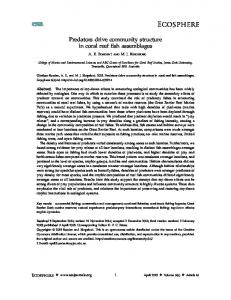
374 T. Pereira et al. Fig. 1. Confocal microscopy images of wild-type S. nodosus encapsulated in alginate and stained with BacLight Live/Dead (green viable, red non-viable). Encapsulated S. nodosus cultured in YMG at (a) day 0 and after incubation at 28°C at (b) day 2, (c) day 7 and (d) day 30. Inserts are magnified differential interference contrast images indicating morphology. Bars represent: 10 mm (a–d) and 5 mm (inserts in a–d).
molecules or autoinducers] (Horinouchi and Beppu, 1992; Chater and Horinouchi, 2003; Takano, 2006). Examples include g-butyrolactones regulating differentiation and antibiotic synthesis in at least seven Streptomyces species (Takano, 2006) and the identification of a novel autoinducer for the antibiotic pimaricin which was used to increase yield (Recio et al., 2004). Little is known about the regulation of amphotericin synthesis other than putative regulatory genes being clustered with the genes encoding amphotericin polyketide synthases [(PKS) AmphA, B, C, I, J and K] (Caffery et al., 2001; Aparicio et al., 2003). Antibiotic production from immobilized Streptomyces in liquid cultures is difficult to demonstrate as a detached free-dwelling population often contributes to production (Bandyopadhyay et al., 1993; Asanza Teruel et al., 1997; Yang and Yueh, 2001; Bhattacharyya and Sen, 2002). Exposure of immobilized organisms to medium with endogenous QS molecules and reduced nutritional status could permit synthesis without the formation of this co-population. This study reports the community structure, viability and antibiotic production of alginateencapsulated S. nodosus in growth permissive and non-permissive conditions. In addition sporulation of this industrially important organism in liquid cultures was demonstrated by manipulation of quorum size.
Results Morphology and viability of encapsulated wild-type S. nodosus in YMG Streptomyces nodosus wild-type spores and mycelia at late log and early stationary phase (48 and 72 h incuba-
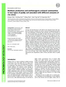
tion of spores in YMG) were immobilized in 2% (w/v) alginate forming uniformly spherical capsules of diameter 2.88 ⫾ 0.04 mm (n = 5). The viability and morphology of organisms within these capsules were determined using Live/Dead stains and confocal laser scanning microscopy (CLSM). Figure 1 shows smears of capsules containing sheared mycelia (48 h) encapsulated at day 0 (Fig. 1a, 98% viable) increasing in biomass after 2 days of fermentation in YMG (Fig. 1b, 76% viable). After 7 days of culturing, 60% (n = 5) of the biomass was viable (Fig. 1c). Although total biomass appeared to be lower, after 30 days of fermentation, 85% (n = 5) of the existing population was still viable (Fig. 1d). At all times hyphae appeared to have infrequent branching and no sporulation was evident by fluorescence and differential interference contrast microscopy. Encapsulated spores germinated after 24 h and biomass remained as hyphae for up to 30 days with no indication of spore formation. Coexistence of a freedwelling population was visible after 2 days of fermentation. The mycelia masses showed less than 32% (n = 5) viability after 30 days and no indication of sporulation. Scanning electron microscopy (SEM) was used to visualize the morphology of the immobilized community (Fig. 2A). After 48 h in YMG, the surface of the capsules showed regular protrusions of mycelial masses emerging from the matrix (Fig. 2A, i and ii) with the surface of these projections having erect and branching hyphae (Fig. 2A, iii and iv). At higher magnification, the branching hyphae appeared to have regular constrictions on the surface altering the direction of the hyphae. In contrast, the organisms sampled from within the capsules had longer hyphae with infrequent branching (Fig. 2A, v–viii) consistent with hyphae in the deeper layers of the protrusions.
© 2008 The Authors Journal compilation © 2008 Society for Applied Microbiology and Blackwell Publishing Ltd, Microbial Biotechnology, 1, 373–381
Streptomyces bioreactors: community structure and antibiotic production 375 sporogenic hyphae after 2 days (Fig. 2B, ii) and 4 days of fermentation (results not shown). Morphology and viability of encapsulated wild-type S. nodosus in conditioned YMG
Fig. 2. Electron microscopy of alginate capsules containing wild-type S. nodosus and cultured in YMG for 48 h. A. (i–iv) SEM of mycelia associated with surface structures protruding into media; (v–viii) SEM of mycelia embedded in capsules. Arrows indicate branching hyphae. B. (i and ii) TEM of immobilized S. nodosus hyphal forms. Bars represent (A): 500 mm (insert), 100 mm (i and v), 20 mm (ii and vi), 5 mm (iii and vii) and 1 mm (iv and viii); (B): 0.5 mm (i), 0.2 mm (ii).
Transmission electron microscopy (TEM) of capsular associated hyphae was used to assess cell size, septa frequency and structure, as well as degree of branching in cultures (Fig. 2B). Hyphae (0.5 mm diameter) with regular septation at 3–4 mm intervals were observed (Fig. 2B, i). Infrequent branching was noted with no basal septation at the base of hyphal branch points (Fig. 2B, i). Septa (15– 25 nm thickness) of these hyphae showed no evidence of the characteristic double-walled structure expected for
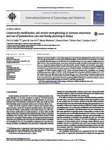
Free-dwelling S. nodosus wild type was cultured in YMG at 28°C for up to 48 h. After filter sterilization, the conditioned medium was used to culture encapsulated S. nodosus wild type (either spores or mycelia) analogous to the previous experiments. Conditioned media generated after 12–36 h growth decreased the rate of the emergence of the free-dwelling population; however, 48 h growth was required to suppress it and subsequently this was used in all further experiments. Capsules were cultured in 48 h conditioned media for up to 30 days. Biomass increased in the capsules up to 48 h (Fig. 3a) with the extent of growth comparable to that observed in capsules exposed to non-conditioned medium (Fig. 1b). Morphologically, the hyphae were indistinguishable between the two samples. On assessment of viability, 54% (n = 5) of hyphae were non-viable when cultured in the conditioned medium compared with 24% (n = 5) for those in capsules exposed to YMG. Transmission electron microscopy images of immobilized cultures in conditioned media showed a variety of hyphal morphologies (Fig. 3b–d). Long narrow cells (4 ¥ 0.4 mm) with thick septa (25 nm) and large inclusions were observed after 2 days (Fig. 3c). These hyphae had extracellular polymers associated with cell walls, something not observed for cells derived from cultures fermented in non-conditioned medium. Other capsular sections contained hyphae with basal septation at branch points with the intracellular space almost entirely consisting of inclusions (Fig. 3d). After 4 days of exposure to conditioned media, capsules developed a grey coloration consistent with spores on solid media. Confocal laser scanning microscopy indicated that the immobilized organisms included chains of spores (Fig. 3e). This was in contrast to capsules cultured in YMG which did not develop any coloration and the organisms continued growing displaying high viability and no evidence of sporulation after 30 days of culturing (Fig. 1d). Transmission electron microscopy images of these cultures showed thicker hyphae with more frequent septation resulting in reduced cell length (0.8 ¥ 0.6 mm, Fig. 3f). The septa displayed the double-walled morphology of sporulation septa, indicative of reproductive hyphae (Fig. 3f and g). Structures resembling spores with rounded morphology and without a sheath for attachment to neighbouring cells were also evident (Fig. 3h). To determine whether sporulation was dependent on a solid surface, after 48 h of growth 6% (v/v) of a free-
© 2008 The Authors Journal compilation © 2008 Society for Applied Microbiology and Blackwell Publishing Ltd, Microbial Biotechnology, 1, 373–381
376 T. Pereira et al. dwelling culture was returned to its own sterilized conditioned medium and cultured for up to 9 days. Confocal laser scanning microscopy revealed extensive sporulation (Fig. 4b) whereas control experiments with unperturbed free-dwelling populations grown in YMG for 9 days showed no evidence of sporulation (Fig. 4a). To assess whether nutrient limitation was involved in promoting sporulation yeast extract (4 g l-1), malt extract (10 g l-1) and glucose (4 g l-1) were added to 48 h conditioned medium. The returned reduced biomass did not sporulate when cultured for up to 9 days.
Construction of S. nodosus MAWhyg1: An amphA mutant for production of conditioned media deficient in antibiotics
Fig. 3. Immobilized wild-type S. nodosus cultured in 48 h conditioned medium from S. nodosus. Confocal laser scanning microscopy images of hyphae and spore chains associated with capsules cultured for 2 days (a) and 4 days (e). Transmission electron micrographs of hyphae at day 2 (b–d) and day 4 (f–h). Bars represent: 10 mm (a), 5 mm (e, and inserts in a and e), 0.5 mm (b, d and f), 0.2 mm (c, g and h).
Double-cross-over homologous recombination was used to inactivate amphA and make S. nodosus MAWhyg1 (Fig. 5a). A disruption plasmid pMAWhyg1 (Fig. 5a) was made with amphA providing homology sequences of 3.4 kb and 0.8 kb flanking a hygromycin resistance cassette and introduced into S. nodosus spores [1 ¥ 107 colony-forming units (cfu)] by conjugation with Escherichia coli. After 14 days of growth with hygromycin selection, colonies were tested for apramycin sensitivity as HygRAprR colonies originate from a single cross whereas HygRAprS colonies result from a double-cross-over recombination event. Streptomyces nodosus MAWhyg1, (HygRAprS), was propagated through four generations on non-selective media and retained HygR, indicative of stable genomic integration of the cassette. Southern blot analysis of NcoIdigested gDNA using 2.8 kb amphA DNA as a probe (Fig. 5a) demonstrated the integration of the cassette. The probe hybridized to a single NcoI band, 7.2 kb in size in MAWhyg1 compared with S. nodosus wild type which hybridized to a 5 kb band (Fig. 5b), a size shift consistent with the integration of 2.2 kb HygR cassette within amphA gene. Streptomyces nodosus wild type and MAWhyg1 were grown in YMG and PYG with no difference in growth rate or biomass yield. After 5 days, culture broths were extracted and examined for the presence of polyene chromophores by UV/visible spectral analysis. Streptomyces nodosus wild type grown in both media indicated the presence of amphotericins A and B (Fig. 5c). Amphotericin A is identified by absorptions at 280, 292, 305 and 318 nm, while amphotericin B chromophore gives UV absorptions at 346, 364, 382 and 405 nm (McNamara et al., 1998). In contrast, the absorption spectrum from MAWhyg1 culture fluids (Fig. 5c) clearly indicated absence of both compounds. This was also confirmed by HPLC analyses.
© 2008 The Authors Journal compilation © 2008 Society for Applied Microbiology and Blackwell Publishing Ltd, Microbial Biotechnology, 1, 373–381
Streptomyces bioreactors: community structure and antibiotic production 377 Fig. 4. Confocal microscopy images of free-dwelling populations stained with BacLight viability stain. S. nodosus grown in YMG after 9 days showing presence of hyphae and absence of spores (a) and S. nodosus cultured in conditioned medium from S. nodosus after 9 days showing presence of spores chains (b). Inserts are differential interference contrast images. Bars represent 10 mm.
Antibiotic production using sporulating cultures in liquid environments Amphotericin A and B production was monitored by HPLC of culture fluid from immobilized wild-type organisms (both spores and mycelia). In biphasic cultures grown in unconditioned YMG, amphotericin B was produced at 0.6 mg ml-1 with no amphotericin A detected. Immobilized S. nodosus wild type was induced to sporulate by culturing in 48 h conditioned media from S. nodosus MAWhyg1 (results not shown). This condi-
tioned medium would have been devoid of the antibiotics and thus the presence of antibiotics in this conditioned culture fluid during fermentation would have originated from the wild-type immobilized organisms. These sporulated cultures showed amphotericin B production of 0.5 mg ml-1. Free-dwelling wild-type organisms, induced to sporulate with this conditioned medium produced amphotericin B at 0.5 mg ml-1. Free-dwelling populations of S. nodosus in YMG produced 14 mg ml-1 amphotericin B with no amphotericin A detected.
Fig. 5. Generation and characterization of S. nodosus MAWhyg1. a. Inactivation of the chromosomal copy of amphA by double-cross-over homologous recombination. Plasmid pMAWhyg1 containing amphA with HygR inserted was introduced to the wild-type organism to cause replacement and a chromosomal insertion of a HygR in amphA. b. Southern blot of NcoI-digested S. nodosus wild type (1) and MAWhyg1 (2) gDNA using a probe hybridizing to amphA. c. UV spectra of butanol-extracted culture fluids from S. nodosus wild type (1) and MAWhyg1 (2). Amphotericin A shows absorbance bands at 280, 292, 305 and 318 nm, whereas amphotericin B has specific UV absorptions at 346, 364, 382 and 405 nm.
© 2008 The Authors Journal compilation © 2008 Society for Applied Microbiology and Blackwell Publishing Ltd, Microbial Biotechnology, 1, 373–381
378 T. Pereira et al.
Fig. 6. Model of Streptomyces at a solid–air or solid–liquid interface. Vegetative or substrate hyphae associated with a solid surface show infrequent branching and septation. Hyphae emerging from this biomass (termed ‘aerial hyphae’ at a solid–air interface) have infrequent septa and an indeterminate fate. These hyphae can develop into branched reproductive/sporogenic hyphae with frequent double-walled septa or continue growing forming branched hyphae, infrequent septation and no commitment to sporulation (reconnoitring hyphae).
Discussion The biochemical pathways of Streptomyces differentiation are still emerging with many genetic products assessing environmental parameters, transducing and mitigating the transition to aerial hyphae with separate pathways postulated to sense aerial growth and control sporulation (Chater, 2001; Chater and Horinouchi, 2003; Claessen et al., 2006). Streptomyces have difficulty undergoing this complete life cycle in liquid media and as some secondary metabolites are only produced in significant yield on solid cultivation, studies into the regulation of this life cycle are of industrial importance (Chater, 1989; Kieser et al., 2000). The alginate-encapsulated Streptomyces model provided the opportunity to study the development of this organism at a solid–liquid interface. When given the opportunity to proliferate in unconditioned media, the encapsulated S. nodosus formed a structured community. The protrusions observed are similar to those reported for other organisms at solid–liquid interfaces and are thought to facilitate flow and increase the surface area of the biofilm (Pasmore and Costerton, 2003). Vegetative/substrate hyphae left the surface of the matrix to form these projections. The branched and curving hyphae on the surface of the protrusions were morphologically indistinguishable from S. nodosus aerial hyphae formed at a solid–air interface. There were no basal or double-walled septa in these hyphae, indicating no commitment to sporulation. On solid media, it has been postulated that aerial hyphae emerge from substrate mycelial masses not only to aid dispersion of spores but also to explore new environments for growth opportunities (Yeo and Chater, 2005). These reconnoitring hyphae are consistent with the hyphal forms seen on the surface of the protrusions associated with the surface of the capsules (Fig. 6). It is not known whether their formation is to scout for new environments or to increase the surface area of the biomass exposed to the liquid environment for nutrient uptake and translocation to the rest of the community.
The free-dwelling hyphae in biphasic cultures had lower viability compared with immobilized organisms. This difference in the physiology of the two populations could be due to the large number of phenotypic changes reported when cells associate with a surface or the protection alginate affords to microorganisms in unfavourable environments (Pasmore and Costerton, 2003). The low viability of the immobilized cells destined for sporulation was consistent with reproductive hyphae transition where rounds of substrate hyphae cell death are required (Miguelez et al., 1999; Manteca et al., 2005). Removal of 94% of the biomass after 48 h of growth in liquid YMG and a further 5 days of incubation induced sporulation. This occurred whether the biomass was encapsulated or not. These environment conditions usually occur with higher quorum size and include metabolic waste, cell signalling molecules and nutrient depletion of the medium. The commitment to sporulate in this model included an assessment of the nutritional status of the environment as additional nutrients inhibited sporulation. The antibiotic production from the immobilized population, albeit low, is encouraging for the use of bacteria for drug delivery. In vitro diffusion studies of capsules containing amphotericin confirmed that release from the polymer was possible (results not shown). Augmentation of the rate of sporulation maybe required as antibiotic synthesis would not be expected when spores develop dormancy. This model will allow investigation into the biochemistry of cell signalling, differentiation and antibiotic production in this industrially important organism in a liquid environment which affords the advantage of a tightly controlled system allowing addition or analysis of nutrients, growth factors and labelled substrates (Nguyen et al., 2005). This article also reports the characterization of S. nodosus MAWhyg1 deficient in amphotericin A and B production. AmphA must be required for synthesis of both compounds supporting a biosynthetic model with the structural differences of the antibiotics are due to an inefficient
© 2008 The Authors Journal compilation © 2008 Society for Applied Microbiology and Blackwell Publishing Ltd, Microbial Biotechnology, 1, 373–381
Streptomyces bioreactors: community structure and antibiotic production 379 Table 1. Plasmids and strains used in study. Strain or vector
Relevant characteristic
Reference or source
Streptomyces nodosus ATCC14899 Streptomyces nodosus MAWhyg1 Escherichia coli JM109 Escherichia coli ET12567 pGEM-T pHP45Whyg pOJ260 pOJ26A pKC1138 pGA pGAWhyg pMAWhyg1
Wild-type amphotericin producer amphA insertional disruption, HygR General cloning host dam, dcm, hsdM containing non-transmissible oriT plasmid (pUZ8002) Vector for cloning PCR products, CarbR Source of hygromycin cassette oriT RK2, AprR pOJ260 derivative with 1.6 kb internal amphA fragment (EcoRV) oriT RK2, AprR pGEM-T derivative containing 4.2 kb amphA pGEM-T derivative containing the 6.4 kb DNA fragment pKC1138 derivative containing the 7.2 kb pGAWhyg (PstI)
ATCC This work Stratagene MacNeil et al. (1992) Promega Blondelet-Rouault et al. (1997) Bierman et al. (1992) This work Bierman et al. (1992) This work This work This work
HygR, CarbR, AprR: hygromycin, carbenicillin and apramycin resistance markers.
enoyl reductase (ER) in AmphC (Caffery et al., 2001). AmphA is a putative substrate loading protein for the rest of the enzyme having the domain structure ketosynthase(s)acyl transferase-dehydratase-acyl carrier protein (KSs-ATDH-ACP). The homologous protein (NysA) in the nystain PKS is required for the initiation of synthesis and decarboxylates malonyl groups loaded onto the protein via the AT (Brautaset et al., 2000). The lack of amphotericin synthesis in the mutant established that the first extension module (AmphB) cannot be loaded directly by a starter carboxylic acid unlike the erythromycin PKS (Pereda et al., 1998; Jacobsen et al., 1998). As initiation is governed by a distinct loading protein (AmphA), it may provide a useful target for genetic manipulations towards synthesis of novel amphotericin products.
Experimental procedures Bacterial strains, maintenance and growth Spore suspensions of S. nodosus (ATCC 14899) (Kieser et al., 2000) were resuscitated on YMG (yeast extract 4 g l-1, malt extract 10 g l-1, glucose 4 g l-1, agar 15 g l-1) for 5 days at 28°C. Spores (1 ¥ 108 cfu) were grown in YMG or PYG (glucose 10 g l-1, peptone 5 g l-1, yeast extract 5 g l-1, NaCl 5 g l-1, casamino acid 1 g l-1) media (50 ml) at 28°C for up to 10 days for analyses of biomass or amphotericin production.
extension step at 65°C for 10 min. The 4.2 kb fragment was purified and cloned into pGEM-T to obtain plasmid pGA. A hygromycin resistance cassette was excised from pHP45Whyg (Table 1) by BamHI and cloned into BamHI restriction site of pGA. The amphA disruption vector, pMAWhyg1, was constructed by linearizing pGAWhyg with PstI and subcloning the 9.4 kb fragment into pKC1138 PstI site. pMAWhyg1 was introduced into S. nodosus by intergeneric conjugation from E. coli ET12567 (pUZ8002) (Nikodinovic et al., 2003b) and incubated with hygromycin (25 mg ml-1) for 14–21 days at 28°C. Resistant colonies were screened by replica plating on MS medium (soy flour 20 g l-1, mannitol 20 g l-1, agar 20 g l-1) with apramycin (50 g l-1) and nalidixic acid (25 g l-1) to restrict the growth of plasmid containing E. coli. NcoI-digested gDNA of S. nodosus wild type and mutant MAWhyg1 (10 mg each) were separated on 0.7% (w/v) agarose gel and immobilized onto Hybond N+ nylon membrane (Amersham Pharmacia) under vacuum in 0.4 M NaOH and fixed (120°C, 30 min). Pre-hybridization at 55°C using hybridization solution (3 ml, 30 min, Roche Molecular Biochemicals) was carried out and then the same solution containing 100 ng of digoxigenin-dUTP-labelled PCR-amplified amphA fragment as a probe (3 ml, Fig. 5a) was used for hybridization at 55°C for 20 h. After washing (2¥ SSC buffer, 300 mM NaCl, 30 mM sodium citrate) then further washed in the same buffer supplemented with 0.1% SDS (v/v) (2 ¥ 5 ml, 5 min, 25°C), the membrane was further washed in 0.5¥ SSC supplemented with 0.1% SDS (v/v) (2 ¥ 5 ml, 15 min, 68°C). Detection of the probe was carried out according to the manufacturer (Roche).
Generation of S. nodosus amphA disruption mutant MAWhyg1
Preparation and fermentation of Streptomyces capsules
Table 1 shows strains and vectors used for the inactivation of amphA (Accession No. AF357202, bases 66081–70319) in S. nodosus. A DNA fragment containing amphA was PCR amplified (20 ml) using S. nodosus gDNA (Nikodinovic et al., 2003a) and 5′-TGAAACTTCATATGACGATCGGTGCCAA CGAC-3′ (10 pmol) and 5′-GACGCGGCTTAGGAGATT TCGAACTCTTC-3′ (10 pmol) primers, dNTPs (200 pmol), PCR buffer, Taq DNA polymerase (2 U) and 10% (v/v) DMSO using a Gradient 96 hot-top Robocycler(tm) (Stratagene). Reactions were held at 94°C for 2 min then 30 cycles at 94°C for 50 s, 53°C for 1 min and 65°C for 5 min, and a final
Streptomyces nodosus mycelia produced after fermentation of spores for 48 h in YMG (log phase, 6.8 g wet weight) or spores (2 ¥ 106 cfu) were suspended in saline (10 ml) and added to 2% (w/v) alginate (90 ml), medium viscosity (Sigma). After mixing, the solution was extruded into droplets using a peristaltic pump falling into 100 mM CaCl2 from a constant height of 6 cm. Capsules were cured in the calcium chloride solution for 30 min at 20°C and washed twice with saline. Capsules (10 g) containing spores or mycelia were transferred to YMG (50 ml), conditioned medium produced by mutant MAWhyg1 (50 ml) or conditioned medium from
© 2008 The Authors Journal compilation © 2008 Society for Applied Microbiology and Blackwell Publishing Ltd, Microbial Biotechnology, 1, 373–381
380 T. Pereira et al. wild-type S. nodosus (50 ml) in a 250 ml flask and incubated at 28°C at 60 r.p.m. for up to 30 days. Conditioned medium was prepared by filter sterilizing medium (0.22 mm) after 48 h growth of the mutant MAWhyg1 or wild-type S. nodosus. Capsules were removed at defined intervals for viability and morphology assessment.
Microscopy For CLSM, capsules were stained in 1:1 (v/v) SYTO 9 plus propidium iodide (BacLight bacterial viability kit L-13152, Molecular Probes) for 20 min at 20°C in the dark. Samples were rinsed in water and a smear made by crushing capsules between a slide and a coverslip (Pereira et al., 2005). Samples were examined using a Fluoview 300 laser scanning confocal system equipped with an IX70 inverted microscope under a ¥100 oil immersion objective (numerical aperture = 1.35). SYTO 9 fluorescence was detected with an Argon laser (488 nm laser excitation) with a 515 nm interference emission filter, and propidium iodide fluorescence was detected with a HeNe green laser (543 nm laser excitation) and a long pass 565 nm emission filter. Sequential dual channel scanning was used to display green and red fluorescence. For triple channel imaging, a transmitted light photomultiplier tube was used in conjunction with Normaski optics. Quantitative viability assessments were determined using analysis performed on a Windows™ computer using the public domain ImageJ program (developed at the US National Institutes of Health and available on the Internet at http://rsb.info.nih.gov/ij/). Capsules for SEM and TEM were fixed in 4% (w/v) paraformaldehyde and 3% (w/v) glutaraldehyde in 0.1 M PIPES, pH 7.2 at room temperature for 4 h. Capsules were washed (3¥ PIPES buffer) and post-fixed in 1% (w/v) osmium tetroxide in the same buffer at room temperature for 1 h. The samples were rinsed (3¥ H2O) and dehydrated through a graded series of ethanol (50%, 70%, 80%, 90%, 95%, 2¥ 100%) for 15 min each at room temperature. After dehydration, the samples for SEM were dried to the critical point (Emitech K850 CPD) with liquid carbon dioxide, mounted on a metal stub with a carbon tab and sputter coated (Emitech K550) with ~20 nm gold. Observations were made on JEOL scanning electron microscope (JSM 6480LA) at an acceleration voltage of 5 or 15 kV. After dehydration in ethanol, the samples for TEM were infiltrated in 50% (v/v) resin in ethanol (LR White resin, medium grade, Proscitech, C023) for 1 h followed by infiltration with 100% resin for a further 1 h. Capsules were then added to a fresh aliquot of 100% resin and incubated overnight at 4°C before being embedded in gelatine capsules and polymerized at 60°C for 20 h. Ultrathin sections (60 nm) were prepared using an ultramicrotome (Reichert Ultracut S) with a glass knife, collected on pioloform-coated copper grids (300 mesh). The sections were treated in a humid chamber with saturated aqueous uranyl acetate (7.7% w/v) (35 min), rinsed with water (30 s), stained with Reynolds’s lead citrate (5 min, Reynolds, 1963) and washed in water (5 ¥ 30 s each). The grids were dried and examined under a Philips CM-10 transmission electron microscope.
Assays for amphotericin production For UV spectroscopy, DMSO (1 ml) was added to culture
broth (1 ml) from which cells had been removed by centrifugation (5000 g, 10 min, 25°C). The solution was vortexed and incubated for 5 min before centrifugation (5000 g, 10 min, 25°C). The supernatant was diluted 10-fold with methanol and the absorption spectrum recorded from 250–500 nm using a Beckman DU-7500 spectrophotometer. For HPLC, culture fluids were centrifuged and filtered (0.22 mm) before analysis using a C-18 reverse-phase LiChrospher® 100 column (Merck) and a isocratic mobile phase [MeOH and 0.1% TFA (85:15, v/v)] with a flow rate of 1 ml min–1 at 25°C and a HP 1100 system equipped with diode array detector (Agilent Technologies). Samples were directly injected (20 ml) and signals monitored at 408, 386 and 366 nm. The detection limit of this assay was 100 ng ml-1. AmB was pre-concentrated from culture fluids using solidphase extraction (SPE) to increase the sensitivity of the assay. Oasis HLB cartridge (sorbent weight of 60 mg, particle size 30 mm, pore size 80 Å and syringe barrel size 3 cc, Waters, Milford, MA, USA) was conditioned with methanol (3 ml), equilibrated with water (2 ml) and the filter-sterilized sample (10–50 ml) was introduced into the cartridge under vacuum (1.5 psig). AmB was eluted with methanol (1 ml), evaporated to dryness, re-suspended in methanol (1 ml), before being spiked with an internal standard (10 mg ml–1, biphenyl) and injected (20 ml) into the HPLC system. Using this assay, a detection limit of 6 ng ml–1 was achieved.
Acknowledgements We would like to acknowledge the use of the Electron Microscopy Facility, Macquarie University, NSW, Australia. T.P. would like to acknowledge the receipt of a University of Western Sydney Priority Postgraduate Scholarship.
References Al-mohsen, I., and Hughes, W.T. (1998) Systemic antifungal therapy: past, present and future. Ann Saudi Med 18: 28–38. Aparicio, J.F., Caffrey, P., Gil, J.A., and Zotchev, S.B. (2003) Polyene antibiotic biosynthesis gene clusters. Appl Microbiol Biotechnol 61: 179–188. Asanza Teruel, M.L., Gontier, E., Bienaime, C., Nava Saucedo, J.E., and Barboti, J.-N. (1997) Response surface analysis of chlortetracycline and tetracycline production with K-carrageenan immobilized Streptomyces aureofaciens. Enzyme Microb Technol 21: 314–320. Bandyopadhyay, A., Das, A.K., and Mandal, S.K. (1993) Erythromycin production by Streptomyces erythreus entrapped in calcium alginate beads. Biotechnol Lett 15: 1003–1106. Bhattacharyya, B.K., and Sen, S.K. (2002) Ester antibiotic accumulation by Streptomyces hygroscopicus. Microbiologica 25: 477–484. Bierman, M., Logan, R., O’Brien, K., Seno, E.T., Rao, R.N., and Schoner, B.E. (1992) Plasmid cloning vectors for the conjugal transfer of DNA from Escherichia coli to Streptomycas spp. Gene 116: 43–49. Blondelet-Rouault, M., Weiser, J., Lebrihi, A., Branny, P. , and Pernodet, J. (1997) Antibiotic resistance gene cassettes derived from the W interposon for use in E. coli and Streptomyces. Gene 190: 315–317.
© 2008 The Authors Journal compilation © 2008 Society for Applied Microbiology and Blackwell Publishing Ltd, Microbial Biotechnology, 1, 373–381
Streptomyces bioreactors: community structure and antibiotic production 381 Brautaset, T., Sekurova, O.N., Sletta, H., Ellingsen, T.E., Strom, A.R., Valla, S., and Zotchev, S. (2000) Biosynthesis of the polyene antifungal antibiotic nystatin in Streptomyces noursei ATCC 11455: analysis of the gene cluster and deduction of the biosynthetic pathway. Chem Biol 7: 395– 403. Caffery, P., Lynch, S., Flood, E., Finnan, S., and Oliynyk, M. (2001) Amphotericin biosynthesis in Streptomyces nodosus: deductions form analysis of polyketide synthase and late genes. Chem Biol 8: 713–723. Chandramouli, V., Kailasapathy, K., Peiris, P., and Jones, M. (2004) An improved method for microencaspulation and its evaluation to protect Lactobacillus spp. in simulated gastric conditions. J Microbiol Methods 56: 27–35. Chater, K.F. (1989) Multilevel regulation of Streptomyces differentiation. Trends Genet 5: 372–377. Chater, K.F. (2001) Regulation of sporulation in Streptomyces coelicolor A3(2): a checkpoint multiplex? Curr Opin Microbiol 4: 667–673. Chater, K.F., and Chandra, G. (2006) The evolution of development in Streptomyces analysed by genome comparisons. FEMS Microbiol Rev 30: 651–672. Chater, K.F., and Horinouchi, S. (2003) Signalling and early developmental events in two highly diverged Streptomyces species. Mol Microbiol 48: 9–15. Claessen, D., de Jong, W., Dijkhuizen, L., and Wosten, H.A.B. (2006) Regulation of Streptomyces development: reach for the sky. Trends Microbiol 14: 313–319. Colton, C.K. (1996) Engineering challenges in cell encapsulation technology. Trends Biotechnol 14: 158–162. Elliot, M., Damji, F., Passantino, R., Chater, K., and Leskiw, B. (1998) The bldD gene of Streptomyces coelicolor A3(2): a regulatory gene involved in morphogenesis and antibiotic production. J Bacteriol 180: 1549–1555. Horinouchi, S., and Beppu, T. (1992) Autoregulatory factors and communication in actinomycetes. Annu Rev Microbiol 46: 377–398. Jacobsen, J.R., Cane, D.E., and Khosla, C. (1998) Spontaneous priming of a downstream module in 6-deoxyerythronolide B synthase leads to polyketide biosynthesis. Biochemistry 37: 4928–4934. Kieser, T., Bibb, M.J., Buttner, M.J., Chater, K.F., and Hopwood, D.A. (2000) Practical Streptomyces Genetics. Norwich, UK: The John Innes Foundation. Kwak, J., Dharmatilake, A.J., Jiang, H., and Kendrick, K.E. (2001) Differential regulation of ftsZ transcription during septation of Streptomyces griseus. J Bacteriol 183: 5092– 5101. Liu, C., McDaniel, L.E., and Schaffner, C.P. (1975) Factors affecting the production of candicidin. Antimicrob Agents Chemother 7: 196–202. MacNeil, D.J., Gewain, K.M., Ruby, C.L., Dezeny, G., Gibbons, P.H., and MacNeil, T. (1992) Analysis of Streptomyces avermitilis genes required for avermectin biosynthesis utilizing a novel integration vector. Gene 111: 61–68. Manteca, A., Fernandez, M., and Sanchez, J. (2005) A death round affecting a young compartmentalized mycelium precedes aerial mycelium dismantling in confluent surface cultures of Streptomyces antibioticus. Microbiology 151: 3689–3697.
McNamara, C.M., Box, S., Crawforth, J.M., Hickman, B.S., Norwood, T.J., and Rawlings, B.J. (1998) Biosynthesis of amphotericin B. J Chem Soc Perkin Trans I 1: 83–87. Miguelez, E.M., Hardisson, C., and Manzanal, M.B. (1999) Hyphal death during colony development in Streptomyces antibioticus: morphological evidence for the existence of a process of cell deletion in a muticellular prokaryote. J Cell Biol 145: 515–525. Nguyen, L.D., Kalachova, L., Novotna, J., Holub, M., Kofronova, O., Benada, O., et al. (2005) Cultivation system using glass beads immersed in liquid medium facilitates studies of Streptomyces differentiation. Appl Environ Microbiol 71: 2848–2852. Nikodinovic, J., Barrow, K.D., and Chuck, J. (2003a) High yield preparation of genomic DNA from Streptomyces. BioTechniques 35: 932–934, 936. Nikodinovic, J., Barrow, K.D., and Chuck, J. (2003b) High frequency transformation of the amphotericin-producing bacterium Streptomyces nodosus. J Microbiol Methods 55: 273–277. Ohnishi, Y., Seo, J., and Horinouchi, S. (2002) Deprogrammed sporulation in Streptomyces. FEMS Microbiol Lett 216: 1–7. Orive, G., Hernandez, R.M., Gascon, A.R., Calafiore, R., Chang, P., De Vos, P., et al. (2003) Cell encapsulation: promise and progress. Nat Med 9: 104–107. Pasmore, M., and Costerton, J.W. (2003) Biofilms, bacterial signalling, and their ties to marine biology. J Ind Microbiol Biotechnol 30: 407–413. Pereda, A.S., Stassi, R.G., Ruan, D.L., and Katz, L. (1998) The loading domain of the erythromycin polyketide synthase is not essential for erythromycin biosynthesis in Saccharopolyspora erythraea. Microbiology 144: 543–553. Pereira, T., Millar, T.J., and Chuck, J. (2005) Viability analysis of alginate encapsulated micro-organisms using fluorescent stains. J Microencapsul 22: 787–792. Prakash, S., and Chang, T.M.S. (1996) Microencapsulated genetically engineered live E. coli DH5 cells administered orally to maintain normal plasma urea levels in uremic rats. Nat Med 2: 883–887. Recio, E., Colinas, A., Rumbero, A., Aparicio, J.J., and Martin, J.F. (2004) PI factor, a novel type of quorumsensing inducer elicits pimaricin production in Streptomyces natalensis. J Biol Chem 279: 41586–41593. Reynolds, E.S. (1963) The use of lead citrate at high pH as an electron-opaque stain in electron microscopy. J Cell Biol 17: 208–212. Takano, E. (2006) Butyrolactones: Streptomyces signalling molecules regulating antibiotic production and differentiation. Curr Opin Microbiol 9: 1–8. Torrado, J.J., Espada, R., Ballesteros, M.P., and TorradoSantiago, S. (2007) Amphotericin B formulations and drug targeting. J Pharm Sci. Early View (Article online in advance of print). Yang, S., and Yueh, C. (2001) Oxytetracyclin production by immobilised Streptomyces rimosus. J Microbiol Immunol Infect 34: 235–242. Yeo, M., and Chater, K. (2005) The interplay of glycogen metabolism and differentiation provides an insight into the developmental biology of Streptomyces coelicolor. Microbiology 151: 855–861.
© 2008 The Authors Journal compilation © 2008 Society for Applied Microbiology and Blackwell Publishing Ltd, Microbial Biotechnology, 1, 373–381