Pediatr Neonatol 2008;49(6):223−229
O R I G I N A L A RT I C L E
Comparison between Bubble CPAP and Ventilator-derived CPAP in Rabbits Wen-Chin Huang1, Yi-Ming Hua1*, Chuen-Ming Lee1, Chun-Cheng Chang2, Yeong-Seng Yuh3 1
Department of Pediatrics, Tri-Service General Hospital and National Defense Medical Center, Taipei, Taiwan 2 Department of Pediatrics, Tauyuan Armed Forces General Hospital, Tauyuan, Taiwan 3 Department of Pediatrics, Cathay General Hospital, Taipei, Taiwan
Received: Dec 25, 2007 Revised: Jun 13, 2008 Accepted: Sep 19, 2008 KEY WORDS: blood gas analysis; blood pressure; continuous positive airway pressure (CPAP); heart rate; rabbits
Background: Continuous positive airway pressure (CPAP) is used in infants with respiratory distress and apnea. Bubble CPAP (B-CPAP) and ventilator-derived CPAP (V-CPAP) are two of the most popular CPAP modes, and use different pressure sources. However, few studies have been performed to compare their differences and effectiveness. This study was to determine whether B-CPAP and V-CPAP would have different effects on vital signs and arterial blood gas analysis. Methods: We performed a randomized crossover study to measure vital signs, including mean blood pressure (MBP), heart rate (HR), and respiratory rate (RR), in 12 ketamineanesthetized healthy rabbits receiving endotracheal intubation by tracheostomy with B-CPAP or V-CPAP. Arterial blood was also sampled and analyzed for PaO2, PaCO2, HCO3¯ and pH. Results: We observed statistically significant decreases in RR, pH and PaO2 with corresponding incrases in PaCO2 and HCO3¯ during the V-CPAP; however, no significant changes from baseline were observed for B-CPAP. Neither modality resulted in statistically significant changes in MBP or HR. Both forms of CPAP altered vital signs and arterial blood gases in a similar manner. There was a trend towards a lower percentage of change from baseline in all variables in B-CPAP compared with V-CPAP. Conclusions: Our results suggest that B-CPAP seems to be superior to V-CPAP in terms of its effect on arterial blood gases and vital signs. We speculate that B-CPAP could have certain protective effects that better preserve both arterial blood gases and vital signs when compared to V-CPAP. However, the results of this study still need to be tested by clinical study.
1. Introduction Continuous positive airway pressure (CPAP) was first used to support the breathing of neonates in the early 1970s.1 Neonatal applications of CPAP include the prevention of extubation failure2 and the apnea
of prematurity,3 and offers an alternative to intubation and ventilation in respiratory distress syndrome.4 There are several techniques for CPAP generation; however, little evidence exists demonstrating the superiority of one technique over another.5−7
*Corresponding author. Department of Pediatrics, Tri-Service General Hospital, National Defense Medical Center, 325 Section 2, Cheng-Kung Road, Neihu District, Taipei City 114, Taiwan. E-mail:
[email protected] ©2008 Taiwan Pediatric Association
224 For the past 30 years, CPAP has been primarily delivered to neonates using certain devices connected to a conventional mechanical ventilator (ventilator-derived CPAP, V-CPAP). In V-CPAP, a variable resistance in a valve is adjusted to provide resistance to the flow of air. Underwater bubble CPAP (B-CPAP) has provided an alternative to pressure derived from conventional ventilators and has remained in use since first devised in the early 1970s.1 The positive pressure in the circuit is achieved by simply immersing the distal expiratory tubing in a water column to a desired depth rather than using a variable resistor. Although these two different pressure sources for CPAP delivery have been used for three decades, surprisingly few studies have been performed to compare their differences and effectiveness.8,9 The objective of this study was to determine whether vital signs and arterial blood gas analyses were influenced by the application of CPAP in spontaneously breathing healthy rabbits, and if there were an impact, whether B-CPAP and V-CPAP would differ in this regard.
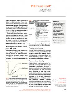
W.C. Huang et al Ventilator
Exhalation hose Pressure tube Bottle with sterile water
CPAP circuit
Figure 1 Schematic representation of the CPAP circuit. For V-CPAP, the exhalation hose was connected to the exhalation valve of the ventilator; for B-CPAP, the exhalation hose was submerged under 5 cm of water.
for the continuous registration of arterial blood pressure and heart rate (PowerLab hardware, Chart version 4.2 software; AD Instruments, Castle Hill, Australia).
2. Materials and Methods
2.2. CPAP setup
The experiments were approved by the local animal ethics committee of the National Defense Medical Center. Twelve pathogen-free, female, juvenile New Zealand White rabbits (mean body weight 1.86 ± 0.22 kg) were used in this study.
CPAP was achieved by using two modes: V-CPAP and B-CPAP. The circuit structures for both modes were almost identical. The ventilator (Sechrist Industries) provided gas flow at a rate of 8 L/min. The only difference between the modes was the site to which the end of the exhalation hose connected. For V-CPAP, the hose was connected to the exhalation valve of the ventilator. The pressure tube was connected to the Y-piece and the pressure was adjusted at 5 cmH2O. For B-CPAP, the hose was submerged under 5 cm of water to obtain 5 cmH2O of CPAP (Figure 1).
2.1. Surgical preparation Animals were anesthetized by means of an intramuscular injection of a mixture of ketamine (23 mg/kg), acepromazine (0.58 mg/kg), and xylazine (0.8 mg/kg). The left marginal ear vein was cannulated to administer a continuous infusion of 5% dextrose at a rate of 5 mL/kg/h, and a catheter was inserted in the right auricular artery to obtain samples for blood gas analysis. The animals were placed in the supine position and given a tracheotomy just below the cricoid cartilage. An endotracheal tube of 3.0 mm inner diameter was then inserted. The tracheal walls were tightened with 3−0 nylon suture around the endotracheal tube to avoid accidental extubation and leakage of air. The endotracheal tube was connected to a ventilator (Sechrist Infant Ventilator, model IV-100B, Sechrist Industries, Anaheim, CA, USA), and the rabbits were allowed to breathe spontaneously without assisted ventilation (ventilation rate = 0, FiO2 = 21%, flow = 8 L/min). The end of the exhalation hose was open to make the circuit at a CPAP of 0 cmH2O. A catheter primed with normal saline containing heparin was introduced into the left carotid artery
2.3. Anesthesia maintenance During the surgical preparation, anesthesia was maintained with repeated intravenous injections of ketamine (20 mg/kg). After instrumentation, the rabbits received another dose of ketamine to prevent them from awakening during the experiment. The injection of ketamine decreased the animals’ blood pressure and heart rate, though the measures returned to baseline, usually within 5 minutes. In all animals, we allowed a delay of at least 10 minutes after the injection of ketamine and before experimentation to obtain stable circulatory conditions.
2.4. Experimental protocol In a crossover design, each rabbit received two courses (I, II) of the experimental procedure (Figure 2). Each
Comparison between two different modes of CPAP
225
Ketamine BL-V 0 cmH2O
V-CPAP 5 cmH2O
BL-B 0 cmH2O
B-CPAP 5 cmH2O
Sequence A 0
Randomization
6
12
Course I
0
6
0
Washout period
12
6
12 (min)
Course II
0
6
12 (min)
Sequence B BL-B 0 cmH2O
B-CPAP 5 cmH2O
BL-V 0 cmH2O
Ketamine
V-CPAP 5 cmH2O
Figure 2 Study protocol. Each rabbit was randomly assigned to one of two sequences—A or B. BL = baseline period.
course was comprised of two 6-minute periods. In the first period, which was a baseline period (BL-V in sequence A; BL-B in sequence B), the rabbits received 0 cmH2O of CPAP by keeping the exhalation hose open. In the second period, the rabbits were randomly assigned to receive V-CPAP (sequence A) or B-CPAP (sequence B) by connecting the end of the exhalation hose to the exhalation valve of the ventilator or submerging it into water. Randomization was performed using a computer program that generated random numbers to determine the mode of CPAP to be first used. After completing course I of the experiment, CPAP was returned to 0 cmH2O by disconnecting the exhalation hose from the ventilator (in sequence A) or pulling it out of the water (in sequence B). The rabbits then received another dose of ketamine, and a second course was performed at least 10 minutes later when the rabbits’ circulatory conditions were stable. In the first period of course II, the CPAP mode was still 0 cmH2O as a baseline (BL-B in sequence A; BL-V in sequence B). In the second period of course II, the CPAP mode was changed to the CPAP mode not used in the second period of course I.
2.5. Data collection Vital signs data, including mean blood pressure (MBP), heart rate (HR) and respiratory rate (RR), were collected during the 2.5- to 4.5-minute timeframe of each period, and were used for statistical analysis. RR was measured by a manual count of chest movements over this 120-second timeframe. The mean of each variable was used to represent the result for the period. Arterial blood was sampled for blood gas analysis at 5 minutes into each period. Arterial blood samples were analyzed for PaO2, PaCO2, HCO3¯, and pH by a blood gas analyzer (ABL 510, Radiometer, Copenhagen, Denmark).
2.6. Statistical analysis The Wilcoxon signed ranks test was used for comparisons of vital signs (MBP, HR and RR) and arterial blood gas (PaO2, PaCO2, HCO3¯ and pH) between periods (BL-V and V-CPAP, BL-B and B-CPAP, and BL-V and BL-B) and between the percentage change from baseline of each CPAP modality. To test for “carryover” and “period” effects, we used nonparametric Mann-Whitney tests (two-tailed) on the sums and the differences, respectively, of these variables for the V-CPAP and B-CPAP. Statistical analysis was performed using SPSS software for Windows, version 11.0 (SPSS, Inc., Chicago, IL, USA). A p-value ≤ 0.05 was considered statistically significant.
3. Results In total, we performed 24 courses of experiments. Six rabbits were randomly assigned to receive sequence A, and six received sequence B. Accordingly, there were twelve V-CPAP periods and twelve B-CPAP periods in this study. The effect of the two different modes of CPAP on vital signs (RR, MBP and HR) and arterial blood gas (PaO2, PaCO2, HCO3¯ and pH) are shown in Figures 3 and 4. When we compared data from the two baseline periods, we found no significant differences except in MBP, which was significantly higher during BL-V than BL-B (p = 0.018). We observed statistically significant decreases in RR, pH and PaO2 with corresponding increases in PaCO2 and HCO3 during the V-CPAP; however, no significant changes from baseline were observed for B-CPAP. Neither modality resulted in statistically significant changes in MBP or HR. The mean percentage of change in variables from baseline of these two CPAP modalities is shown in Figure 5. Both forms of CPAP altered vital signs and
226
W.C. Huang et al p = 0.005
C
Heart rate (beat/min)
250
p = 0.109
B
120 110 100 90 80 70 60 50
V-CPAP
p = 0.083
BL-B
B-CPAP
BL-V
V-CPAP
BL-B
B-CPAP
p = 0.157
240 230 220 210 200 190 180
7.50
BL-V
V-CPAP
p = 0.041
BL-B
B-CPAP
p = 0.637
Figure 3 Effect of modes of CPAP on vital signs. Median values are indicated with horizontal lines.
B
PaCO2 (mmHg)
pH
7.40 7.35 7.30
p = 0.023
p = 0.060
35 30 25
7.25
20 BL-V
V-CPAP
p = 0.050
C
45 40
7.45
BL-B
B-CPAP
p = 0.638
BL-V
28
140
26 HCO3− (mmol/L)
120 110 100 90 80 70
V-CPAP
p = 0.031
D
150 130 PaO2 (mmHg)
p = 0.937
130
BL-V
A
p = 0.109
80 75 70 65 60 55 50 45 40 35 30 25
Mean blood pressure (mmHg)
Respiratory rate (breaths/min)
A
BL-B
B-CPAP
p = 0.099
24 22 20 18 16 14
60
12
50 BL-V
V-CPAP
BL-B
B-CPAP
BL-V
V-CPAP
BL-B
B-CPAP
Figure 4 Effect of modes of CPAP on arterial blood gas measures. Median values are indicated with horizontal lines.
Comparison between two different modes of CPAP
− 21.6
− 9.18
*
RR
227
*
− 0.25 − 3.92
MBP
1.53 2.07
HR − 0.038 − 0.16
pH
5.49
PaCO2
9.75 − 0.14 − 6.32
PaO2 B-CPAP
HCO3−
4.63 7.37
V-CPAP − 20
− 15
− 10 −5 0 Change from baseline (%)
5
10
Figure 5 Mean percentage change in variables from baseline of each CPAP modality. * = mean percentage change from baseline has reached significance.
arterial blood gases in a similar manner. The changes tended to be higher in all of these variables during V-CPAP than B-CPAP, and the differences between these two CPAP modalities in mean percentage change from baseline in RR and MBP were significant (p = 0.023 and 0.050, respectively). There were no significant carryover or period effects, except in MBP (carryover effect, p = 0.01).
4. Discussion In this study, we demonstrated that V-CPAP and B-CPAP both affected vital signs and arterial blood gases in a similar manner in the healthy rabbit model. Moreover, a striking finding in our study was that B-CPAP had no significant effect on vital signs and arterial blood gases, but V-CPAP significantly influenced both arterial blood gases and the respiratory rate. In addition, B-CPAP had a smaller percentage change from baseline than V-CPAP, although the difference between them was only significant in the respiratory rate and mean blood pressure. These findings suggest that B-CPAP is superior to V-CPAP in terms of its effect on arterial blood gases and vital signs. B-CPAP, in addition to providing positive airway pressure, delivers mechanical oscillatory vibrations that are transmitted into the chest secondary to the non-uniform flow of gas bubbles across a downstream underwater seal. These vibrations simulate waveforms produced by high-frequency ventilation (HFV).8,10 Accordingly, B-CPAP may possess the characteristics of CPAP and HFV at the same time, and this may make B-CPAP to different V-CPAP. Hemodynamics has
been reported to be better preserved during HFV than during conventionally controlled mechanical ventilation.11,12 However, the effect of B-CPAP on circulation is, to our knowledge, untested. Whether or not our results were related to the bubbleassociated high-frequency oscillation needs further exploration. In general, the application of CPAP improves gas exchange, increases PaO2 and decreases PaCO2 due to increases in functional residual capacity. However, surpassing optimum CPAP levels can result in a fall in tidal volume and a rise in dead space ventilation, leading to a rise in PaCO2. Furthermore, high CPAP levels can lead to a reduction in venous return, compromised cardiac output, increased pulmonary vascular resistance (PVR), and enhanced ventilationperfusion mismatch, resulting in a lower PaO2.13,14 In addition, an increase in PVR results in a shift of the intraventricular septum to the left and inhibits left ventricular filling, further decreasing cardiac output. In our study, the CPAP level we used (5 cmH2O) may have been too much for healthy rabbit lungs, resulting in a PaO2 decrease and a PaCO2 increase. Consequently, the cardiac output might have been reduced, producing an insignificantly mild increase in heart rate and a slight decrease in blood pressure. The decreases in the respiratory rate during CPAP, both ventilator-derived (significant) and bubble (insignificant), are not surprising and have been reported previously. CPAP can assist the respiratory muscles,15,16 thereby reducing inspiratory work, as indicated by a decrease in the respiratory rate.17 Furthermore, it has been shown that both inspiratory and expiratory times are increased with CPAP,18,19 causing a decrease in the respiratory rate.
228 One of the primary goals in neonatal medicine today is to decrease pulmonary injury and avoid chronic lung disease (CLD) while preserving brain function. Studies have shown that the “Columbia approach”20,21 in which B-CPAP is used early in the course of respiratory distress in both premature and term-gestation infants, can effectively lower the incidence of CLD.22−24 At Columbia University, the early initiation of nasal prong B-CPAP in combination with a tolerance to elevated PCO2 levels has been shown to reduce the incidence of CLD to < 5% in infants weighing less than 1500 g.25 However, the mode of pressure generation for the delivery of nasal CPAP which most effectively reduces the need for additional respiratory support is still uncertain. It is an issue of significant clinical interest as to whether B-CPAP, as opposed to CPAP only is one of the keys to reducing the incidence of CLD. A comparison of underwater B-CPAP with V-CPAP in preterm neonates suggested that the bubbling contributed to gas exchange which decreases the infant’s required work of breathing.8 The results from that study also suggest that B-CPAP may decrease the need for tidal ventilation, resulting in decreased fatigue and a decreased need for endotracheal intubation and positive pressure ventilation, which may help to reduce the incidence of CLD. Another study compared early B-CPAP with “conventional” (ventilator-derived) CPAP9 and showed that B-CPAP, but not conventional CPAP, reduced the use of postnatal steroids and produced a directional, but insignificant, decrease in the incidence of CLD. However, this was not a randomized study. Our study showed that B-CPAP had no significant adverse effects on vitals signs and arterial blood gases, but V-CPAP did, which suggests that B-CPAP might have certain protective effects that contribute to a lower incidence of CLD. Further study is needed to investigate these ideas. There is an important limitation in this study in that we did not measure the actual airway enddistending pressure in either CPAP modality. Courtney et al26 showed that in three different nasal CPAP devices, there were no significant differences in the respiratory rate and the oxygen changes from baseline among the groups at CPAP of 8, 6, and 4 cmH2O. Coddazzi’s study27 also showed that CPAP increments (from 0 to 10 cmH2O) did not significantly alter PaCO2, respiratory rate, heart rate, or systolic blood pressure. Accordingly, we believe that the failure to demonstrate equivalence between the two modalities with respect to the mean delivered distending pressure may be not so important. Moreover, in current clinical practice, the desired CPAP level is usually achieved just by placing the tubing in a water column to a specific depth in B-CPAP, or adjusting the pressure measured at the Y-piece in V-CPAP, and the actual
W.C. Huang et al airway end-distending pressure is usually not measured. We believed that reflecting the true clinical situation was more practical than measuring the actual airway end-distending pressure.
5. Conclusion In conclusion, from our preliminary results, B-CPAP seems to be superior to V-CPAP in terms of its effect on arterial blood gases and vital signs. We speculate that B-CPAP could have certain protective effects that better preserve arterial blood gases and vital signs than V-CPAP. Because the lungs in the rabbits in our study functioned normally, further study is needed to assess the value of B-CPAP in the treatment of immature lungs or more compromised lung conditions; a larger number of subjects and a longer study duration are also suggested. Since it is always difficult to extrapolate from findings in animal models to humans, the results of this study can only give an indication of what may occur in humans and results still need to be attained by a clinical study.
Acknowledgments We would like to thank Dr. An-Tsz Hsieh for his assistance in the statistical analysis and Wei-Na Hsiao for her laboratory assistance.
References 1.
2.
3.
4.
5.
6.
7.
Gregory GA, Kitterman JA, Phibbs RH, Tooley WH, Hamilton WK. Treatment of the idiopathic respiratory distress syndrome with continuous positive airway pressure. N Engl J Med 1971; 284:1333−40. Davis PG, Henderson-Smart DJ. Nasal continuous positive airways pressure immediately after extubation for preventing morbidity in preterm infants. Cochrane Database Syst Rev 2003;(2):CD000143. Speidel BD, Dunn PM. Use of nasal continuous positive airway pressure to treat severe recurrent apnoea in very preterm infants. Lancet 1976;2:658−60. Subramaniam P, Henderson-Smart DJ, Davis PG. Prophylactic nasal continuous positive airways pressure for preventing morbidity and mortality in very preterm infants. Cochrane Database Syst Rev 2000;(2):CD001243. De Paoli AG, Davis PG, Faber B, Morley CJ. Devices and pressure sources for administration of nasal continuous positive airway pressure (NCPAP) in preterm neonates. Cochrane Database Syst Rev 2002;(4):CD002977. Ahluwalia JS, White DK, Morley CJ. Infant Flow Driver or single prong nasal continuous positive airway pressure: shortterm physiological effects. Acta Paediatr 1998;87:325−7. Stefanescu BM, Murphy WP, Hansell BJ, Fuloria M, Morgan TM, Aschner JL. A randomized, controlled trial comparing two different continuous positive airway pressure systems for the successful extubation of extremely low birth weight infants. Pediatrics 2003;112:1031−8.
Comparison between two different modes of CPAP 8.
9.
10.
11.
12.
13.
14.
15.
16.
17.
Lee KS, Dunn MS, Fenwick M, Shennan AT. A comparison of underwater bubble continuous positive airway pressure with ventilator-derived continuous positive airway pressure in premature neonates ready for extubation. Biol Neonate 1998;73:69−75. Narendran V, Donovan EF, Hoath SB, Warner BB, Streichen JJ, Jobe AH. Comparison between early bubble CPAP and conventional CPAP in reducing the incidence of chronic lung disease. Pediatr Res 2002;51:337A. Nekvasil R, Kratky J, Penkova Z, Stejskal J. High frequency “bubble” oscillation ventilation in the neonatal period. Cesk Pediatr 1992;47:465−70. [In Czech] Lucking SE, Fields AI, Mahfood S, Kassir MM, Midgley FM. High-frequency ventilation versus conventional ventilation in dogs with right ventricular dysfunction. Crit Care Med 1986;14:798−801. Chiaranda M, Rubini A, Fiore G, Giron G, Carlon GC. Hemodynamic effects of continuous positive-pressure ventilation and high-frequency jet ventilation with positive end-expiratory pressure in normal dogs. Crit Care Med 1984;12:750−4. Shaffer TH, Koen PA, Moskowitz GD, Ferguson JD. Positive end expiratory pressure: effects on lung mechanics of premature lambs. Biol Neonate 1978;34:1−10. Hobelmann CF Jr, Smith DE, Virgilio RW, Peters RM. Mechanics of ventilation with positive end-expiratory pressure. Ann Thorac Surg 1977;24:68−76. Baratz DM, Westbrook PR, Shah PK, Mohsenifar Z. Effect of nasal continuous positive airway pressure on cardiac output and oxygen delivery in patients with congestive heart failure. Chest 1992;102:1397−401. Lenique F, Habis M, Lofaso F, Dubois-Rande JL, Harf A, Brochard L. Ventilatory and hemodynamic effects of continuous positive airway pressure in left heart failure. Am J Respir Crit Care Med 1997;155:500−5. Anonymous. AARC (American Association for Respiratory Care). Application of continuous positive airway pressure to neonates
229
18.
19.
20. 21.
22.
23.
24.
25. 26.
27.
via nasal prongs or nasopharyngeal tube. Respir Care 1994; 39:817−23. British Thoracic Society Standards of Care Committee. Non-invasive ventilation in acute respiratory failure. Thorax 2002;57:192−211. Hird MF, Greenough A. Influence of increasing postnatal age on respiratory timing and reflex activity in preterm ventilated infants. J Perinat Med 1992;20:73−7. Wung JT, Driscoll JM Jr, Epstein RA, Hyman AI. A new device for CPAP by nasal route. Crit Care Med 1975;3:76−8. Wung JT, Koons AH, Driscoll JM Jr, James LS. Changing incidence of bronchopulmonary dysplasia. J Pediatr 1979; 95:845−7. Avery ME, Tooley WH, Keller JB, et al. Is chronic lung disease in low birth weight infants preventable? A survey of eight centers. Pediatrics 1987;79:26−30. De Klerk AM, De Klerk RK. Nasal continuous positive airway pressure and outcomes of preterm infants. J Paediatr Child Health 2001;37:161−7. Van Marter LJ, Allred EN, Pagano M, et al. Do clinical markers of barotrauma and oxygen toxicity explain interhospital variation in rates of chronic lung disease? The Neonatology Committee for the Developmental Network. Pediatrics 2000; 105:1194−201. Polin RA, Sahni R. Newer experience with CPAP. Semin Neonatal 2002;7:379−89. Courtney SE, Pyon KH, Saslow JG, Arnold GK, Pandit PB, Habib RH. Lung recruitment and breathing pattern during variable versus continuous flow nasal continuous positive airway pressure in premature infants: an evaluation of three devices. Pediatrics 2001;107:304−8. Codazzi D, Nacoti M, Passoni M, Bonanomi E, Sperti LR, Fumagalli R. Continuous positive airway pressure with modified helmet for treatment of hypoxemic acute respiratory failure in infants and a preschool population: a feasibility study. Pediatr Crit Care Med 2006;7:455−60.