[Downloaded free from http://www.jiaomr.in on Thursday, February 15, 2018, IP: 212.252.57.234]
Original Article
Comparison Between Conventional Radiography and 3D Volumetric Imaging for Location of Mandibular Canal: In Vivo Study Rakashree Chakraborty, Aarti Panchbhai, Rahul R Bhowate, Sourav Sen1 Department of Oral Medicine and Radiology, and 1Department of Public Health Dentistry, Sharad Pawar Dental College, Datta Meghe Institute of Medical Sciences (Deemed University), Wardha, Maharashtra, India
Abstract Aim: The aim of the study was to evaluate the visibility and location of mandibular canal in the mandibular posteriors using orthopantomography and digitalized volumetric tomography. Materials and Methods: Twenty‑five patients were included in the study with mandibular posterior edentulous areas, where visibility and location of mandibular canal was assessed using orthopantomography and digitalized volumetric tomography. Both parameters were assessed at the mental foramen, at 1 cm, 2 cm, and 3 cm posterior to mental foramen. Results: The results obtained showed that the mandibular canal was more clearly visible at the mental foramen at 1 cm and 2 cm posterior to the mental foramen using digitalized volumetric tomography, which was statistically significant (P = 0.000). At 3 cm posterior to the mental foramen the visibility of the mandibular canal did not differ among the techniques (P = 0.297). The location of the mandibular canal was obtained in superoinferior dimension in orthopantomography and superoinferior and buccolingual dimension was measured in digitalized volumetric tomography where the mandibular canal was located more inferiorly and lingually in mandible. Conclusion: Digitalized volumetric tomography (DVT) is advantageous than orthopantomography (OPG) for visibility and location of mandibular canal. The precise location of the mandibular would help in planning for implant placement and other interventions in mandibular posteriors. Keywords: Digital volumetric tomography, implant, mandibular canal, mental foramen, orthopantomogram
Introduction The placement of dental implants has revolutionized our ability as an oral health practitioner to manage and restore partially or completely edentulous states in patients. The modern era of dental implantology was guided by the pioneering work of Branemark.[1] Despite the remarkable progress made in the field of implant dentistry, the mandibular posterior regions present unique challenging conditions in rehabilitation as compared to other regions of the jaw.[2] The preoperative assessment of dental implant site in the posterior segment of the mandible requires an accurate localization of the mandibular canal.[2] Anatomically, the mandibular canal begins within the mandible at mandibular foramen on the medial surface of the ascending ramus and runs obliquely downward and then horizontally forward in the body till mental foramen. The precise location of mandibular canal in three dimensions is necessary in the 2nd and 3rd molar regions and in extreme Access this article online Quick Response Code:
Website: www.jiaomr.in
DOI: 10.4103/jiaomr.jiaomr_62_17
posterior region of mandible for the placement of implants, orthognathic surgeries, third molar impactions, and other procedures. There have been very few reports published that evaluated the mandibular canal in 3 dimensions and also at 3 cm posterior to the mental foramen. The localization of the mandibular canal at these areas is equally important because the course of the mandibular canal is not uniform throughout all regions and is present with variations.[3] The radiographic evaluation plays a key role in localization of mandibular canal. Conventional panoramic radiography is still Address for correspondence: Dr. Rakashree Chakraborty, Department of Oral Medicine and Radiology, Sharad Pawar Dental College, Datta Meghe Institute of Medical Sciences (Deemed University), Paloti Road, Sawangi (Meghe), Wardha, Maharashtra, India. E‑mail:
[email protected] This is an open access article distributed under the terms of the Creative Commons Attribution-NonCommercial-ShareAlike 3.0 License, which allows others to remix, tweak, and build upon the work non-commercially, as long as the author is credited and the new creations are licensed under the identical terms. For reprints contact:
[email protected]
How to cite this article: Chakraborty R, Panchbhai A, Bhowate RR, Sen S. Comparison Between Conventional Radiography and 3D Volumetric Imaging for Location of Mandibular Canal: In Vivo Study. J Indian Acad Oral Med Radiol 2017;29:267-72. Received: 12-07-2017 Accepted: 18-01-2018 Published: 15‑02‑2018
© 2018 Journal of Indian Academy of Oral Medicine & Radiology | Published by Wolters Kluwer - Medknow
267
[Downloaded free from http://www.jiaomr.in on Thursday, February 15, 2018, IP: 212.252.57.234] Chakraborty, et al.: Panoramic radiography vs digitalized volumetric tomography
the most commonly used imaging modality in the treatment planning for implant placement and other interventions in mandibular canal posterior region. [4] Digital volume tomography (DVT) has been introduced in the field of dentistry since the late 1990s as a 3D radiographic technique. The basic advantage of DVT is its low radiation exposure particularly, when just a small volume is examined. It provides high quality thin slice images. The serial cross‑sectional views can be made in the axial, sagittal, and coronal planes.[5] Dentascan is a computed tomography (CT) software that facilitate the image construction in three dimensional plane that is presently used widely preoperatively for implant surgery as it provides a comprehensive assessment and analysis of the morphology and measurement of quality as well as the quantity of the bone prior to placement of implants. Jeopardizing the mandibular canal is a major complication of implant surgery and other interventions in the mandible. Hence, pre‑surgical planning is the best precaution in order to avoid sensory disturbances of the lower lip and mandibular posterior region. The best mode of pre‑surgical planning to visualize the vital structures in the mandible is through appropriate radiographic examination, which seems to be mandatory prior to every mandibular intervention.[6] With these considerations, the present study was conducted with the purpose to evaluate the visibility and precise location of mandibular canal in mandibular posterior region using OPG and DVT.
Material and Methods The Institutional Ethical Committee approved study included 25 subjects recruited from patients attending the outpatient department. The inclusion criteria were: • Age group of patients between 15 to 65 years • Patients with edentulous areas in the posterior region mandible. The exclusion criteria were: • Patients with systemic disease (rheumatoid arthritis, osteomalacia, osteogenesis imperfecta, osteoporosis), which can affect the bone integrity • Patients with pathologies in the upper and lower jaws and oral mucous membrane • Patients on I.V. bisphosphonates.
Visibility of the mandibular canal
The visibility of the mandibular canal was interpreted at mental foramen, 1 cm, 2 cm, and 3 cm posterior to the foramen [Figures 1 and 2]. The distinctness of the canal was rated from 0 to 2 as follows.[7] • 0 ‑ The mandibular canal could not be identified ‑ ‘not visible’ • 1 ‑ The mandibular canal was visible, but had diffuse borders ‑ ‘visible with diffuse borders’ • 2 ‑ The mandibular canal was ‘clearly visible.’
Location of the mandibular canal Measurement in superoinferior dimension of mandible
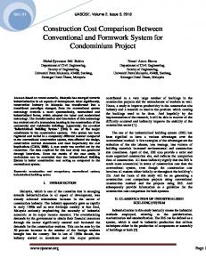
Location of mandibular canal in the jaw bone was evaluated from superior and inferior cortex of mandible at the mental foramen, at 1 cm, 2 cm, and 3 cm posterior to the mental foramen. The distances were measured from superior border of jawbone to the upper border of mandibular canal and from the inferior border of jaw bone to the lower border of mandibular canal using OPG [Figure 3], DVT [Figure 4] and again OPG [Figure 5].
Measurement in buccolingual dimension of mandible
Location of mandibular canal in the jaw bone was evaluated with respect to buccolingual dimension of jaw bone using DVT at the mental foramen, at 1 cm, at 2 cm, and 3 cm posterior to the mental foramen. The measurement was made for the distance from the buccal cortex and lingual cortex to the mandibular canal using DVT [Figures 4 and 6].
Results After following strict inclusion and exclusion criteria, we included 25 subjects having edentulous areas in the mandibular posterior region of jaw. Out of the 25 patients, 9 were males and 16 were females. The age of patients ranged from 15 to 65 years with the mean age of 35.48 (±12.69) years. In the present study, it was observed that the mandibular canal was not visible at the mental foramen, visible with diffuse borders at 1 cm and 2 cm posterior to the mental foramen and clearly visible at 3 cm posterior to the mental foramen using OPG. The mandibular canal was more clearly visible at the mental foramen and 1 cm and 2 cm posterior to the mental foramen in DVT as compared to OPG, which was statistically significant (P = 0.000), while at
For 3D volumetric imaging, Phillips Allura Xper FD20 3D RA, Digital Subtraction Angiography unit (Netherlands) was used. OPG was taken using Orthopantomogram Machine (Planmeca Proline CC Panoramic X‑ray, Planmeca OY Helsinki, Finland).
Methodology
A thorough case history, oral, and general examinations of participants were carried out and findings were noted. The radiographic evaluation of mandibular canal in posterior region was done using Orthopantomography and DVT. The mandibular canal was evaluated using the below criteria. 268
Figure 1: Visibility of mandibular canal using orthopantomography
Journal of Indian Academy of Oral Medicine & Radiology ¦ Volume 29 ¦ Issue 4 ¦ October‑December 2017
[Downloaded free from http://www.jiaomr.in on Thursday, February 15, 2018, IP: 212.252.57.234] Chakraborty, et al.: Panoramic radiography vs digitalized volumetric tomography
Figure 3: Location of mandibular canal using orthopantomography
Figure 2: Visibility of mandibular canal using digitalized volumetric tomography
Figure 5: Location of mandibular canal using orthopantomography with respect to superoinferior dimension
Table 1: Age and gender distribution of study subjects Age in years
Gender Male n
Figure 4: Location of mandibular canal using digitalized volumetric tomography
3 cm posterior to the mental foramen it was clearly visible in both OPG and DVT, which was statistically not significant (P = 0.297) [Table 1 and Graph 1]. Gender‑wise comparison revealed no difference between males and females for visibility of the mandibular canal in both OPG and DVT. The location of the mandibular canal was more accurately evaluated in DVT because it was possible to determine the location of the mandibular canal in both superoinferior and buccolingual dimensions of the jaw bone, whereas the buccolingual dimension could not be evaluated in OPG. The maximum mean distance at the superior aspect was at the mental foramen using DVT and OPG was 1.57 ± 0.20 cm and 1.93 ± 0.34 cm, respectively. The maximum mean distance
χ2
Total
P
Female %
n
%
n
%
≤33 05 20 08 32 13 52 >33 04 16 08 32 12 48 Total 09 36 16 64 25 100 Mean±SD 35.67±13.49 35.37±12.67 35.48±12.69 SD: Standard deviation, χ2: Chi square, P: Probability
0.071 0.790
at the inferior aspect at 3 cm posterior to the mental foramen using DVT and OPG was 0.84 ± 0.16 cm and 1.70 ± 0.21 cm, respectively [Table 2]. The buccal (0.50 ± 0.07 cm) and lingual aspect (0.41 ± 0.09 cm) was maximum at 3 cm posterior to the mental foramen. Overall, the mandibular canal was located more towards the inferior cortex and lingual cortex in the jaw bone [Table 3]. Gender‑wise comparisons revealed that there was no significant difference for location of mandibular canal between males and females using both OPG and DVT.
Discussion The present study has included the evaluations of mandibular canal in all 3‑dimensions involving the buccolingual,
Journal of Indian Academy of Oral Medicine & Radiology ¦ Volume 29 ¦ Issue 4 ¦ October‑December 2017
269
[Downloaded free from http://www.jiaomr.in on Thursday, February 15, 2018, IP: 212.252.57.234] Chakraborty, et al.: Panoramic radiography vs digitalized volumetric tomography
Table 2: Comparison between OPG and DVT for the mean distances of mandibular canal in the superoinferior dimension of jaw bone Parameters
Distances measured
OPG
At mental foramen
Superior border of jaw bone to the upper border of mandibular canal Lower border of mandibular canal to the inferior cortex of jaw bone At 1 cm posterior Superior border of jaw bone to the upper border of mandibular canal to mental foramen Lower border of mandibular canal to the inferior cortex of jaw bone At 2 cm posterior Superior border of jaw bone to the upper border of mandibular canal to mental foramen Lower border of mandibular canal to the inferior cortex of jaw bone At 3 cm posterior Superior border of jaw bone to the upper border of mandibular canal to mental foramen Lower border of mandibular canal to the inferior cortex of jaw bone SD: Standard deviation, t: Unpaired t‑test, P: Probability, * Statistically significant
DVT
Mean
±SD
Mean
±SD
1.93 1.36 1.78 1.22 1.60 1.27 1.58 1.70
0.34 0.32 0.41 0.38 0.46 0.40 0.45 0.21
1.57 0.68 1.54 0.69 1.40 0.76 1.18 0.84
0.20 0.20 0.26 0.18 0.29 0.13 0.27 0.16
t
P
4.50 8.90 2.44 6.14 1.83 5.95 3.82 15.70
0.000* 0.000* 0.019* 0.000* 0.075 0.000* 0.000* 0.000*
Table 3: Comparison between OPG and DVT for the mean distances of mandibular canal in the buccolingual dimension of jaw bone Distances measured
Location
Mean
SD
F
P
Mandibular canal to the buccal cortex
At mental foramen 0.40 0.07 8.469 0.000* At 1 cm 0.43 0.07 At 2 cm 0.46 0.07 At 3 cm 0.50 0.07 Mandibular At mental foramen 0.33 0.14 3.698 0.014* canal to the At 1 cm 0.34 0.10 lingual cortex At 2 cm 0.40 0.08 At 3 cm 0.41 0.09 SD: Standard deviation, F: ANOVA test, P: Probability, *Statistically significant
anteroposterior and superoinferior dimensions. The superoinferior dimension allows the evaluation of mandibular canal from the alveolar crest to the inferior cortex of mandible in all the four sections. The anteroposterior dimensions allow the evaluation of mandibular canal at the mental foramen, at 1 cm, 2 cm and 3 cm posterior to the mental foramen and buccolingual dimension from the mandibular canal to the buccal and lingual cortex. The present cross‑sectional study was conducted on 25 subjects with edentulous posterior region of jaw with the mean age of 35.48(±12.69) years. The sample size was corresponding to some previous studies.[7–12] The mean age was approximately corresponding to the study by Oliveira‑Santos C et al.[13] and Afkhami F et al.[14] The present study included 9 males and 16 females where the gender‑wise differences were not significant, which was similar to study by other authors.[7,8,11–13]
Visibility of the mandibular canal
The radiographic evaluation for visibility was done using radiographic techniques. The visibility criteria used in our study was given by Lindh C et al.[7] In our study, it was observed that the mandibular canal was not visible at the mental foramen, visible with diffuse borders at 1 cm and 2 cm posterior to the mental foramen and clearly visible at 3 cm posterior to the mental foramen using OPG [Figure 1]. Dharmar[15] examined 270
Figure 6: Location of mandibular canal using digitalized volumetric imaging with respect to buccolingual dimension
subjects for clear visualization of the mandibular canal with OPG and observed that mandibular canal was poorly visible from mental foramen to 2nd molar because of porosity of the mandibular canal walls and clearly visible posteriorly from 2nd molar to the mandibular foramen. In DVT, it was observed that the mandibular canal was visible with diffuse borders at the mental foramen and was clearly visible at 1 cm, 2 cm, and at 3 cm posterior to the mental foramen using DVT [Figure 2], which was similar to the results obtained by Dharmar.[15] When OPG and DVT was compared, it was found that the mandibular canal was more clearly visualized using DVT compared to OPG at the mental foramen, at 1 cm, and at 2 cm posterior to the mental foramen. Similar studies were conducted by Lindh et al.[7] where it was observed that mandibular canal was clearly visualized in volumetric imaging compared to the OPG similar to the results of our study. Tomography revealed significantly higher scores for visibility than OPG in the region of the mental foramen and 1 cm posterior to the mental foramen. CT was reported to visualize mandibular canal more clearly at 1 cm posterior to mental foramen than OPG. Similar results were obtained by Klinge et al.,[16] Angelopoulos et al.,[17] Juodzbalys et al.,[18] Santos et al.,[19] and Angelopoulous et al.[20]
Journal of Indian Academy of Oral Medicine & Radiology ¦ Volume 29 ¦ Issue 4 ¦ October‑December 2017
[Downloaded free from http://www.jiaomr.in on Thursday, February 15, 2018, IP: 212.252.57.234] Chakraborty, et al.: Panoramic radiography vs digitalized volumetric tomography
>33 48%
≤33 52%
Graph 1: Age distribution of study subjects
Oliveira‑Santos et al.[13] stated that the visibility decreased towards the mental foramen compared to the posterior segment of the mandibular canal due to the lack of definite walls in the anterior portion of the canal. Wadu et al.[21] studied mandibular canal appearance on the panoramic radiographs and found that in number of cases the radiopaque border of canal was either disrupted or even absent. The superior border was more prone to disruption than the inferior border. While the study by Lindh et al.[7] and Kim[8] were in disagreement, where they observed no differences between OPG and DVT at 2 cm posterior to the mental foramen, the study conducted by Kim[8] on 11 patients observed that mandibular canal could not be identified in 10% of the regions on conventional panoramic radiographs and 7% on Dentascan. The present study also estimated the visibility of canal at 3 cm posterior to mental foramen, where the mandibular canal was clearly visible in both OPG and DVT, which was similar to Dharmar.[15] Ylikontiola et al.[9] also reported that the canal cortication was clearly visible at the posterior region of mandible. Gender‑wise comparison in the present study revealed no significant difference between males and females for the visibility of mandibular canal. This has been disagreed by the study conducted by Kim et al.,[12] whereas the mandibular canal is more clearly visible in males compared to females because the resorption of the superior border of the mandibular canal was detected more often in females (32.6%) than in males (9.8%).
Location of the mandibular canal
In the present study, the mean distances of mandibular canal from superior border of jaw bone to the upper border of mandibular canal and the lower border of mandibular canal to the inferior cortex of jaw bone were evaluated using OPG and DVT. There was a significant difference in mean distances between the two radiographic techniques for the superoinferior dimension. Mean distances in OPG were more compared to DVT, which may be attributed to the magnification in OPG. The mandibular canal was observed to be more inferiorly placed with respect to superoinferior dimension in the
mandible. A study conducted by Gowgiel[22] demonstrated that the neurovascular bundle was located about 10 mm above the inferior border in the body of the mandible. In the molar region, the neurovascular bundle was located 0.5 mm to 1.5 mm closer to the inferior border than in the premolar region. Levine et al.[23] reported that the measurement at the inferior aspect in relation to the second molar region measured around 17.4 mm. Similar study by Nkenke et al.[24] measured the distance from the alveolar crest to the inferior alveolar canal ‑ 11(±2.2) mm[25] ranging from 15.3 to 17.4 mm in 79 Japanese patients using CT. Kamburoglu et al.[26] observed that CBCT had a high accuracy for locating the position of the inferior alveolar canal. The canal was observed to be more lingually placed in the mandible [Figure 4]. Ylikontiola et al.[9] reported that the mandibular canal was better visualized buccolingually on CT scans than on conventional radiographs. The mean distances from the mandibular canal to the buccal cortex of the specimen mandibles were 2.5 mm and to the lingual cortex of the mandible was 0.6 mm. CT revealed 7% of the mandibular canal was in direct contact (1 mm) with the buccal cortex and 67% in contact with the lingual cortex. Similar to our study, the mandibular canal was closer to the lingual than to the buccal cortex of the mandible. Dalili et al.[27] also reported that the mandibular canal is most often positioned lingually (60.5%) than buccally. Similar results were obtained by Shahidi et al.,[28] Gowgiel,[22] Jung et al.,[29] Katakam et al.,[30] and Li N et al.[31] Kamburoglu et al.[32] observed that the inferior alveolar canal was about 4.9 mm from the buccal cortical bone. Levine et al.[23] reported the mean buccal width measured 4.9 mm, which was approximately similar to our study. Kwon et al.[33] evaluated the distance from the buccal cortex to the mandibular canal was 5.39 (±1.56) mm. Rajchel et al.[34] also reported the mean lingual width was 2 mm. Comparing males and females, in our study it was observed that there were no significant differences in mean distances in the superoinferior and buccolingual dimensions between males and females. This was similar to the result obtained by Kwon et al.[33]
Conclusion In the present study, comparative evaluation of the mandibular canal for visibility and location has been done in posterior edentulous area of mandible using OPG and DVT, where it was observed that DVT had some added advantages over OPG regarding the visibility and location of mandibular canal. In addition, it is possible to evaluate both superoinferior and buccolingual dimension of jaw bone in DVT. The volumetric imaging is the best indicator to determine the precise anatomical relationship between the edentulous area and the mandibular canal. It is recommended that the visibility and location should also be evaluated at 3 cm posterior to the mental foramen, as some patients need replacement of missing teeth in the 3rd molar or extreme posterior region of mandible. The results of the present study may provide useful guidelines
Journal of Indian Academy of Oral Medicine & Radiology ¦ Volume 29 ¦ Issue 4 ¦ October‑December 2017
271
[Downloaded free from http://www.jiaomr.in on Thursday, February 15, 2018, IP: 212.252.57.234] Chakraborty, et al.: Panoramic radiography vs digitalized volumetric tomography
for locating the precise location of the mandibular canal in planning for implant placement and other interventions in mandibular posterior regions. A future study might be planned including larger sample size, so that, the study results could be generalized and applicable to larger population.
Financial support and sponsorship Nil.
Conflicts of interest
There are no conflicts of interest.
References 1. Misch CE, editor. Contemporary Implant Dentistry. 2nd ed. St Louis, Missouri: Mosby; 1999. p. 3‑12. 2. Kraut RA. A case for routine CT imaging of dental alveolus before implant placement. J Oral Maxillofac Radiol 2001;59:64‑7. 3. Bhatia HP, Goel S, Srivastava B. Dental Scan. J Health Community Dent 2012;6:25‑7. 4. Williams MY, Mealey BL, Hallmon WW. The role of computerized tomography in dental Implantology. Int J Oral Maxillofac Implants 1992;7:373‑80. 5. Raichur PS, Setty SB, Thakur SL, Naikmasur VG. Comparison of radio‑visiography and digital volume tomography to direct surgical measurements in the detection of infrabony defects. J Clin Exp Dent 2012;4:43‑7. 6. Chandel S, Agarwal A, Singh N, Singhal A. Dentascan: A Diagnostic Boon. J Dent Sci Res 2013;1:13‑7. 7. Lind C, Petersson A. Radiologic examination for location of the mandibular canal: A comparison between panoramic radiography and conventional tomography. Int J Oral Maxillofac Implants 1989;4:249‑53. 8. Kim EK. Comparison of different radiographic methods for the detection of the mandibular canal. Korean J Oral Maxillofac Radiol 2003;33:199‑205. 9. Ylikontiola L, Moberg K, Huumonen S, Soikkonen K, Oikarinen K. Comparison of three radiographic methods used to locate the mandibular canal in the buccolingual direction before bilateral sagittal split osteotomy. Oral Surg Oral Med Oral Pathol Oral Radiol Endod 2002;93:736‑42. 10. Martin H. Three‑dimensional digital radiography in dental application. Int J Digital Dental News 2009;3:1863‑7957. 11. Lara JS, Quezada AS, Valenzuela JS. Mandibular canal duplication prevalence, digital panoramic radiography analysis. Int J Odontostomat 2010;4:207‑13. 12. Kim YK, Park JY, Kim SG, Kim JS, Kim JD. Magnification rate of digital panoramic radiographs and its effectiveness for pre‑operative assessment of dental implants. Dentomaxillofac Radiol 2011;40:76‑83. 13. de Oliveira‑Santos C, Souza PH, Berti‑Couto SA, Stinkens L, Moyaert K, Rubira‑Bullen IR, et al. Assessment of variations of the mandibular canal through cone beam computed tomography. Clin Oral Investig 2012;16:387‑93. 14. Afkhami F, Haraji A, Boostani HR. Radiographic localization of the mental foramen and mandibular canal. J Dent 2013;10:436‑42. 15. Dharmar S. Locating the mandibular canal in panoramic radiograph. Int J Oral Maxillofac Implants 1997;12:113‑7. 16. Klinge B, Petersson A, Maly P. Location of the mandibular canal: Comparison of macroscopic findings, conventional radiography, and computed tomography. Int J Oral Maxillofac Implants 1989;4:327‑32.
272
17. Angelopoulos C, Scarfe WC, Fracd S, Farman AG. Comparison of maxillofacial CBCT and medical CT. Atlas Oral Maxillofac Surg Clin North Am 2012;20:1‑17. 18. Juodzbalys G, Wang HL. Guidelines for the identification of the mandibular vital structures: Practical clinical applications of anatomy and radiological examination method. J Oral Maxillofac Res 2010;1:1‑15. 19. Santos CO, Capelozza AL, Dezzoti M, Fischer C, Poleti ML, Bullen IR. Visibility of the mandibular canal on CBCT cross‑sectional images. J Appl Oral Sci 2011;19:240‑3. 20. Angelopoulous C, Thomas SL, Hechler S, Parissis N, Hlavacek M. Comparison between digital panoramic radiography and cone‑beam computed tomography for the identification of the mandibular canal as part of pre‑surgical dental implant assessment. J Oral Maxillofac Surg 2008;66:2130‑5. 21. Wadu SG, Penhall B, Townsend GC. Morphological variability of the human inferior alveolar nerve. Clin Anat 1997;10:82‑7. 22. Gowgiel JM. The position and course of the mandibular canal. J Oral Implantol 1992;18:383‑5. 23. Levine MH, Goddard AL, Dodson TB. Inferior alveolar nerve canal position: A clinical and radiographic study. J Oral Maxillofac Surg 2007;65:470‑4. 24. Nkenke E, Radespiel‑Troger M, Wiltfang J, Schultze‑Mosgau S, Winkler G, Neukam FW. Morbidity of harvesting of retromolar bone grafts: A prospective study. Clin Oral Implants Res 2002;13:514‑21. 25. Watanabe H, Mohammad AM, Kurabayashi T, Aoki H. Mandible size and morphology determined with CT on a premise of dental implant operation. Surg Radiol Anat 2010;32:343‑9. 26. Kamburoglu K, Kilic C, Ozen T, Yuksel SP. Measurements of mandibular canal region obtained by cone‑beam computed tomography: A cadaveric study. Oral Surg Oral Med Oral Pathol Oral Radiol Endod 2009;107:e34‑42. 27. Dalili Z, Mahjoub P, Sigaroudi AK. Comparison between cone beam computed tomography and panoramic radiography in the assessment of the relationship between the mandibular canal and impacted class C mandibular third molars. Dent Res J 2011;8:203‑10. 28. Shahidi S, Zamiri B, Bronoosh P. Comparison of panoramic radiography with cone beam CT in predicting the relationship of the mandibular third molar roots to the alveolar canal. Imaging Sci Dent 2013;43:105‑9. 29. Jung YH, Nah KS, Cho BH. Correlation of panoramic radiographs and cone beam computed tomography in the assessment of a superimposed relationship between the mandibular canal and impacted third molars. Imaging Sci Dent 2012;42:121‑7. 30. Katakam SK, Shankar U, Thakur D, Reddy TP, Hari KR, Jang D. Comparison of orthopantomography and computed tomography image for assessing the relationship between impacted mandibular third molar and mandibular canal. J Contemp Dent Pract 2012;13:819‑23. 31. Li N, Zhao B, Tan C. Intramandibular course and anatomic structure of the inferior alveolar nerve canal. Zhonghua Kou Qiang Yi Xue Za Zhi 2001;36:446‑7. 32. Kamburoglu K, Kilic C, Ozen T, Yuksel SP. Measurements of mandibular canal region obtained by cone‑beam computed tomography: A cadaveric study. Oral Surg Oral Med Oral Pathol Oral Radiol Endod 2009;107:34‑42. 33. Kwon KH, Sim KB, Lee JM. Evaluation of the course of the inferior alveolar canal in the mandibular ramus using cone beam computed tomography. J Korean Assoc Oral Maxillofac Surg 2012;38:231‑9. 34. Rajchel J, Ellis E 3rd, Fonseca RJ. The anatomical location of the mandibular canal: Its relationship to the sagittal ramus osteotomy. Int J Adult Orthodon Orthognath Surg 1986;1:37‑47.
Journal of Indian Academy of Oral Medicine & Radiology ¦ Volume 29 ¦ Issue 4 ¦ October‑December 2017