Dentomaxillofacial Radiology (2008) 37, 228–231 ’ 2008 The British Institute of Radiology http://dmfr.birjournals.org
RESEARCH
New software: comparison between three software programs for root canal length measurement D Goodarzi Pour*,1, H Razmi1, S Jabedar Maralani2 and S Zeighami1 1
Dental School, Tehran University of Medical Science and Health Service, Iran; 2Dentist, Tehran University of Medical Science and Health Service, Iran
Objectives: The purpose of this study was to design a software program to estimate the curved root canal working length by using curved line software and comparing its accuracy with Trophy and Cygnus software programs. Methods: 120 simulated curved canals were divided into 12 groups based on the radius and the angle of the curvature. The true canal length was measured from the orifice to the terminus. Canals were filled with urographin and were imaged by a digital radiographic system. Canal length was measured with newly developed software that uses a curved line, as well as with Cygnus and Trophy software, both of which use a sequence of straight lines, and their accuracies were compared. Results: The proposed software was significantly more accurate than other software with respect to the gold standard (P , 0.001). However, the error was less than 0.5 mm in over 99% of the cases with all software with three clicks, and over 96% of the cases with six clicks in Trophy, which was not statistically significant (P 5 0.99). Conclusions: The studied software programs were not significantly different and their statistically significant difference compared with the gold standard is not clinically significant. The suggested software has to be studied more regarding its capabilities in the utilization of curved lines in measuring curved canals and calibration of the measurements. Dentomaxillofacial Radiology (2008) 37, 228–231. doi: 10.1259/dmfr/16010494 Keywords: root canal; software; length; measurement; comparison Introduction In treating teeth that require endodontic therapy, dentists face three different situations – vital pulp, necrotic pulp and therapeutic cases.1 Preparation of an ideal coneshaped canal warrants a preliminary estimation of the length and curvature of the canal.2 The goal of determining the working length is the determination of the length of the canal that has to be prepared and filled. Any error and miscalculation in estimation of the working length, especially in curved canals, can result in complications during and after the treatment of the canal.1 There are various methods available to determine the length of the canal, the most common of which is conventional radiography.3 The most important limitation of conventional radiography is its lack of precision in the measurement of the length of curved canals, which
becomes more prominent with increase in the curvature of the canal.1,4 The introduction of digital radiography enabled us to measure the curvature angle and length of curved canals.3 This method’s accuracy is less influenced by the curvature of the canal.2 Despite its appropriate abilities and performance, digital imaging still has certain limitations in measurement of the length of curved canals since most commonly used software utilizes sets of consecutive straight lines. The purpose of this study was to introduce software to draw the curvature of the canals, to accurately measure the length of curved canals in digital X-rays and to compare the accuracy with commonly utilized software.
Materials and methods *Correspondence to: Daryoush Goodarzi Pour, No. 17, Ostad Shahriar Alley, Western Sarv Avenue, Sadat Abad, Tehran, Iran 1981657354; E-mail:
[email protected] Received 24 December 2006; revised 22 June 2007; accepted 8 July 2007
After approval of the proposal by the research deputy of the dentistry faculty of Tehran University of Medical
Software for root canal length measurement D Goodarzi Pour et al
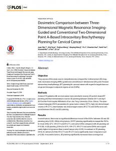
Sciences, 120 simulated polyester canals were prepared in 12 equally distributed groups with curvature radii of 5 mm, 7 mm and 9 mm, and curvature angles of 5–15 ˚, 15–30 ˚, 30–45 ˚ and 45–60 ˚ (Figure 1). The curvature radius is defined as the length of the curved line connecting the beginning point of the curve and the end point of the canal. The curvature angle is defined as the angle between the line which is parallel to the longitudinal axis of the canal in the coronal third, and the line which connects the apical foramen to the junction of the above-mentioned line and the longitudinal axis of the canal. The curvatures of the curved canals were measured using the Schneider method.5 The length of canals were different and their diameters were the same as a no. 40 endodontics spreader. After the canals were prepared, a nickel–titanium size 15 file was inserted into the canal until its tip was visible at the end point of the canal. It was then extracted and the length of the canal was measured using a digital caliper with a standard error of 0.02 mm. This was repeated after 1 week and the measurements were redone from the beginning in cases that differed by more than 0.1 mm in the results of the two measurements. These measurements were made by a postgraduate student of prosthodontics who had been trained by raters (raters are discussed below).
229
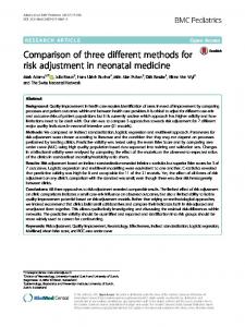
Measurements were checked randomly by raters. The canals were then filled with urographin (Darupakhsh Co., Tehran, Iran) using insulin syringes and files. After that, digital imaging was done by means of Planmeca Proline radiography equipment (Planmeca Oy, Helsinki, Finland) (exposure of 0.3 s with 8 mA and 50 kVp). The images were taken using RadioVisioGraphy (RVG) (Trophy 2000, Paris, France) and the parallel technique. The object was adjacent to the sensor, the distance between the sensor and the tube was 3 cm and the central beam was vertical to the plane aligned to the curvature. A calibration wire was included. The images were evaluated using three software programs: Cygnus (Cygnus Media 3.0.1.391, Fort Pierce, FL), Trophy (Trophy Windows v4.2f Paris, France) with three and six clicks of the mouse during measurement and drawing consecutive straight lines, and our proposed software (designed by the medical engineering team of Tehran University of Medical Science and Health Service, Iran) (Figure 2). Our software measures the canal length estimating curved line length instead of the conventional use of consecutive straight lines by Cygnus and Trophy software. In the proposed software, the image is opened using the ‘‘File’’ tab. Then, one of the radio buttons of the ‘‘Line mode’’ tab can be selected in terms of the root form. In order to measure the canal or calibration of the software program, the ‘‘Measure mode’’ tab should be selected. It is possible to set the colour and width of the brush by clicking on ‘‘Tools’’. Measurement can be performed by drawing a line from the reference point to the end of the working length. Each measurement by each software program was carried out twice. Raters consisted of a maxillofacial radiologist with 8 years’ experience and an endodontist with 22 years’ experience. The frequency of error compared with the true length of the canal was calculated with an accuracy of ¡0.5 mm and ¡1 mm. The frequencies of error of each software program were calculated generally (without considering angles and radiuses of curvatures) and were compared using Fischer’s exact test. Also, the Pearson correlation coefficient was calculated between the figures determined by each software program and the gold standard. Finally, the absolute error values for each software program and the gold standard were calculated in each measurement and were compared using ANOVA and Tukey’s post hoc tests. The data were analysed utilizing SPSS for Windows v10.05 (SPSS Inc., Chicago, IL). A P-value below 0.05 was considered significant.
Results
Figure 1
Simulated canal model used in this study
The frequencies of measurement errors with different accuracies (¡0.5 mm and ¡1 mm) in measurements carried out by the different software are displayed in Table 1. Dentomaxillofacial Radiology
Software for root canal length measurement D Goodarzi Pour et al
230
Overall, Cygnus overestimated all measurements while Trophy with three and six clicks overestimated all samples except one, which was underestimated.
However, the proposed software mostly underestimated the values but overestimation occurred in a minority of cases. The Pearson correlation coefficients between Cygnus, Trophy with three clicks and our proposed software, and the gold standard were significant and equal (r 5 0.998, P , 0.001). This coefficient for Trophy with six clicks was lower (r 5 0.997, P , 0.001), but this was not statistically significant (P 5 0.12). The comparison of each software error from the gold standard with each other is summarized in Table 2. Based on the results, it is obvious that our proposed software was significantly more accurate than other software with respect to the gold standard (P , 0.001). Assuming ¡0.5 mm as the clinically acceptable error, the accuracy of Cygnus, Trophy with three clicks and our proposed software was 99.2% in comparison with an accuracy of 95.9% for Trophy with six clicks, which was not statistically significant (P 5 0.99 with Fischer’s exact test).
Discussion
Figure 2 Curved canal measurement using (a) Cygnus, (b) Trophy and (c) our software Dentomaxillofacial Radiology
The results of our study revealed that allowing a ¡0.5 mm error, the accuracies of Cygnus, Trophy with three clicks and the proposed software are equal to 99.2%, which is not significantly different from the results of Trophy with six clicks. It is also shown that the Pearson correlation coefficients of the software with the gold standard are remarkably high and do not yield any significant difference with each other. These results are in congruence with the results of Burger et al,3 who indicated that there is no significant difference between the four digital methods of measuring the canal length and that they all overestimated the real length of the canal. We also observed that the results of Cygnus and Trophy mostly overestimated the real values and were compatible with the results of Burger et al3 and Mentes et al.2 However, our proposed software mostly underestimated the real values. As mentioned above, measurement of working length, especially in curved canals, is of great importance in endodontic treatments. In this field there have been extensive studies and numerous methods investigated in both the anatomy of the apical area to determine the end point of the treatment1 and the measurement of the working length of the canal in endodontic treatments.6 Moreover, there have been various studies to compare the results of conventional and digital methods of radiography in measurement of working length.2,4,6,7 Our results showed a significant difference between all software and the gold standard. Burger et al3 have also shown the same difference in all imaging methods with the real length of the canals. However, the data from the present study revealed that the results of the proposed software have a significantly smaller difference with the gold standard in comparison with other
Software for root canal length measurement D Goodarzi Pour et al
231
Table 1 The frequency of the measurement error of the three software programs
Trophy with three clicks Trophy with six clicks Cygnus Proposed software
Accuracy of 21 mm to 20.51 mm
Accuracy of 20.5 mm to 0 mm
Accuracy of 0 mm to 0.5 mm
Accuracy of 0.51 mm to 1 mm
0 0 0 1
1 1 0 86
118 115 119 33
1 4 1 0
(0%) (0%) (0%) (0.8%)
(0.8%) (0.8%) (0%) (71.7%)
software (P , 0.001). It can be concluded that statistically the proposed software is significantly more accurate than the other software. However, since an error of less than 0.5 mm is not clinically significant,7 this statistical difference is not clinically remarkable and all softwares yield enough ability to measure the working length in curved canals. Of note, the proposed software is the only Iranian-made software available for this purpose. This software should be further evaluated especially in other root forms (C and J root forms).The next logical step is to evaluate this software using natural extracted teeth without contrast media, as well as to determine whether the new software has any advantages in ease of use over the other commercially available software.
(98.4%) (95.9%) (99.2%) (27.5%)
(0.8%) (3.3%) (0.8%) (0%)
Table 2 The comparison of each software error from gold standard and with each other Mean ¡ SD* Trophy 3 Clicks{,** Trophy 6 Clicks{,** Cygnus{,** Proposed Software1,{,{
0.23¡.11 0.30¡.14 0.22¡.11 0.11¡.10
*Significant difference (ANOVA, P 5 0.0001). Significant difference with Trophy 6 Clicks (Tukey’s post hoc highly significant difference (HSD) test, P 5 0.0001). **Significant difference with proposed software (Tukey’s post hoc HSD test, P 5 0.0001). { Significant difference with Cygnus (Tukey’s post HSD test, P 5 0.0001). 1 Significant difference with Trophy three clicks (Tukey’s post hoc HSD test, P 5 0.0001) {
References 1. Walton RE. Access preparation and length determination. In: Walton RE, Torabinejad M (eds). Principles and practice of endodontics (3rd edn). Philadelphia, PA: WB Saunders, 2002. pp: 200–204. 2. Mentes A, Gencoglu N. Canal length evaluation of curved canals by direct digital or conventional radiography. Oral Surg Oral Med Oral Pathol Oral Radiol Endod 2002; 93: 88–91. 3. Burger CL, Mork TO, Hutter JW, Nicoll B. Direct digital radiography versus conventional radiography for estimation of canal length in curved canals. J Endod 1999; 25: 260–263.
4. Kim-Park MA, Baughan LW, Hartwell GR. Working length determination in palatal roots of maxillary molars. J Endod 2003; 29: 58–61. 5. Schneider SW. A comparison of canal preparations in straight and curved root canals. Oral Surg Oral Med Oral Pathol 1971; 32: 271–275. 6. Subramanian P, Konde S, Mandanna DK. An in vitro comparison of root canal measurement in primary teeth. J Indian Soc Pedod Prev Dent 2005; 23: 124–125. 7. Lamus F, Katz JO, Glaros AG. Evaluation of a digital measurement tool to estimate working length in endodontics. J Contemp Dent Pract 2001; 2: 24–30.
Dentomaxillofacial Radiology
Copyright of Dentomaxillofacial Radiology is the property of British Institute of Radiology and its content may not be copied or emailed to multiple sites or posted to a listserv without the copyright holder's express written permission. However, users may print, download, or email articles for individual use.