JOURNAL OF CLINICAL MICROBIOLOGY, Aug. 2010, p. 2897–2901 0095-1137/10/$12.00 doi:10.1128/JCM.00136-10 Copyright © 2010, American Society for Microbiology. All Rights Reserved.
Vol. 48, No. 8
Comparison of an Automated Repetitive-Sequence-Based PCR Microbial Typing System with Pulsed-Field Gel Electrophoresis for Molecular Typing of Vancomycin-Resistant Enterococcus faecium䌤† Yu-Chung Chuang,1 Jann-Tay Wang,1,2 Mei-Ling Chen,2 and Yee-Chun Chen1,2,3* Department of Internal Medicine, National Taiwan University Hospital, Taipei, Taiwan1; Center for Infection Control, National Taiwan University Hospital, Taipei, Taiwan2; and Department of Medicine, National Taiwan University College of Medicine, Taipei, Taiwan3 Received 27 January 2010/Returned for modification 9 April 2010/Accepted 10 June 2010
Vancomycin-resistant Enterococcus faecium (VRE) has become an important health care-associated pathogen because of its rapid spread, limited therapeutic options, and possible transfer of vancomycin resistance to more-virulent pathogens. In this study, we compared the ability to detect clonal relationships among VRE isolates by an automated repetitive-sequence-based PCR (Rep-PCR) system (DiversiLab system) to pulsedfield gel electrophoresis (PFGE), the reference method for molecular typing of VRE. Two sets of VRE isolates evaluated in this study were collected by active microbial surveillance at a large teaching hospital in Taiwan during 2008. The first set included 90 isolates randomly selected from the surveillance cohort. The first set consisted of 34 pulsotypes and 10 Rep-PCR types. There was good correlation between the two methods (P < 0.001). The second set included 68 VRE isolates collected from eight clusters of colonization. A dominant clone was detected in five out of eight clusters by both methods. Two clusters were characterized by Rep-PCR as being caused by a dominant clone, whereas PFGE showed polyclonal origins. One cluster was shown to be polyclonal by both methods. A single Rep-PCR clone type was detected among 12 of 14 vancomycin-intermediate enterococci, whereas PFGE detected six pulsotypes. In conclusion, the Rep-PCR method correlated well with PFGE typing but was less discriminative than PFGE in defining clonal relationships. The ease of use and more rapid turnaround time of Rep-PCR compared to PFGE offers a rapid screening method to detect outbreaks of VRE and more rapidly implement control measures. PFGE remains the preferred method to confirm clonal spread. noncoding repetitive sequences in bacterial genomes (28). Recently, Healy et al. described an automated Rep-PCR technology and its performance characteristics (10). RepPCR has been used in VRE outbreak investigations (20, 23), but we are unaware of any comparative field tests of RepPCR and PFGE. This study was designed to compare the ability of the two methods to distinguish clonal relationships among a panel of epidemiologically well-defined isolates of VRE. We also evaluated the association of specific drug resistance patterns with selected genotypes.
Vancomycin-resistant Enterococcus faecium (VRE) was first isolated in England in 1986 (26). It has become an important health care-associated pathogen because of its rapid spread, significant attributable mortality, limited options for therapy, and the possible transfer of vancomycin resistance to morevirulent pathogens (3, 4, 14). VRE organisms are now commonly isolated in hospitals around the world. Molecular typing methods are needed to distinguish prevalent strains from epidemic strains. Pulsed-field gel electrophoresis (PFGE) is the accepted reference method for molecular typing of VRE chromosomes because of its high discriminative power and established interpretative guidelines (25, 29). PFGE is labor-intensive and skill dependent, and the turnaround time is 2 to 3 days (7, 27). VRE isolates collected during outbreak investigations at our hospital during the past 2 years consisted of a wide variety of PFGE types. Polyclonal outbreaks have also been reported by other investigators (8, 12). It is not clear whether this represents multiple introductions of different strains or microevolutionary changes in native VRE or whether PFGE is overly discriminatory. Repetitive-sequence-based PCR (Rep-PCR) methods are rapid typing procedures that amplify the regions between the
MATERIALS AND METHODS Hospital infection control program for VRE. The National Taiwan University Hospital (NTUH) is a 2,200-bed major teaching hospital in Taipei, Taiwan. The VRE surveillance program was initiated in 1995 (29). The perirectal swab surveillance cultures (22) were obtained from patients in rooms located near or in the same room as the infected or colonized index case. The surveillance program was expanded in January 2008 to include selected wards where VRE continued to spread despite reinforced standard precautions and compliance with contact precautions for VRE carriers. Perirectal surveillance cultures were obtained from patients admitted to these wards and weekly thereafter until discharge. Bacterial isolates. VRE isolates were screened by bile esculin azide broth containing 8 g/ml of vancomycin (BEAV) and chromogenic agar medium (chromID VRE; bioMe´rieux, Marcy l’Etoile, France). Colonies on the chromID VRE plates with purple or green pigmentation were presumptively identified as vancomycin-resistant Enterococcus faecium or Enterococcus faecalis, respectively. Presumptive VRE colonies were then subcultured onto Trypticase soy agar plates supplemented with 5% sheep blood (BBL) for confirmatory identification using the Vitek2 system (bioMe´rieux, Marcy l’Etoile, France). The microorganisms were identified to the species level using the API 20 Strep system (bioMe´rieux Vitek, Hazelwood, MO) and the API 32 Strep system (bioMe´rieux Vitek). In vitro susceptibility against vancomycin of these isolates was
* Corresponding author. Mailing address: Department of Internal Medicine, National Taiwan University Hospital, No. 7 Chung-Shan South Road, Taipei, Taiwan 100. Phone: 886-2-23123456, ext. 65054. Fax: 886-2-23971412. E-mail:
[email protected]. † Supplemental material for this article may be found at http://jcm .asm.org/. 䌤 Published ahead of print on 16 June 2010. 2897
2898
CHUANG ET AL.
J. CLIN. MICROBIOL.
TABLE 1. Relationship between Rep-PCR type, PFGE pulsotypes, and antimicrobial susceptibility testing patterns among 90 isolates of vancomycin-resistant Enterococcus faecium Rep-PCR type (no. of isolates)
PFGE pulsotype(s) (no. of isolates)
Antimicrobial susceptibility testing pattern (no. of isolates)a
a (34)
F (1), K (3), M (1), NA2b (1), NA5 (1), NA6 (1), NA8 (1), NA9 (1), P (10), Q (5), S (2), T (2), U (1), V (2), Y (2) H (1), I (1) N (2) M (1), Q (1) NA4 (1) J (1) E (1) C (2), H (2), I (2), W (2), X (2) A (13), B (6), D (2), E (1), F (1), J (1), L (5), NA1 (1), NA3 (1), NA7 (1), O (2), R (2) NA (1)
AST 1 (5), AST 2 (9), AST 3 (3), AST 6 (14), AST 7 (2), AST 8 (1) AST 2 (1), AST 3 (1) AST 3 (2) AST 3 (1), AST 6 (1) AST 2 (1) AST 2 (1) AST 1 (1) AST 1 (5), AST 2 (4), AST 3 (1) AST 1 (27), AST 2 (1), AST 3(5), AST 5 (2), AST 6 (1) AST 5 (1)
b (2) c (2) d (2) e (1) f (1) g (1) h (10) i (36) j (1) a b
Antimicrobial susceptibility testing patterns AST 1 to AST 8 are shown. NA, nontypeable. The numbers associated with “NA” in some of the entries represent different nontypeable examples.
determined by a disk diffusion susceptibility test (6) and confirmed by the Vitek2 system (bioMe´rieux, Marcy l’Etoile, France). Molecular typing was performed by pulsed-field gel electrophoresis (PFGE) during each of the outbreak investigations. The first set of VRE isolates for repetitive-sequence-based PCR (Rep-PCR) typing included 90 isolates randomly selected from a cohort of surveillance in 2008. The second set of VRE isolates for Rep-PCR typing included 68 VRE isolates collected from eight clusters of colonization with time and place clustering for external validation of PFGE and for Rep-PCR typing. Pulsed-field gel electrophoresis. The electrokaryotypes of VRE isolates digested by the SmaI restriction enzyme were analyzed by pulsed-field gel electrophoresis according to the methods previously described (5, 29). The PFGE patterns were determined by using the Pearson product-moment correlation coefficient, with the software package Gel Compare II (Applied Maths BVBA, Austin Texas). Unweighted-pair group method using average linkages (UPGMA) dendrograms were constructed from these data. Isolates that exhibited similarity of ⬎86.7% of their banding patterns (⬍3-band difference) were considered to belong to the same pulsotypes (13). Repetitive-sequence-based PCR. The genetic relatedness of the VRE isolates was determined by Rep-PCR typing as previously described (10). Briefly, VRE organisms were cultured overnight and the DNA was extracted using the UltraClean microbial DNA isolation kit (Mo Bio Laboratories, Carlsbad, CA). RepPCR was performed using the DiversiLab Enterococcus kit (Bacterial BarCodes, Spectral Genomics, Houston, TX). The Rep-PCR products were analyzed by the DiversiLab system and software (Bacterial BarCodes, Spectral Genomics, Houston, TX). The DNA fingerprint patterns were presented as virtual electropherograms and scatter plot analysis. The analysis was performed with DiversiLab software version 3.3 using the Pearson correlation coefficients to determine genetic similarities to create dendrograms. Samples were classified into the same Rep-PCR group if the similarity was ⬎97% (10). Antimicrobial susceptibility. In vitro susceptibility against penicillin G, ampicillin, gentamicin, teicoplanin, tetracycline, and ciprofloxacin of these isolates was determined by a disk diffusion susceptibility test (6) and confirmed by the Vitek2 system (bioMe´rieux, Marcy l’Etoile, France). These isolates were classified according to the following antimicrobial susceptibility testing patterns (ASTs): antimicrobial susceptibility testing pattern 1 (AST 1), VRE isolates resistant to all six agents evaluated; AST 2, AST 3, or AST 4, VRE isolates susceptible only to tetracycline, high-dose gentamicin, or teicoplanin, respectively; AST 5, VRE isolates susceptible to teicoplanin and high-dose gentamicin; AST 6, VRE isolates susceptible to teicoplanin and tetracycline; AST 7, VRE isolates susceptible to tetracycline and high-dose gentamicin; and finally AST 8, VRE isolates susceptible to teicoplanin, tetracycline, and high-dose gentamicin. Isolates susceptible to one or more antibiotics were defined as a more-susceptible isolate. Statistical analysis. The concordance between Rep-PCR type and PFGE types was calculated using EpiCompare v.1.00 (Ridom GmbH, Wurzburg, Germany), and Wallace’s coefficient was determined (2). Wallace’s coefficient offered an asymmetric view of concordance in which the agreement of clustering A with B may be different to the agreement of B with A. The correlation between the antimicrobial susceptibility pattern and the molecular typing pattern was
determined by Pearson’s chi-square test using SPSS software, version 10.0 (SPSS Inc., Chicago, IL), for Windows. A P value of ⬍0.05 was considered statistically significant. The results of PFGE and Rep-PCR typing were classified as type 1, when both methods showed a dominant clone and were concordant with time and place clustering. They were classified as type 2, discordant, when the typing results for PFGE revealed polyclonal patterns and Rep-PCR showed only a dominant clone.
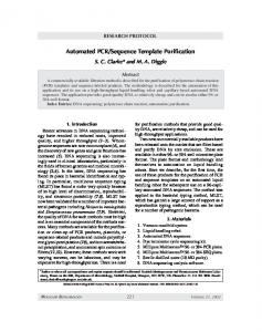
RESULTS Differentiation of VRE isolates by PFGE and Rep-PCR. Molecular typing with PFGE was used to define the clonal patterns of 304 VRE isolates collected during 2008. Ninety of these isolates (88 isolates from perirectal swabs, one isolate from a blood sample, and one isolate from a urine sample) were selected for Rep-PCR typing. All of these 90 VRE isolates were E. faecium. These 90 VRE isolates included 34 different pulsotypes and 10 different Rep-PCR types (Table 1). Up to 80 isolates (88.9%) belonged to the three Rep-PCR types (types a, h, and i). There were discrepancies between the two typing methods. For example, two pulsotype F isolates belonged to Rep-PCR type a and type i. Overall, there was a significant correlation between PFGE and Rep-PCR (P ⬍ 0.001). Wallace’s coefficient was 0.93 when comparing RepPCR with PFGE, and it was 0.14 when comparing PFGE with Rep-PCR. Relationship of Rep-PCR types and antimicrobial susceptibility. The relationships of Rep-PCR types and antimicrobial susceptibility patterns are shown in Table 1 and in the supplemental material. Thirty-eight of the VRE isolates were resistant to all antibiotics tested (AST 1), and 71.1% of these isolates (27/38) belonged to Rep-PCR type i (Table 1). That is, Rep-PCR type i was highly correlated with pandrug-resistant VRE isolates (P ⬍ 0.001). The most common pulsotypes were pulsotype A (13/38 [34.2%]) and pulsotype B (5/38 [13.2%]). Of the 20 teicoplanin-susceptible isolates, 15 (75%) were Rep-PCR type a and they were susceptible to tetracycline (AST 6 and AST 8). Pulsotype P (six isolates), pulsotype Q (five isolates) were the predominate pulsotypes of these teicoplanin-susceptible isolates. Of the 90 E. faecium isolates evaluated, 14 isolates were vancomycin intermediate. Twelve of these isolates (85.7%), including six pulsotypes, belonged to
VOL. 48, 2010
Rep-PCR FOR VRE
2899
FIG. 1. Dendrogram showing similar Rep-PCR types for 12 of 14 vancomycin-intermediate Enterococcus faecium (VRE) isolates. Sample ID, sample identification.
Rep-PCR type a (P ⬍ 0.001) (Fig. 1). In addition, 29 of 34 isolates of Rep-PCR type a (85.3%) were susceptible to one or more antibiotics. There was a strong correlation between RepPCR type a and the more-susceptible isolates (P ⬍ 0.001). In contrast, there were 24 PFGE pulsotypes among the 52 moresusceptible isolates. Comparison of Rep-PCR with PFGE typing of VRE during cluster of colonization investigations. The two methods were compared for 68 isolates collected during eight clusters of colonization. Five of the clusters of colonization were classified as type 1 (showing a dominant clone concordant with time and place clustering). Two clusters of colonization were classified as type 2 (discordant). In one cluster of colonization, there was no dominant clone by either Rep-PCR or PFGE. Thus, RepPCR and PFGE provided similar results in six of the eight clusters of colonization. A representative type 1 cluster of colonization is shown in Fig. 2. In this cluster of colonization, isolates 1 to 8 belonged to the same Rep-PCR type, and seven isolates belonged to pulsotype B. Samples VRE2844 and VRE2816 were isolated from the same patient. They were considered nontypeable by PFGE typing but were shown to be related by Rep-PCR. A representative type 2 cluster of colonization is shown in Fig. 3. In this cluster of colonization, isolates 1 to 4 belonged to the same Rep-PCR type, but were polyclonal by PFGE (pulsotypes A, I, and W). DISCUSSION Prospective active surveillance of VRE during a 1-year period that included eight well-defined clusters of colonization
provided us with the opportunity to compare the ability of Rep-PCR and PFGE typing of VRE to distinguish clonal colonization from multiple-source colonization. Although Rep-PCR was less discriminative than PFGE in defining clonal relationships, there was good correlation between the two methods in defining a dominant clone that was concordant in time and place clustering and a polyclonal outbreak. Rep-PCR type a was significantly more likely to be associated with intermediate vancomycin resistance and antibiotic-susceptible strains. Rep-PCR type i was highly correlated with isolates resistant to the six antibiotics evaluated (P ⬍ 0.001). Rep-PCR has been reported to have good typeability and reproducibility (10) and has been used to investigate several nosocomial outbreaks (1, 24). Comparable findings have been reported for Rep-PCR and PFGE for Acinetobacter baumannii, Streptococcus pneumoniae, and methicillin-resistant Staphylococcus aureus (MRSA) (9, 17, 18, 21). Concordance and good reproducibility of Rep-PCR with PFGE for VRE was found in a laboratory-based study (16). To the best of our knowledge, there is only one study that used Rep-PCR to investigate a VRE outbreak, but it did not include a comparison with PFGE (20, 23). The costs of instrumentation, including the hardware and software are slightly higher for Rep-PCR than for PFGE ($35,000 versus $33,500, respectively) (16). The cost of the reagents per reaction is $27 for Rep-PCR and $8 for PFGE. Nevertheless, the turnaround time for Rep-PCR is much shorter. Furthermore, health care-associated infections increase medical costs and hospital stay (19). It has been proposed that implementation of rapid typing systems coupled with prompt inter-
2900
CHUANG ET AL.
J. CLIN. MICROBIOL.
FIG. 2. A representative type 1 cluster of colonization showing the concordance of genetic relatedness of vancomycin-resistant Enterococcus faecium isolates determined by Rep-PCR and PFGE typing. Both methods showed a dominant clone and were concordant with time and place clustering.
vention could reduce health care-associated infection rates by up to 23% (15), and therefore, cost savings are anticipated. As Rep-PCR is insufficiently discriminatory and might overestimate the genetic relatedness of VRE, Rep-PCR may be more suitable as a rapid screening method to exclude the possibility of clonal spread and to facilitate prompt intervention for outbreaks. PFGE could be reserved for confirmation. There may be a concern that if PFGE is required to “confirm” the relatedness in most situations, performing both Rep-PCR and PFGE results in significantly increased costs and imposes further delays in the definite assessment of relatedness, especially in areas where VRE is endemic. However, in those areas with low VRE prevalence and low clonal spread, Rep-PCR may be an ideal quick screening tool (20). The current study has the following strengths. The isolates were selected from a relatively large epidemiologically defined sample size based on prospective surveillance during an entire year. All the isolates were analyzed by Rep-PCR and PFGE, and the methods were shown to have good reproducibility.
Antimicrobial susceptibility was included in the analysis of the data. The study has the following limitations. It was conducted in a single large medical center in Southeast Asia that may not be representative of similar institutions in Western countries. This study did not calculate the discriminative index and compared the discriminatory power between Rep-PCR and PFGE. This is because surveillance data of outbreaks were used, which means that those isolates may not be unrelated, and the discriminatory power of a typing method is the ability to distinguish between unrelated strains (11). In conclusion, we found that Rep-PCR was highly correlated with PFGE typing to evaluate the clonal and nonclonal spread of VRE in a large prospective surveillance study. Rep-PCR has the advantage of a much shorter turnaround time, but it is less discriminative and might overestimate the presence of clonal outbreaks. PFGE remains the preferred method to define clonality. Rep-PCR appears to be a good screening method for preliminary evaluation of outbreaks of VRE to determine the need to implement more intensive control measures.
FIG. 3. A representative type 2 cluster of colonization showing discordance of genetic relatedness of vancomycin-resistant Enterococcus faecium isolates determined by Rep-PCR and PFGE typing. The typing results for PFGE revealed polyclonal patterns, and the Rep-PCR results showed only a dominant clone.
VOL. 48, 2010
Rep-PCR FOR VRE ACKNOWLEDGMENTS
We are grateful to members of the NTUH Center for Infection Control and the hospital staff for their commitment to improve patient safety and reduce health care-associated infections. We are indebted to Calvin Kunin for suggestions and critical review of the manuscript. Y.-C. Chen received a grant (DOH97-DC-1005) from the Center for Disease Control, Department of Health, Taiwan. J.-T. Wang received a grant (NSC98-2314-B-002-066-MY3) from the National Science Council of Taiwan. None of the authors had a conflict of interest. REFERENCES 1. Bertin, M. L., J. Vinski, S. Schmitt, C. Sabella, L. Danziger-Isakov, M. McHugh, G. W. Procop, G. Hall, S. M. Gordon, and J. Goldfarb. 2006. Outbreak of methicillin-resistant Staphylococcus aureus colonization and infection in a neonatal intensive care unit epidemiologically linked to a healthcare worker with chronic otitis. Infect. Control Hosp. Epidemiol. 27: 581–585. 2. Carrico, J. A., C. Silva-Costa, J. Melo-Cristino, F. R. Pinto, H. de Lencastre, J. S. Almeida, and M. Ramirez. 2006. Illustration of a common framework for relating multiple typing methods by application to macrolide-resistant Streptococcus pyogenes. J. Clin. Microbiol. 44:2524–2532. 3. Centers for Disease Control and Prevention. 1993. Nosocomial enterococci resistant to vancomycin–United States, 1989–1993. MMWR Morb. Mortal. Wkly. Rep. 42:597–599. 4. Centers for Disease Control and Prevention. 1995. Recommendations for preventing the spread of vancomycin resistance. Recommendations of the Hospital Infection Control Practices Advisory Committee (HICPAC). MMWR Recommend. Rep. 44:1–13. 5. Chen, M. L., S. C. Chang, H. J. Pan, P. R. Hsueh, L. S. Yang, S. W. Ho, and K. T. Luh. 1999. Longitudinal analysis of methicillin-resistant Staphylococcus aureus isolates at a teaching hospital in Taiwan. J. Formos. Med. Assoc. 98:426–432. 6. Clinical and Laboratory Standards Institute. 2007. Performance standards for antimicrobial susceptibility testing: 17th informational supplement, M100-S17. Clinical and Laboratory Standards Institute, Wayne, PA. 7. Davis, M. A., D. D. Hancock, T. E. Besser, and D. R. Call. 2003. Evaluation of pulsed-field gel electrophoresis as a tool for determining the degree of genetic relatedness between strains of Escherichia coli O157:H7. J. Clin. Microbiol. 41:1843–1849. 8. Donskey, C. J., J. R. Schreiber, M. R. Jacobs, R. Shekar, R. A. Salata, S. Gordon, C. C. Whalen, F. Smith, and L. B. Rice. 1999. A polyclonal outbreak of predominantly VanB vancomycin-resistant enterococci in northeast Ohio. Northeast Ohio Vancomycin-Resistant Enterococcus Surveillance Program. Clin. Infect. Dis. 29:573–579. 9. Harrington, S. M., F. Stock, A. L. Kominski, J. D. Campbell, J. C. Hormazabal, S. Livio, L. Rao, K. L. Kotloff, S. O. Sow, and P. R. Murray. 2007. Genotypic analysis of invasive Streptococcus pneumoniae from Mali, Africa, by semiautomated repetitive-element PCR and pulsed-field gel electrophoresis. J. Clin. Microbiol. 45:707–714. 10. Healy, M., J. Huong, T. Bittner, M. Lising, S. Frye, S. Raza, R. Schrock, J. Manry, A. Renwick, R. Nieto, C. Woods, J. Versalovic, and J. R. Lupski. 2005. Microbial DNA typing by automated repetitive-sequence-based PCR. J. Clin. Microbiol. 43:199–207. 11. Hunter, P. R., and M. A. Gaston. 1988. Numerical index of the discriminatory ability of typing systems: an application of Simpson’s index of diversity. J. Clin. Microbiol. 26:2465–2466. 12. Kim, W. J., R. A. Weinstein, and M. K. Hayden. 1999. The changing molecular epidemiology and establishment of endemicity of vancomycin resistance in enterococci at one hospital over a 6-year period. J. Infect. Dis. 179:163– 171. 13. Mascini, E. M., A. Troelstra, M. Beitsma, H. E. Blok, K. P. Jalink, T. E. Hopmans, A. C. Fluit, R. J. Hene, R. J. Willems, J. Verhoef, and M. J.
14.
15.
16.
17.
18.
19.
20.
21.
22.
23.
24.
25.
26. 27.
28.
29.
2901
Bonten. 2006. Genotyping and preemptive isolation to control an outbreak of vancomycin-resistant Enterococcus faecium. Clin. Infect. Dis. 42:739–746. Noble, W. C., Z. Virani, and R. G. Cree. 1992. Co-transfer of vancomycin and other resistance genes from Enterococcus faecalis NCTC 12201 to Staphylococcus aureus. FEMS Microbiol. Lett. 72:195–198. Peterson, L. R., and G. A. Noskin. 2001. New technology for detecting multidrug-resistant pathogens in the clinical microbiology laboratory. Emerg. Infect. Dis. 7:306–311. Pounder, J. I., C. K. Shutt, B. J. Schaecher, and G. L. Woods. 2006. Clinical evaluation of repetitive sequence-based polymerase chain reaction using the Diversi-Lab System for strain typing of vancomycin-resistant enterococci. Diagn. Microbiol. Infect. Dis. 54:183–187. Ross, T. L., W. G. Merz, M. Farkosh, and K. C. Carroll. 2005. Comparison of an automated repetitive sequence-based PCR microbial typing system to pulsed-field gel electrophoresis for analysis of outbreaks of methicillin-resistant Staphylococcus aureus. J. Clin. Microbiol. 43:5642–5647. Saeed, S., M. G. Fakih, K. Riederer, A. R. Shah, and R. Khatib. 2006. Interinstitutional and intrainstitutional transmission of a strain of Acinetobacter baumannii detected by molecular analysis: comparison of pulsed-field gel electrophoresis and repetitive sequence-based polymerase chain reaction. Infect. Control Hosp. Epidemiol. 27:981–983. Sheng, W. H., J. T. Wang, D. C. Lu, W. C. Chie, Y. C. Chen, and S. C. Chang. 2005. Comparative impact of hospital-acquired infections on medical costs, length of hospital stay and outcome between community hospitals and medical centres. J. Hosp. Infect. 59:205–214. Sherer, C. R., B. M. Sprague, J. M. Campos, S. Nambiar, R. Temple, B. Short, and N. Singh. 2005. Characterizing vancomycin-resistant enterococci in neonatal intensive care. Emerg. Infect. Dis. 11:1470–1472. Shutt, C. K., J. I. Pounder, S. R. Page, B. J. Schaecher, and G. L. Woods. 2005. Clinical evaluation of the DiversiLab microbial typing system using repetitive-sequence-based PCR for characterization of Staphylococcus aureus strains. J. Clin. Microbiol. 43:1187–1192. Siegel, J. D., E. Rhinehart, M. Jackson, and L. Chiarello. 2007. Management of multidrug-resistant organisms in health care settings, 2006. Am. J. Infect. Control 35:S165–S193. Singh, N., M. M. Leger, J. Campbell, B. Short, and J. M. Campos. 2005. Control of vancomycin-resistant enterococci in the neonatal intensive care unit. Infect. Control Hosp. Epidemiol. 26:646–649. Tam, V. H., K. T. Chang, M. T. LaRocco, A. N. Schilling, S. K. McCauley, K. Poole, and K. W. Garey. 2007. Prevalence, mechanisms, and risk factors of carbapenem resistance in bloodstream isolates of Pseudomonas aeruginosa. Diagn. Microbiol. Infect. Dis. 58:309–314. Tenover, F. C., R. D. Arbeit, R. V. Goering, P. A. Mickelsen, B. E. Murray, D. H. Persing, and B. Swaminathan. 1995. Interpreting chromosomal DNA restriction patterns produced by pulsed-field gel electrophoresis: criteria for bacterial strain typing. J. Clin. Microbiol. 33:2233–2239. Uttley, A. H., C. H. Collins, J. Naidoo, and R. C. George. 1988. Vancomycinresistant enterococci. Lancet i:57–58. van Belkum, A., W. van Leeuwen, M. E. Kaufmann, B. Cookson, F. Forey, J. Etienne, R. Goering, F. Tenover, C. Steward, F. O’Brien, W. Grubb, P. Tassios, N. Legakis, A. Morvan, N. El Solh, R. de Ryck, M. Struelens, S. Salmenlinna, J. Vuopio-Varkila, M. Kooistra, A. Talens, W. Witte, and H. Verbrugh. 1998. Assessment of resolution and intercenter reproducibility of results of genotyping Staphylococcus aureus by pulsed-field gel electrophoresis of SmaI macrorestriction fragments: a multicenter study. J. Clin. Microbiol. 36:1653–1659. Versalovic, J., T. Koeuth, and J. R. Lupski. 1991. Distribution of repetitive DNA sequences in eubacteria and application to fingerprinting of bacterial genomes. Nucleic Acids Res. 19:6823–6831. Wang, J. T., Y. C. Chen, S. C. Chang, M. L. Chen, H. J. Pan, Y. Y. Chang, C. C. Sun, L. H. Wang, S. H. Wang, H. C. Lin, S. F. Chien, and M. S. Tseng. 2004. Control of vancomycin-resistant enterococci in a hospital: a five-year experience in a Taiwanese teaching hospital. J. Hosp. Infect. 58:97–103.