International Food Research Journal 16: 21-30 (2009)
Comparison of DNA extraction efficiencies using various methods for the detection of genetically modified organisms (GMOs) Tung Nguyen, C. T., 2Son, R., 3Raha, A. R., 3Lai, O. M. and 4*Clemente Michael, W. V. L.
1
Department of Genetics and Agricultural Breeding, College of Agriculture and Applied Biology, Cantho University, Vietnam 2 Department of Food Science, Faculty of Food Science and Technology, Universiti Putra Malaysia, 43400 UPM Serdang, Selangor. Malaysia 3 Department of Bioprocess Technology, Faculty of Biotechnology and Biomolecular Sciences, Universiti Putra Malaysia, 43400 UPM Serdang, Selangor, Malaysia 4 Biotechnology Research Institute, Universiti Malaysia Sabah, Locked Bag 2073, 88999 Kota Kinabalu, Sabah, Malaysia 1
Abstract: The ability to detect the presence of transgenes in crop-derived foods depends on the quantity and quality of DNA obtained from a product to be analyzed. The efficiency of DNA extraction protocols differs due to the nature of each food product. In this paper, we described two main DNA extraction protocols and their modifications that have been applied and evaluated for DNA extraction from raw and processed food as well as animal feed. The yield and quality for five categories of food and feed samples namely, raw soybean, raw maize, animal feed, smooth tofu and soymilk are discussed. The statistical interaction analyses showed that the cetyltrimethyl ammonium bromide (CTAB) method was proven to be the best method to extract DNA from raw soybean, maize and animal feed samples which not only obtained high DNA yield of 32.7, 28.4 and 33.4 ng DNA/mg sample respectively, but also produced high quality DNA with the absorbance A260/A280 ratio of 1.9, 1.9 and 2.0, respectively. These DNA were suitable for PCR amplification which produced a 164 bp DNA fragment of the lectin gene from soybean, and a 277 bp DNA fragment of the zein gene from maize. In the processed food category, the Wizard isolation method was found to be the best for the extraction of DNA from smooth tofu and soymilk with the yield of 13.2 and 3.4 ng DNA/mg sample, and the quality of the DNA at the absorbance A260/A280 ratio ranged from 1.9 to 1.7. These DNA were successfully amplified using primers specific to the lectin gene of soybean. Keywords: DNA extraction, CTAB method, Wizard method, soybean, maize
Introduction Genetically modified organisms (GMOs) contain specific traits which have been added to the organisms to improve their properties that have not occurred by mating or natural combination (Anklam et al., 2002; Taverniers et al., 2004). The addition of foreign genes has often been used in plants to produce novel protein that confer pest and disease tolerance and, more recently, to improve the chemical profile of process product, for example vegetable oils (Hemmer, 1997). In the European Community, foods and food ingredients derived from GMOs are strictly regulated and are labeled mandatory to keep under control the possible impact of GMOs both on public health and the environment (EC/258/97; EC/1139/98; *Corresponding author E-mail:
[email protected] Tel: +6088-320000 ext. 5596 ; Fax: +6088-320993
EC/49/2000; EC/50/2000; EC/1829/2003). In most cases, the identification of GMOs is carried out based on the presence or absence of the introduced gene(s) at the DNA level in the sample matrix (Allmann et al., 1993; Meyer, 1995; 1996; 1999; 2003). Among DNA-based methods, polymerase chain reaction (PCR) technology is preferred by many analytical laboratories interested in detection of GMOs because of its high sensitivity, specificity and wide range of gene constructs (Ahmed, 2002; Anklam et al., 2002; Giovanini and Concilio, 2002; Holst-Jensen et al., 2003). In addition, any physical or chemical treatment of food samples, such as heat, pH or shear forces results in a decrease on the average size of genomic DNA due to random cleavage of these macro molecules (Hupfer et al., 1998; Kakihara et al.,
© All Right Reserved
22
Tung Nguyen, C. T., Son, R., Raha, A. R., Lai, O. M. and Clemente Michael, W. V. L.
2005). All of these factors make DNA isolation from foods difficult and challenging (Holden et al., 2003; Kakihara et al., 2005). DNA extraction protocol must be developed on a case-by-case basis for different food matrices, because of the differences on the type, composition and the level of processing of each food product (Gryson et al., 2004). In addition, Zimmermann et al. (1998) stated that although many DNA extraction protocols are available, they had been rarely compared in a comprehensive manner. The prerequisite to successfully identify GMO based on PCR depends on the ability to obtain enough DNA for amplification. Hence, there is a need to compare the efficiency of different extraction methods for each type of food matrix to ascertain which method is the most suitable to yield good and high quality DNA. In this study, two of main protocols based on the Wizard method (Hemmer, 1997) and CTAB method (Jankiewicz et al., 1999), and three sub-protocols are examined for DNA extraction. The qualities of the DNA are determined by using spectrophotometer and later subjected to PCR amplification. In addition, the role of beta-mercaptoethanol (BME) in the lysis buffer is elucidated. These methods are used to isolate DNA from food samples, namely raw soybean, raw maize, animal feed, smooth tofu and soymilk. Materials and methods Sample collection Raw soybean and raw maize samples were obtained randomly from supermarkets in Kuala Lumpur, Malaysia while animal feed sample was collected from Seri Kembangan, Selangor, Malaysia. Smooth tofu and soymilk were purchased from Seri Serdang, Selangor, Malaysia. DNA extraction methods Two of main protocols, and three sub-protocols for DNA extraction were evaluated, the Wizard method (protocol 1), the modified Wizard method by an addition of 1% BME in lysis buffer (protocol 2), the combination method based on the pre-incubation of samples with TNE buffer (protocol 3), the CTAB method (protocol 4) and the modified CTAB method by an addition of 1% BME in lysis buffer (protocol 5). The Wizard method (Hemmer, 1997) – protocol 1 Homogenized food sample (350 mg) was mixed with 860 µl TNE buffer [10 mM Tris-HCl (pH 8),
150 mM NaCl, 2 mM EDTA, 1% SDS] and 40 µl proteinase K (20 mg/ml). The sample was incubated for 3h at 55°C in a water bath. After centrifugation at 13,000 rpm for 10 min, 500 µl of supernatant was transferred in a new 1.5 ml tube and added with the same volume of chloroform. The mixture was centrifuged at 13,000 rpm at 10 min and the upper phase was transferred into a new 1.5 ml tube. The chloroform extraction was repeated twice to get a clear interface. 500 µl of the supernatant was added with 15 µl of 3M sodium acetate (pH 5.2) and 50 µl of absolute ethanol to precipitate the remaining starch and polysaccharides. The mixture was kept on ice for 15 min and centrifuge at 13,000 rpm for 7 min. The supernatant were transferred into a new 1.5 ml tube, and 5 µl of 3M sodium acetate (pH 5.2) and 500 µl of absolute ethanol were added. The mixture was incubated on ice for 15 min to allow the DNA to precipitate, and later centrifuged for 7 min at 12,000 rpm. The DNA pellet was washed again with 500 µl of 70% ethanol, centrifuged at 12,000 rpm for 10 min and air-dried. The pellet was dissolved in 100 µl of distilled water and stored at -18°C until use. The modified Wizard method – protocol 2 The protocol was similar to the Wizard method except that 1% BME was added to the TNE buffer. The combination method – protocol 3 Homogenized sample (350 mg) was mixed with 860 µl of TNE buffer [10 mM Tris-HCl (pH 8), 150 mM NaCl, 2 mM EDTA, 1% SDS], 40 µl proteinase K (20 mg/ml) and vortexed vigorously. The mixture was incubated for 1 h and 30 min at 55°C in a water bath. Then 500 µl of CTAB buffer [20 g/l CTAB, 1.4 M NaCl, 100 mM TrisHCl (pH 8), 20 mM EDTA] was added in the mixture and further incubated at 65°C for 30 min. After centrifugation at 13,000 rpm for 10 minutes, 650 µl of supernatant was transferred into a new 1.5 ml tube, and gently mixed with the same volume of chloroform. The mixture was centrifuged at 13,000 rpm at 10 min and the upper phase was transferred in a new 1.5 ml tube. The chloroform extraction was repeated twice to get a clear interface. Then 500 µl of the supernatant was added with 15 µl of 3M sodium acetate (pH 5.2) and 50 µl of absolute ethanol to precipitate the remaining starch and polysaccharides. The
International Food Research Journal 16: 21-30
Comparison of DNA extraction efficiencies using various methods for the detection of genetically modified organisms (GMOs)
23
Table 1. Sequences of oligonucleotides used in this study Primer LEC1
LEC2
ZE03 ZE04
Sequence 5’-GTG CTA CTG ACC AGC AAG GCA AAC TCA GCG-3’ 5’-GAG GGT TTT GGG GTG CCG TTT TCG TCA AC-3’ 5’-AGT GCG ACC CAT ATT CCA G-3’ 5’-GAC ATT GTG GCA TCA TCA TTT-3’
Gene specificity Soybean lectin
Amplicon (bp) 164
277
References Vollenhofer et al., 1999
Pauli et al., 2000
Maize zein
Table 2. PCR amplification conditions Step Pre-denaturation Denaturation Annealing Extension Final extension
LEC1/LEC2 12 min, 95°C 1 min, 95°C 30 sec, 72°C 30 sec, 72°C 10 min, 72°C
mixture was kept on ice for 15 min and centrifuge at 13,000 rpm for 7 min. Then the supernatant were transferred into a new 1.5 ml tube and was added with 5 µl of 3M sodium acetate (pH 5.2) and 500 µl of absolute ethanol. The mixture was incubated on ice for 15 min to allow the DNA to precipitate, and later centrifuged for 7 min at 12,000 rpm. The DNA pellet was washed again with 500 µl of 70% ethanol, centrifuged at 12,000 rpm for 10 min and air-dried. The pellet was dissolved in 100 µl of distilled water and stored at -18°C until use. The CTAB method (Jankiewicz et al., 1999) – protocol 4 Homogenized samples of up to 350 mg were mixed with 500 µl CTAB buffer [20 g/l CTAB, 1.4 M NaCl, 100 mM Tris-HCl (pH 8), 20 mM EDTA] and incubated at 65°C for 30 min. The samples were then centrifuged for 10 min at 13,000 rpm. The supernatant was transferred to a new 1.5 ml tube, extracted with 200 µl chloroform and centrifuged for 10 min at 13,000 rpm. The upper phase was transferred into a new 1.5 ml tube, precipitated with 1 volume of isopropanol and centrifuged for 10 min at 13,000 rpm. The supernatant was discarded and the pellet was washed once with 500 µl of 70% ethanol and air-dried for approximately 45 min. The pellet was dissolved in 100 µl distilled water and stored at -18°C until use.
ZE03/ZE04 4 min 30 sec, 95°C 1 min 45sec, 96°C 2 min, 60°C 1 min 50 sec, 72°C 4 min 50 sec, 72°C
The modified CTAB method – protocol 5 The protocol was similar to the CTAB method except that 1% BME was added to the lysis buffer. Methods for DNA Quantification DNA concentration from all DNA stocks were determined by using a spectrophotometer at the absorbance of 260 nm (A260) and 280 nm (A280) in an Eppendorf Biophotometer 6131 spectrophotometer. The purity of extracted DNA was determined by using A260/ A280 ratio and later tested by PCR amplification. Polymerase chain reaction (PCR) PCR amplification was carried out in a PCR mix of 25 µl on a PTC-200 thermal cycler (MJ Research, Watertown, MA). The final concentrations of each PCR reaction were as follows: 2.5 µl of 10 x PCR buffer (Finnzymes, Finland); 100 ng of genomic DNA; 0.5 M of each primers; 200 M of dNTPs mix; 0.625 unit/reaction of DyNAzyme II DNA polymerase. Oligonucleotide primers Oligonucleotide primers were synthesized by Research Biolabs Sdn Bhd. (Malaysia) at the final concentration of 100 mM. All oligonucleotide primers were diluted to working concentration of 10 pmol/l with sterilized deionized water and stored at -18°C until use. The sequences and amplification conditions are presented in Tables 1 and 2.
International Food Research Journal 16: 21-30
24
Tung Nguyen, C. T., Son, R., Raha, A. R., Lai, O. M. and Clemente Michael, W. V. L.
Agarose gel electrophoresis Amplicons were analyzed using 1.8% agarose gel electrophoreses in a 1 x TBE [10 mM Tris-base (pH 8); 2.75 g/l Boric acid; 1mM EDTA (pH 8)] and were made visible under UV transilluminator after staining with 0.5 (g/ml of ethidium bromide). Statistical analysis The data of DNA yield and quality (A260/A280 ratio) were subjected to an analysis of variance for Completely Randomized Design (CRD) using the MSTAT-C program version 1.2 (Michigan State University, 1986). Duncan’s Multiple Range Test was used to compare the mean values among the treatments at 95% probability. Results and discussion Evaluation on DNA yield (ng DNA/mg sample) of two plant DNA extraction protocols and their modification on five categories of samples The efficiency of two main DNA extraction protocols of plant and their modifications involving SDS and CTAB were used as the main detergents in this work. The DNA produced was quantified using a spectrophotometer at the absorbance of 260 and 280
1
2
3
4
5
6
7
8
9
10
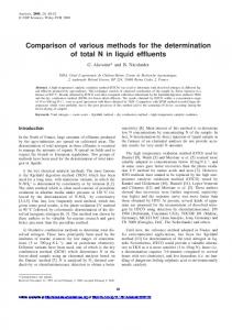
nm. The result in Table 3 shows that the CTAB-based protocols gave good DNA yield. Among CTABderived protocols, the CTAB method (protocol 4) gave the best result with a DNA yield of 19.7 ng DNA/mg sample (Table 3) and produced a clear DNA band on the agarose gel (Figure 1). This finding was in accordance with the result of Chen and Ronald (1999). They have used the CTAB protocol for extracting total DNA from grains of rice and maize, and leaves of other species yielding 2.3 – 5.2 g DNA / 25 – 50 mg fresh leaf tissue. In contrast, the SDSbased or CTAB-based protocols (protocols 2 and 5) containing BME (Table 3) produced the lowest yield of DNA. BME retards the oxidation of biological compounds in solution by breaking disulfide bonds in protein molecules and is also a potential health hazard. Both the highest and the lowest treatments were significantly different (P