Sep 9, 1974 - drogenase (36000) and chymotrypsinogen (257000) were used as marker proteins of known molecular weight. Proteins were incubated at ...
Biochem. J. (1975) 145, 575-579 Printed in Great Britain
575
Comparison of the Subunit and Primary Structures of the Pyruvate Kinases from Rabbit and Sturgeon Muscles By PETER J. ANDERSON and RICHARD F. RANDALL Department of Biochemistry, University ofOttawa, Ont. KlN 6N5, Canada
(Received 9 September 1974) The structures of the pyruvate kinases isolated from rabbit and sturgeon muscles were compared. Both enzymes are composed of subunits of 56000 mol.wt. Amino acid compositions of the two enzymes are similar, but not identical. Examination of the peptides produced by CNBr cleavage demonstrated that there are at least some highly homologous regions in the two proteins. There are only two replacements between an 18-residue portion of the polypeptide chain of rabbit muscle pyruvate kinase and a portion of the polypeptide chain of the enzyme isolated from sturgeon muscle. Purified sturgeon muscle pyruvate kinase has been obtained in sufficient yield (Randall & Anderson, 1975) to permit a comparison ofthe structure with the enzyme from rabbit muscle. The point of interest lies in that whereas the rabbit muscle enzyme gives a hyperbolic Michaelis curve with phosphoenolpyruvate and is not activated by fructose 1,6-diphosphate we have found that the sturgeon enzyme, like that from rabbit liver, gives a sigmoidal Michaelis curve with phosphoenolpyruvate and is activated by fructose 1,6-diphosphate. The origin of this difference may have considerable mechanistic and evolutionary significance.
Experhmental Materials Rabbit muscle pyruvate kinase was obtained from BMC Ltd., Montreal, Canada. The enzyme from sturgeon muscle was prepared as described (Randall & Anderson, 1975). Iodo[2-14C]acetic acid was obtained from Amersham-Searle, Oakville, Ont., Canada. Phenyl isothiocyanate, trifluoroacetic acid and pyridine were Sequanal-grade reagents obtained from Pierce Chemical Co., Rockford, Ill., U.S.A. All other reagents were of the best grade available.
Chemical modification (carboxymethylation) of thiol groups
Enzyme was dialysed against water and freezedried. The protein was dissolved to a concentration of 10mg/ml in 8M-urea containing 0.2M-Tris-HCI, pH8.0, and 5mM-dithiotheitol. After l5min iodoacetic acid was added to a final concentration of 20mM and the mixtures were sealed and placed in the dark for 50min. Reactions were then stopped by the Vol. 145
addition of excess ofdithiothreitol and the protein was purified by dialysis against water. When radioactive iodoacetic acid was used in the modification, it was diluted to a specific radioactivity of 0.5mCi/mmol with carrier iodoacetic acid before use.
Polyacrylamide-gel electrophoresis Electrophoresis was carried out in polyacrylamide gels containing either sodium dodecyl sulphate or urea. For molecular-weight determinations on sodium dodecyl sulphate-containing gels (Weber & Osborn, 1969) bovine serum albumin (68000), aldolase (40000), glyceraldehyde 3-phosphate dehydrogenase (36000) and chymotrypsinogen (257000) were used as marker proteins of known molecular weight. Proteins were incubated at 60°C for 10min in 8M-urea containing 1% sodium dodecyl sulphate, before application to the gels. Polyacrylamide gels containing 8 M-urea were made by the addition of urea to pH8.9 separating-gel solution (Davis, 1964). Carboxymethylated pyruvate kinase was dissolved in 8M-urea before application to the gels. Protein on sodium dodecyl sulphate-containing gels was located by staining with Coomassie Blue and on urea gels by staining with Amido Black. Hydrolysis and amino acid analysis Protein and peptide samples were hydrolysed in 6M-HCI in evacuated sealed tubes for 24h at 106°C. The HCl was made 1 mm in phenol and mm in 2-mercaptoethanol to prevent loss of tyrosine and sulphur-containing amino acids. Amino acid analysis was carried out on a Technicon TSM Amino Acid Analyzer by using a single-column procedure. An Infotronics CRS-210 Automatic Digital Integrator was used to quantify the amino acids.
576
Peptide generation and isolation Carboxymethylated pyruvate kinase was dissolved to a concentration of 10mg/ml in 70% (v/v) formic acid. Solid CNBr was added to a final concentration of 10mg/ml and reactions were allowed to proceed at 220C for 18h. The CNBr peptides were dried in a stream of N2, suspended in a small volume of water and freeze-dried. The peptides (about 60mg) were then solubilized by modification with an excess of citraconic anhydride (Gibbons et al., 1970) and separated on a column (2.5cm x 90cm) of Sephadex G-75 (superfine grade) equilibrated with 0.5 % NH4HCO3. The column was eluted with 0.5 % NH4HCO3 and the eluate was monitored by measuring the E225 and by scintillation counting of 0.1 ml samples of 2.5ml fractions. Separated fractions were concentrated by freeze-drying and the citraconyl groups were removed from the peptides by treatment with 5 % (v/v) acetic acid for 16h. The acetic acid was thenremoved by freeze-drying. Low-molecular-weight CNBr peptides were further purified by high-voltage paper electrophoresis as described (Anderson et al., 1969). Radioactive peptides were located on paper by radioautography. Amino acid sequence determinations The amino acid sequence of purified peptides was determined by using a subtractive Edman procedure. Peptides (about lOOnmol) were dissolved in 100,ul of 50% (v/v) pyridine and a 5,u1 sample was taken for hydrolysis and amino acid analysis. Phenyl isothiocyanate was then added to a final concentration of 5 % and the mixture was left to react for 50min at 50°C. The solutions were then dried in a stream of N2, suspended in lOO,ul of alcohol and dried again. Then lOOjul of trifluoroacetic acid was added and the mixture left at 50°C for 30min. The trifluoroacetic acid was then removed under a stream of N2 and 100jul of water was added to the residue. The aqueous solution was extracted three times with 200,u1 of water-saturated ethyl acetate. The aqueous solution and the combined ethyl acetate extracts were then dried under a stream ofN2. The dried aqueous extract was dissolved in lOO1l of 50% (v/v) pyridine and a 5jpl sample taken for hydrolysis and amino acid analysis. Further rounds of Edman degradations were carried out by repeating the described procedure. The dried ethyl acetate extracts were used to confirm, when necessary, the residues removed by Edman degradation by regeneration of the amino acids from the phenylthiohydantoin derivatives (Smithies et al., 1971). To facilitate sequence determinations smaller peptides were produced from CNBr peptides by digestion with trypsin (diphenylcarbamoyl chloridetreated; Sigma Chemical Co., St. Louis, Mo., U.S.A.). CNBr peptides (10mg/ml) were suspended in 0.5%
P. J. ANDERSON AND R. F. RANDALL
NH4HCO3, trypsin was added to a final concentration of 0.2mg/ml, and digestion was allowed to proceed for 6h at 370C. The resulting peptides were purified by high-voltage electrophoresis (Anderson et al., 1969). Results Gel electrophoresis in sodium dodecyl sulphate indicated that both sturgeon and rabbit muscle pyruvate kinases consisted of a single component of mol.wt. 56000. When samples of the two pyruvate kinases were run on the same gels they were not resolved from each other. Carboxymethylated rabbit muscle and sturgeon muscle pyruvate kinases gave a single protein band on 8M-urea gels. However, on urea gels the mobility of the rabbit muscle enzyme was only one-half that of the sturgeon muscle enzyme. Amino acid analyses of the enzymes are given in Table 1. The results are expressed per subunit and, on the basis of the results obtained by polyacrylamidegel electrophoresis, assume that the pyruvate kinases from rabbit and sturgeon muscles are composed of identical subunits of 56000 mol.wt. Tryptophan was not determined, but the E280 for both enzymes (Kayne, 1973; Randall & Anderson, 1975) indicate that the content of this amino acid must be very low. The compositions of the two enzymes appear to be
Table 1. Amino acid compositions of the pyruvate kinases from rabbit and sturgeon muscles Values are for 24h hydrolysates and are expressed per subunit and assume that the enzymes are composed of identical subunits of 56000 mol.wt. Tryptophan was not determined. Values are the averages of these determinations. Composition (mol/mol of subunit Amino acid Carboxymethylcysteine Aspartic acid Threonine Serine Glutamic acid Proline Glycine Alanine Valine Methionine Isoleucine Leucine Tyrosine
Phenylalanine Histidine Lysine Arginine
Rabbit 8.4 45.5 26.2 29.5 53.3 20.9 39.3 55.6 39.1 17.3 32.1 37.6 9.0 17.2 13.5 33.7 30.8
Sturgeon 8.1 47.1 28.3 32.0 54.2 23.5 42.9 51.9 35.9 13.9 31.7 41.5 10.1 18.1 9.9 31.4 28.0
1975
577
PYRUVATE KINASE STRUCIURE
1-
(a)
0.75
(b)
I'
0.50 I
>
150.25
0
200
400
300
200
300
400
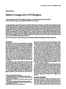
Eluate volume (ml) G-75 1. Elution profiles from Sephadex of sturgeon muscle (a) and rabbit muscke (b) pyruvate kinases Fig. The pyruvate kinases were treated with iodo[2-14C]acetic acid, CNBr and citraconic anhydride before application to the column as described in the text. -~, Radioactivity, ----,E25
Table 2. Amino acid compositions ofpeptides isolatedfrom rabbit and sturgeon muscle pyruvate kinases Peptide RX1 was isolated from carboxymethylated rabbit muscle pyruvate kinase after treatment with CNBr. Peptides RX1TB, RXITNI and RX1TN2 were isolated from tryptic digests of peptide RX1. Peptides SX1, SX1TB, SX1TN1 and SX1TN2 were isolated from sturgeon muscle pyruvate kinase treated in the same way. Hydrolysis was carried out for 24h. Composition (mol/mol of peptide) Amino acid Carboxymethylcysteine Aspartic acid Threonine Glutamic acid Homoserine Proline
Glycine Alanine Valine Isoleucine Lysine
Arginine
RX1
SX1
1.9 1.1 0.9 1.1 0.9 1.1 2.1 2.0 1.2 2.3 1.0 1.9
1.8 1.1 1.0 1.1 0.9 1.1 2.2 2.0
RX1TB
SX1TB
RXlTN1 SXlTN1 RX1TN2 SX1TN2 1.1 1.1
1.2
1.2
0.9 2.1
1.3
1.1
2.0 1.0
1.0
1.0
1.0 1.1
1.0
similar, with the greatest difference being in the smaller number of histidine and methionine residues found in the sturgeon enzyme. As an indication of the degree of homology between the two enzymes, peptide 'maps' were made of tryptic digests of the carboxymethylated proteins. The technique used for peptide 'mapping' has been described (Anderson et al., 1969). An examination of these'maps' indicated that thereweremany differences between the proteins in the peptides produced by tryptic digestion. In both more than 50 peptides were resolved, as might be expected from tryptic Vol. 145
1.0
0.9
1.0 1.1 0.9 1.0 1.1 2.0 1.0 0.7 1.0
1.0 1.0 0.9 1.1 1.1 1.9 0.7 1.0 1.0
1.0
digests of polypeptides containing relatively high contents of lysine and arginine residues (Table 1). This made it difficult to determine the number of homologous peptides generated from the two proteins. It was therefore decided to use a procedure that might be expected to produce a smaller number of peptides. From the methionine content, as determined by amino acid analysis (Table 1), cleavage with CNBr should achieve this. Fig. 1 shows the elution profiles from Sephadex G-75 (superfine grade) of CNBr-treated sturgeon and rabbit muscle pyruvate kinases which had been carboxymethylated with T
378
P. J. ANDERSON AND R. F. RANDALL
Peptide
-X-
T
---(
Th
X
Ile-Ile-Gly-Arg-CmCys-Asn-Arg-Ala(CmCys,Thr,Glu,Gly,Pro,Ala,Val,Ile,Lys)Hse T. x4 T 4----sxi Ile-Gly-Arg-CmCys-Asn-Lys-Ala(CmCys,Thr,Glu,Gly,Pro,Ala,Val,Ile,Lys)Hse Fig. 2. Amino acid sequence data for peptides isolatedfrom rabbit muscle (RX1) and sturgeon muscle (SX2) pyruvate kinases Positions of cleavage by cyanogen bromide (XI) and trypsin C+4) are indicated. - designates a residue released by Edman degradation. CmCys, Carboxynethylcysteine; Hse, homoserine.
RX1
iodo[2-'4C]acetic acid. The elution profiles for the two proteins are different with respect to both the distribution of radioactivity and the amount of 225nm-absorbing material. However, radioactive material eluted in the region 375-425ml was present in the cleavage products of both proteins. Peptides eluted in this region might be expected to be of relatively low molecular weight and therefore amenable to further purification by high-voltage paper electrophoresis. Accordingly, the material eluted from the columns in the volume 375-425m1 was concentrated by freeze-drying, freed of citraconyl groups by exposure to 5 % (v/v) acetic acid for 16h, freeze-dried again, and then subjected to electrophoresis at pH 6.5 for 45min at 60V/cm. Radioautographs of material separated in this way and derived from either rabbit muscle or sturgeon muscle pyruvate kinase which had been carboxymethylated with iodo[2-'4C]acetic acid by treatment with CNBr indicated that in both cases the majority of the radioactivity migrated slightly toward the cathode at pH 6.5. This material was further purified by electrophoresis at pH2.1 and then eluted from the paper. Table 2 gives the amino acid compositions of hydrolysates of this material derived from both rabbit muscle pyruvate kinase (RXl) and from sturgeon muscle pyruvate kinase (SX1). In both cases isoleucine was determined to be the N-terminal residue. Table 2 also contains the compositions of the peptides isolated from tryptic digests of peptides RX1 and SXl. These smaller peptides account for the compositions of peptides RXl and SX1. The sequences of peptides RX1TB, SXlTB, RXlTN1 and SXlTN1 were established by Edman degradations. The peptide RXITB contained an N-terminal isoleucine residue and an additional adjacent isoleucine residue. The presence of a second isoleucine residue was indicated by the finding that three rounds of Edman degradations were necessary to release a glycine residue from this peptide. In addition, it was observed that the mobility of peptide RXITB was less than that of peptide SXITB on electrophoresis at pH6.5. The sequence of peptide RX1TB was therefore deduced to be Ile-Ile-Gly-Arg. Isoleucine was released from
peptide SX1TB by the first round of Edman degradation and glycine by the second round, indicating that the amino acid sequence of this peptide was Ile-Gly-Arg. Since none of the other peptides of tryptic digests of peptides RXl and SXl contained an N-terminal isoleucine residue, it was concluded that peptides RX1TB and SX1TB were derived from the N-terminal regions of peptides RX1 and SX1 respectively. Peptides RXlTNI and SX1TN1 contained N-terminal carboxymethylcysteine residues and the amino acid sequences were established as CmCys-Asn-Arg and CmCys-Asn-Lys respectively by two rounds of Edman degradations. The presence of asparagine was indicated by the neutrality of the peptides on electrophoresis at pH 6.5. The N-terminals of peptides RX1TN2 and SXlTN2 were determined to be alanine residues and the presence of homoserine in these peptides indicated that they were derived from the C-terminal regions of peptides RX1 and SXl. Fig. 2 summarizes the sequence data obtained in these studies. Discussion The results of polyacrylamide-gel electrophoresis suggest that pyruvate kinase is formed from identical subunits. Enzyme isolated from the muscles of both rabbit and sturgeon has an apparent subunit molecular weight of 56000 on sodium dodecyl sulphatepolyacrylamide gels. Although the mobilities of the two S-carboxymethylated pyruvate kinases on 8M-urea-containing polyacrylamide gels were different from each other, in each case only one protein band was present. Since gels of this type are capable of resolving the two types of subunits of rabbit muscle aldolase (Perham & Anderson, 1970), which differ by only one charge change (Lai et al., 1974), if there are any differences between the subunits of a given type of pyruvate kinase, these most probably do not involve any change in net charge. The amino acid compositions of the two pyruvate kinases appear to be similar. The most substantial difference is the lower number of methionine and histidine residues in the enzyme from sturgeon muscle 1975
PYRUVATE KINASE STRUCTURE as compared with that from rabbit muscle. In this respect the sturgeon enzyme is similar to the enzyme from yeast, which also contains substantially less histidine and methionine than rabbit muscle pyruvate kinase (Kayne, 1973). Whether this reflects structural similarities between the sturgeon muscle and the yeast enzymes cannot be decided until more sequence information is available, but the observations suggest that comparative studies of the structures of the rabbit muscle, sturgeon muscle and yeast enzymes may be expected to be of value in the correlation of pyruvate kinase structure and function. Although peptide 'maps' made from tryptic digests of the two enzymes indicated that there were many sequence differences between the two proteins, it is possible that there are substantial regions of high homology. For example, peptide 'maps' of tryptic digests of the enzyme aldolase isolated from the muscles of rabbit and sturgeon are also very different (Anderson et al., 1969). However, further studies have indicated that there are only six replacements between the two aldolases in the N-terminal 39 residues (Anderson & Gibson, 1973; Lai et al., 1974) and only six replacements in a 36-residue portion of the polypeptide chains around the active-site lysine (Gibbons et al.,
1970). The isolation of homologous peptides from the two pyruvate kinases after treatment with CNBr suggests that there are at least some regions of the polypeptide chains that exhibit a high degree of homology. One replacement has been demonstrated (Arg Lys) and a second replacement is suggested (Ile Met) in an 18-residue portion of the polypeptide chain. Both of these variations may be accounted for by single base changes in the amino acid code. Whether the high degree of homology observed between peptides RX1 and SX1 is found in other regions of the polypeptide chains can only be determined by obtaining additional information about the amino acid sequences. It is not known whether residues of
Vol. 145
579 direct or indirect catalytic importance are present in peptides RX1 and SX1. Both lysine residues (Hollenberg et al., 1971; Johnson & Deal, 1970) and cysteine residues (Flashner et al., 1972) have been shown to be of catalytic importance in rabbit muscle pyruvate kinase. The specific residues involved have not been demonstrated, but it is possible that either the cysteine residues or the homologous lysine residues ofpeptides RX1 and SX1 may be involved and that this may account for the high degree of homology. This work was supported by the Medical Research Council of Canada.
References Anderson, P. J. & Gibson, D. (1973) Can. J. Biochem. 51, 514-519 Anderson, P. J., Gibbons, I. & Perham, R. N. (1969) Eur. J. Biochem. 11, 503-509 Davis, B. J. (1964) Ann. N. Y. Acad. Sci. 121, 404 427 Flashner, M., Hollenberg, P. F. & Coon, M. J. (1972) J. Biol. Chem. 247, 8114-8121 Gibbons, I., Anderson, P. J. & Perham, R. N. (1970) FEBS Lett. 10, 49-53 Hollenberg, P. F., Flashner, M. & Coon, M. J. (1971) J. Biol. Chem. 246,946-953 Johnson, G. S. & Deal, W. C. (1970) J. Biol. Chem. 245, 238-245 Kayne, F. J. (1973) Enzymes 8, 353-382 Lai, C. Y., Nakai, N. & Chang, D. (1974) Science 183, 1204-1206 Perham, R. N. & Anderson, P. J. (1970) Biochem. Soc. Symp. 31, 49-58 Randall, R. F. & Anderson, P. J. (1975) Biochem. J. 145, 569-573 Smithies, O., Gibson, D., Fanning, E. M., Goodfiesh, R. M., Gilman, J. G. & Ballantyne, D. L. (1971) Biochemistry 10, 4912-4916 Weber, K. & Osborn, M. (1969) J. Biol. Chem. 244,
4406-4412