Complex I Deficiency Due to Selective Loss of Ndufs4 in the Mouse Heart Results in Severe Hypertrophic Cardiomyopathy Edward T. Chouchani1,2, Carmen Methner1, Guido Buonincontri3, Chou-Hui Hu1, Angela Logan2, Stephen J. Sawiak3, Michael P. Murphy2, Thomas Krieg1* 1 Department of Medicine, University of Cambridge, Cambridge, United Kingdom, 2 MRC Mitochondrial Biology Unit, Cambridge, United Kingdom, 3 Wolfson Brain Imaging Centre, University of Cambridge, United Kingdom
Abstract Mitochondrial complex I, the primary entry point for electrons into the mitochondrial respiratory chain, is both critical for aerobic respiration and a major source of reactive oxygen species. In the heart, chronic dysfunction driving cardiomyopathy is frequently associated with decreased complex I activity, from both genetic and environmental causes. To examine the functional relationship between complex I disruption and cardiac dysfunction we used an established mouse model of mild and chronic complex I inhibition through heart-specific Ndufs4 gene ablation. Heart-specific Ndufs4-null mice had a decrease of ,50% in complex I activity within the heart, and developed severe hypertrophic cardiomyopathy as assessed by magnetic resonance imaging. The decrease in complex I activity, and associated cardiac dysfunction, occurred absent an increase in mitochondrial hydrogen peroxide levels in vivo, accumulation of markers of oxidative damage, induction of apoptosis, or tissue fibrosis. Taken together, these results indicate that diminished complex I activity in the heart alone is sufficient to drive hypertrophic cardiomyopathy independently of alterations in levels of mitochondrial hydrogen peroxide or oxidative damage. Citation: Chouchani ET, Methner C, Buonincontri G, Hu C-H, Logan A, et al. (2014) Complex I Deficiency Due to Selective Loss of Ndufs4 in the Mouse Heart Results in Severe Hypertrophic Cardiomyopathy. PLoS ONE 9(4): e94157. doi:10.1371/journal.pone.0094157 Editor: Tianqing Peng, University of Western Ontario, Canada Received November 27, 2013; Accepted March 11, 2014; Published April 4, 2014 Copyright: ß 2014 Chouchani et al. This is an open-access article distributed under the terms of the Creative Commons Attribution License, which permits unrestricted use, distribution, and reproduction in any medium, provided the original author and source are credited. Funding: This study was supported by the International Society for Heart Research (ISHRES/SERVIER research fellowship to C.M, the Gates Cambridge Trust and the Canadian Institutes of Health Research (doctoral scholarship and postdoctoral fellowship to E.T.C.), the UK Medical Research Council (E.T.C., G.B., M.P.M. and A.L.), and the British Heart Foundation (PG/12/42/29655 to T.K.). The funders had no role in study design, data collection and analysis, decision to publish, or preparation of the manuscript. Competing Interests: The authors have declared that no competing interests exist. * E-mail:
[email protected]
alone. Disruption to complex I activity could contribute to cell dysfunction through a variety of mechanisms. Dysfunction could be driven by decreasing the mitochondrial membrane potential that would in turn lead to defective ATP synthesis and/or calcium homeostasis [13]. Alternatively, complex I disruption could drive pathology through a buildup of mitochondrial NADH that in turn can drive increased superoxide formation from the flavin site of complex I, or from other sources [12,17,18]. To assess the potential primary role of complex I disruption in driving cardiomyopathy through these processes, we chose to disrupt the Ndufs4 gene, which encodes a 18 kDa subunit of complex I that is not directly involved in electron transport but which plays a role in assembly or stability of the entire complex [19]. Ndufs4-null mice have been established as a model for Leigh syndrome; a devastating early onset neurological disorder often associated with mutations to respiratory complexes [19,20]. These mice exhibit increasing ataxia and failure to thrive early in life, and typically die by post-natal day 55 [19,20]. In contrast, a recently developed heart specific Ndufs4-null mouse strain has no reported pathological phenotype up to 1 year of age despite significant inhibition of complex I activity [21]. Therefore, we employed this heart specific Ndufs4-null mouse to explore how persistent
Introduction The heart is particularly susceptible to mitochondrial dysfunction due to its strict dependence on aerobic metabolism [1,2]. Indeed, disruptions in mitochondrial bioenergetics are thought to underlie a variety of chronic myocardial pathologies [3,4]. However, there are a variety of mechanisms by which mitochondrial dysfunction can drive chronic myocardial pathology. A common feature of mitochondrial dysfunction in the heart is disruption to mitochondrial complex I, caused either by mutation in the genes required to generate a fully functional complex or cumulative damage to the complex itself [5–9]. Mitochondrial complex I is the primary entry point for electrons into the mitochondrial respiratory chain and thereby plays an essential role in generating the mitochondrial membrane potential [10], determining the NADH/NAD+ ratio [11], and is also a major source of the reactive oxygen species (ROS) superoxide [12]. Importantly, disruption to all or any of these processes contributes to downstream activation of apoptotic and necrotic cell death pathways [13]. As persistent disruption of complex I activity from a range of causes is frequently observed in cardiomyopathies [5,8,9,14–16], we sought to assess directly the consequence of persistent functional disruption of mitochondrial complex I in the heart PLOS ONE | www.plosone.org
1
April 2014 | Volume 9 | Issue 4 | e94157
Complex I Deficiency Results in Cardiomyopathy
After the acquisition of the standard protocol, MRI was performed using a multi-slice inversion recovery sequence as described previously. For image analysis the papillary muscles and trabeculations were excluded from the delineation of the LV at each phase of the cardiac cycle. The regions from each slice were combined using Simpson’s rule to provide LV mass, end diastolic volume, end systolic volume, stroke volume and ejection fraction using Segment v1.9 [25].
inhibition of mitochondrial complex I activity affected cardiac function specifically. Here we demonstrate that selective disruption of the Ndufs4 gene in the heart leads to a ,50% decrease in complex I activity, that in turn drives severe hypertrophic cardiomyopathy, as determined by magnetic resonance imaging (MRI). However, Ndufs4 gene ablation had no effect on mitochondrial hydrogen peroxide levels, oxidative damage, apoptosis, or tissue fibrosis in the heart. This work suggests that disruption to complex I alone is sufficient to drive myocardial dysfunction. Furthermore, this complex I driven cardiomyopathy occurs through disruption of bioenergetic function and is independent of ROS-dependent pathways.
Measurement of complex I activity Mitochondrial complex I activity was assessed as described previously [7]. Briefly, mitochondria were isolated from entire hearts by differential centrifugation [26] and 100 mg mitochondrial protein was resuspended in a 10 mM HEPES, 120 mM KCl assay buffer containing 100 mM NADH, 10 mM decylubiquinone, 300 nM antimycin, 2 mM potassium cyanide, and 30 mg/ml alamethicin and incubated for 5 min at 32uC while the rate of absorbance change was determined at 340 nm. The background rate following addition of 4 mg/ml rotenone was subtracted from the NADH:decylubiquinone rate. In parallel, the citrate synthase activity of each sample was determined as described previously [7]. The complex I rate was normalized to the citrate synthase rate, and then expressed as a % of the appropriate control.
Materials and Methods Ethics statement All animal experiments were carried out in accordance with the UK Animals (Scientific Procedures) Act 1986 and the University of Cambridge Animal Welfare Policy, and were approved by the UK Home Office under project license 80/2374.
Mouse breeding and maintenance The Ndufs4-null mouse model was a kind gift from the lab of Prof Nils Go¨ran-Larsson, Max Planck Institute for Ageing, Cologne, Germany [21]. Previously, these mice were generated by breeding Ndufs4Loxp/+ mice with mice carrying the heart specific cre recombinase, CKM-NLS-cre and established on a C57Bl/6 background [22,23]. Then double heterozygous offspring were crossed to obtain homozygous Ndufs4LoxP/LoxP mice [21]. In order to generate littermate knockout (CKM-NLS-cre; Ndufs4LoxP/LoxP) and control mice, the male CKM-NLS-cre knockouts (CKMNLS-cre; Ndufs4LoxP/LoxP) were crossed to control females (Ndufs4LoxP/LoxP). Mice with genotype Ndufs4+/LoxP or Ndufs4LoxP/LoxP were used as controls [21]. A mix of CKM-NLS-cre; Ndufs4LoxP/LoxP males and females between the ages of 8–24 weeks were used for all experiments. As described previously, within this age range there was no discernable difference in health, appearance or behaviour between heart-specific Ndufs4-null mice and controls.
Measurement of mitochondrial hydrogen peroxide level in vivo Mitochondrial hydrogen peroxide was measured in vivo using the MitoB mass spectrometric probe as described previously [7,27]. Briefly, 75 nmol MitoB (,3 mmol/kg for 25–30 g mice) in 50 ml saline was administered by tail vein injection to the mouse. 2 hours following injection the entire heart was frozen in liquid nitrogen and then stored at 280uC. For analysis the tissues were homogenized and spiked with deuterated internal standards, extracted, and the content of MitoB and MitoP determined by liquid chromatography and tandem mass spectrometry as described previously [27].
Measurement of protein carbonylation Protein carbonylation levels in the heart were determined by rapid homogenization of entire hearts and protein quantification of heart homogenate, followed by quantification of protein carbonyls using the BioCell Protein Carbonyl Assay Kit according to the manufacturers instructions.
Magnetic resonance imaging of heart function Animals (8–24 weeks of age) were anaesthetized with gaseous isoflurane both for induction (3% in 1 l/min O2) and maintenance (1.25–2% in 1 l/min O2). A pressure sensor for respiration rate was used to monitor anaesthesia depth, breathing rate was maintained in the range 30–60 breaths per minute. Prospective gating of the MRI sequences was achieved with ECG monitoring. Body temperature was monitored using a rectal thermometer and kept constant at 37uC using a water-heated blanket. Cine MRI was performed at 4.7 T with a Bruker BioSpec 47/ 40 system (Bruker Inc., Ettlingen, Germany)[24]. A quadrature birdcage coil of 12 cm was used for signal excitation and a fourchannel cardiac receiver coil for signal reception. Animals were positioned prone. After initial localization images, long axis views were acquired. Using these scans as a reference, short axis slices were arranged perpendicularly to both the long-axis views to cover the LV (FISP, TR/TE 6 ms/2.1 ms, 13–20 frames, 3.5 cm FOV, 2566256 matrix, 1 mm slice thickness, bandwidth 78 kHz, flip angle 20u, NEX 1). Full LV coverage was achieved with no slice gap with 8–10 slices.
PLOS ONE | www.plosone.org
Measurement of apoptosis activation The expression of caspase 3 and cleaved caspase 3 (Cell Signaling Technologies, 1:1000 dilution) was analyzed by standard Western blotting techniques. The band intensity of three independent experiments was measured using SigmaGel software and normalized to Actin (Cell Signaling Technologies, 1:1000 dilution).
Measurement of MnSOD levels The expression of manganese superoxide dismutase (MnSOD) (Cell Signaling Technologies, 1:1000 dilution) was analyzed by standard Western blotting techniques. The band intensity of three independent experiments was measured using SigmaGel software and normalized to Actin (Cell Signaling Technologies, 1:1000 dilution).
2
April 2014 | Volume 9 | Issue 4 | e94157
Complex I Deficiency Results in Cardiomyopathy
Figure 1. Complex I activity in control and Ndufs4-null mouse hearts. Mitochondria isolated from mouse hearts were assessed for rotenonesensitive NADH:decylubiquinone activity. To control for mitochondrial content from heart isolations, complex I activity was expressed as a function of citrate synthase activity from the same hearts. n = 6, ** p,0.01. doi:10.1371/journal.pone.0094157.g001
staining. Finally the slides were dehydrated in ethanol and mounted. The nuclei were stained black, the collagen fibers blue and muscles red
Histological staining for fibrosis Hearts were excised, stored overnight in 10% formalin and embedded in paraffin before 9 mircons thick section were taken throughout the heart. Masson’s Trichrome staining was performed according to the manufacturers specifications (HT15, St. Louis, MO, USA). Briefly, slides were deparaffinized in deionized water and fixed in Bouin’s solution. After washing and incubation in Weigert’s Irono Heamatoxylin solution, the slides were incubated in Biebrich Scarlet Acid Fuchsin and Anilin Blue solution for
Statistics Data are presented as mean 6 standard error from the mean. Statistical analysis was performed using one-way ANOVA. The differences were considered significant at P,0.05.
Figure 2. Cardiac function assessed by MRI. a, Left ventricular ejection fraction (LVEF) of control and Ndufs4-null mouse hearts assessed in vivo by MRI. b, LVEF comparison by sex and c, age. d, left ventricular stroke volume (LVSV) of control and Ndufs4-null mouse hearts assessed in vivo by MRI. n = 2–8, ** p,0.001. doi:10.1371/journal.pone.0094157.g002
PLOS ONE | www.plosone.org
3
April 2014 | Volume 9 | Issue 4 | e94157
Complex I Deficiency Results in Cardiomyopathy
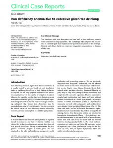
Figure 3. Cardiac morphology assessed by MRI. a, Left ventricular mass (LVM) of control and Ndufs4-null mouse hearts. b, representative longaxis views of the end systole and c, quantification of left ventricular end systolic volume (LVESV) of control and Ndufs4-null mouse hearts. d, representative long-axis views of the end distole and e, left ventricular end diastolic volume (LVEDV) of control and Ndufs4-null mouse hearts. All parameters assessed in vivo by MRI. f, Representative heart slices after Masson trichrome staining of control and Ndufs4-null hearts. n = 4–8, * p,0.05, ** p,0.001. doi:10.1371/journal.pone.0094157.g003
Ndufs4-null mouse that was ,21% of control levels [21], consistent with their assessment of complex I activity using coenzyme Q1 underestimating complex I activity. Therefore, we are confident that the heart selective ablation of the Ndufs4 gene leads to a decrease in complex I activity of about 50%, similar to that found in a number of pathologies [5,8,9,14–16], making this model an effective test for the consequences of complex I disruption on cardiac dysfunction. We next assessed the effects of partial complex I disruption through Ndufs4 ablation on heart function using MRI (Videos S1 and S2). MRI is the current ‘‘gold standard’’ method for clinical assessment of myocardial function and left ventricular mass, which have been established as sensitive predictors of adverse outcomes in hypertrophic cardiomyopathies [29]. Left ventricular ejection
Results and Discussion To study how partial disruption of complex I activity impacted on heart function, we used the recently developed heart-specific Ndufs4-null mouse model [21]. As expected, Ndufs4-null mice exhibited ,50% lower myocardial complex I activity compared to controls (Fig. 1). This activity of complex I in the Ndufs4-null mice was higher than previously reported, which was reported as less than 5% residual activity [21]. This discrepancy is perhaps explained by our use of decylubiquinone as an electron acceptor in contrast to previous assessments which used coenzyme Q1 [21], which is a less effective complex I substrate than decylubiquinone [28]. Supporting this interpretation, the previous study also observed a combined complex I/complex III activity in the
PLOS ONE | www.plosone.org
4
April 2014 | Volume 9 | Issue 4 | e94157
Complex I Deficiency Results in Cardiomyopathy
Figure 4. Assessment of mitochondrial ROS and apoptosis. a, in vivo mitochondrial hydrogen peroxide levels quantified using the ratiometric mass spectrometry probe MitoB. Hearts isolated from control and Ndufs4-null mice injected with MitoB are assessed by MS for MitoB and MitoP levels and the level of mitochondrial hydrogen peroxide are expressed as the ratio of MitoP to MitoB. b, Immunoblot determination and c, densitometry of MnSOD expression in control and Ndufs4-null hearts. d, Quantification of in vivo protein carbonylation in control and Ndufs4-null hearts. e, Immunoblot determination and f, densitometry of Caspase-3 expression and cleavage in control and Ndufs4-null hearts. n = 3–5. doi:10.1371/journal.pone.0094157.g004
mediated hypertrophic cardiomyopathy correlated with changes in mitochondrial ROS levels. To do so we used the recently developed MitoB probe for in vivo quantification of mitochondrial hydrogen peroxide production [27]. Interestingly, Ndufs4-null mouse hearts exhibited mitochondrial hydrogen peroxide levels indistinguishable from controls (Fig. 4a). Importantly, assessment of mitochondrial hydrogen peroxide in vivo is a reliable surrogate for levels of the proximal mitochondrial ROS, superoxide, since mitochondria have micromolar concentrations of MnSOD which reacts with superoxide extremely rapidly (k