Complex Visual Search in Children and Adolescents: Effects of Age and Performance on fMRI Activation Karen Lidzba1*, Kathina Ebner1, Till-Karsten Hauser2, Marko Wilke1 1 Pediatric Neurology and Developmental Medicine and Experimental Pediatric Neuroimaging, Children's Hospital University of Tübingen, Tübingen, Germany, 2 Diagnostic and Interventional Neuroradiology, Radiological Clinic, University of Tübingen, Tübingen, Germany
Abstract Complex visuospatial processing relies on distributed neural networks involving occipital, parietal and frontal brain regions. Effects of physiological maturation (during normal brain development) and proficiency on tasks requiring complex visuospatial processing have not yet been studied extensively, as they are almost invariably interrelated. We therefore aimed at dissociating the effects of age and performance on functional MRI (fMRI) activation in a complex visual search task. In our cross-sectional study, healthy children and adolescents (n = 43, 19 females, 7-17 years) performed a complex visual search task during fMRI. Resulting activation was analysed with regard to the differential effects of age and performance. Our results are compatible with an increase in the neural network's efficacy with age: within occipital and parietal cortex, the core regions of the visual exploration network, activation increased with age, and more so in the right than in the left hemisphere. Further, activation outside the visual search network decreased with age, mainly in left inferior frontal, middle temporal, and inferior parietal cortex. High-performers had stronger activation in right superior parietal cortex, suggesting a more mature visual search network. We could not see effects of age or performance in frontal cortex. Our results show that effects of physiological maturation and effects of performance, while usually intertwined, can be successfully disentangled and investigated using fMRI in children and adolescents. Citation: Lidzba K, Ebner K, Hauser T-K, Wilke M (2013) Complex Visual Search in Children and Adolescents: Effects of Age and Performance on fMRI Activation. PLoS ONE 8(12): e85168. doi:10.1371/journal.pone.0085168 Editor: Daniele Marinazzo, Universiteit Gent, Belgium Received May 8, 2013; Accepted November 24, 2013; Published December 23, 2013 Copyright: © 2013 Lidzba et al. This is an open-access article distributed under the terms of the Creative Commons Attribution License, which permits unrestricted use, distribution, and reproduction in any medium, provided the original author and source are credited. Funding: German Research Foundation: DFG WI3630/1-1 to Marko Wilke (www.dfg.de). European Social Fund and Ministry of Science and the Arts: Margarete-von-Wrangell Programme for Women to Karen Lidzba (www.margarete-von-wrangell.de). The funders had no role in study design, data collection and analysis, decision to publish, or preparation of the manuscript. Competing interests: co-author Marko Wilke is a PLOS ONE Editorial Board member, as is – as of May 2013 - also the author Karen Lidzba. These facts do not alter our adherence to any of the PLOS ONE policies on sharing data and materials. * E-mail:
[email protected]
Introduction
accepted to constitute the ventral and dorsal pathways of visual processing [1,2]. 1.1.1: Basic visual search. Classic visual search paradigms usually require the search for a predefined target in an array of distractors, where subjects perform overt or covert shifts of attention or gaze towards simple visual stimuli (e.g., 3-7). When shifting either attention or gaze, the frontal eye fields (FEF), situated within the posterior superior frontal gyrus (SFG) [8] are invariably activated [9,10-12]. The top-down control of visuo-spatial attention [9,13-18] and the representation of spatial coordinates [19] on the other hand, have been associated with bilateral superior parietal lobule (SPL) and intraparietal sulcus (IPS) in fMRI studies using a number of basic visual search tasks. 1.1.2: Complex visual search. The conscious and planned shifting of attention focus is one of the functions most relevant for the more complex task of visual exploration [20,21]. Only few studies have examined visual search under natural conditions, i.e., the free exploration of a complex visual
Visuospatial processing encompasses a multitude of functions with varying degrees of complexity, such as visual perception, visuospatial attention, object categorization, spatial transformation, occulomotor planning and execution, etc. [1]. Given the broad range of subfunctions which are involved when processing complex visuospatial information, it is not surprising that widely-distributed brain networks are employed to manage these processes.
1.1: Visual Search and Visual Exploration In the present experiment we studied complex visual search, i.e., the wilful exploration of a complex visual stimulus in search for changing targets. This task demands both basic and complex search processes. The successful manipulation of complex visuospatial information is primarily dependent on occipito-temporo-parietal structures, which are commonly
PLOS ONE | www.plosone.org
1
December 2013 | Volume 8 | Issue 12 | e85168
Visual Exploration: Effects of Age and Performance
may also be determined by the cognitive strategies employed [35]. For example, children often have more left-hemispheric activation than adults in fMRI tasks on mental rotation, which has been explained by more piecemeal-like vs. holistic strategies employed by the two groups [39]. In general, however, as children and adolescents mature, cognitive processing increasingly depends on top-down control: Inhibitory control increases and the underlying networks become more efficient and focussed. Thus, task-relevant regions increase in activation, while task-irrelevant regions demonstrate an activation decrease the older the individual gets [40,41]. In addition, the mostly prefrontally represented executive functions, responsible for efficacy and precision, become increasingly available. This can be suspected to be an important explanatory factor for age- and performance related differences in fMRI activation patterns observable during complex visuospatial tasks. 1.2.2: Effects of age and performance in visuospatial tasks. The fronto-striato-parieto-temporal network involved in the allocation of visuospatial attention matures progressively with age [42]. For complex visual search, increasing righthemispheric lateralization with increasing age has been demonstrated by fMRI during late childhood and adolescence [28]. In the embedded figures test (EFT), the activation of typically developing adolescents did not differ from that of adults [43,44], but children demonstrated more left-hemispheric prefrontal activation. This was assumed to reflect verbal strategies and increased effort [45]. Data regarding visuospatial working memory is less clear. While in fMRI, an age-related shift from diffuse to focal activation within the core network has been observed [24,46-48], there were no age-related changes in a study using functional transcranial Doppler sonography [49]. Since performance gains are almost invariably related to normal development, it is both important and difficult to disentangle the two components in studies of age-effects on fMRI activation. The few studies making an attempt to meet this statistical challenge detected either no performance effects (e.g., 50-53), or a performance-related activation-increase in task-related brain regions which was independent of age (e.g., 42,54). With this study, we planned to assess the effects of both physiological maturation and performance in healthy children and adolescents during a complex visual search task, i.e., during the wilful exploration of a complex visual stimulus in search of a changing target. Based on the literature outlined above, we expect (H1) an fMRI activation increase within and an activation decrease outside the core visual attention network (i.e., right SPL, IPS, and FEF) with increasing age, reflecting a focussing of activation; (H2) an age-independent increase of lateralization with increasing performance within the same regions; and (H3) an increase of fMRI activation with age and performance in prefrontal cortex.
scenery. With such a paradigm, Himmelbach and colleagues [22] identified the posterior parietal cortex, IPS, FEF, the insula, the temporo-parietal junction, and STG/STS as areas involved in natural visual search. Complex visual search tasks may, in addition, involve prefrontal networks including superior frontal sulcus (SFS) or ventrolateral / dorsolateral prefrontal cortex. This involvement has been interpreted to reflect a visual working memory component [15,23-25]. From lesion studies, it is well known that visual exploration relies on a predominantly right-hemispheric and densely interconnected perisylvian network (i.e., superior / middle temporal, inferior parietal, and ventrolateral prefrontal cortex of the right hemisphere, including the corresponding fibre-tracts). Disruptions of this network lead to the core symptoms of spatial neglect (i.e., contra-lesionally biased gaze orientation and visual exploration [26]). With the clear preponderance of righthemispheric lesions leading to spatial neglect, it is somewhat puzzling that studies with healthy participants rarely find a strong lateralization of the above-mentioned functions. This may be related to task demand, since task complexity or increased processing demands were associated with a more bilateral activation pattern in complex visual search [27,28] and in mental rotation [29]. The Embedded Figures Task (EFT) can be considered another example of a more complex visual search task. Here, subjects are asked to search a complex figure for a simple geometric shape. The theoretical focus of the EFT lies on the assumption that local details have to be “disembedded” from the global gestalt of the stimulus [30]. Similarly to basic and complex visual search tasks [9,13-18], however, fMRI studies with the EFT highlight a bilateral parietooccipital network comprising mainly SPL and precuneus [31].
1.2: Development of Visuospatial Functions It is well known that - like most cognitive abilities performance in visuospatial tasks improves during childhood and adolescence, i.e., as a function of normal brain maturation (e.g., visuospatial attention [32]; visuospatial analysis [33]; visuospatial memory [34]). However, the underlying neuronal mechanisms are not yet entirely clear. 1.2.1: Developmental changes in task-related fMRI activation. In the last decade, an increasing number of fMRI studies have provided evidence for two trends: On the one hand, brain regions may become more functionally specialized with age (for reviews, see 35,36). On the other hand, a “frontalization” of activation seems to reflect the ongoing maturation of the frontal cortex [37]. Functional specialization can be inferred from an increase in focus and (in some cases) in lateralization in older as compared to younger children [35,36]. Such a shift from diffuse to focal activation with increasing age is thought to be caused by the combination of an activation increase in task-critical areas and a decrease of activation in areas less relevant for the task [35]. However, unspecific changes in vascular coupling or in data quality (e.g., movement artefacts) may have to be taken into account when interpreting age-related localization of fMRI activation [38]. Apart from maturation of the relevant neural networks, the variation in activation patterns between children and adults
PLOS ONE | www.plosone.org
Materials and Methods 2.1: Participants Fourty-three neurologically healthy children and adolescents (24 male, 19 female; mean age 12 years; range 7 to 17 years;
2
December 2013 | Volume 8 | Issue 12 | e85168
Visual Exploration: Effects of Age and Performance
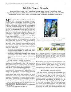
Figure 1. Examples for search condition (left) and control condition (right) of the complex visual search task. doi: 10.1371/journal.pone.0085168.g001
native German speakers) participated in this study. According to the Edinburgh Handedness Inventory [55], 38 subjects were right-handed, 4 were left-handed, and 1 ambidextrous. Exclusion criteria were common MR-contraindications, prematurity and pre-existing neurological or psychiatric disorders. Data of three subjects was excluded due to technical problems in the recording of performance data, and one further subject (11 year old boy) was excluded due to extremely low performance (hit rate more than 2 standard deviations below the mean). All included subjects completed the German adaptation of the Wechsler Intelligence Scale for Children (HAWIK-IV) to ensure a normal level of cognitive abilities (mean full scale IQ = 110.02, SD = 8.02). 2.1.1: Ethics Statement. The study was approved by the ethics committee of the Medical Faculty of the University of Tübingen. All participants and their parents gave written informed consent. Subjects were compensated for their participation according to the time they spent on the study.
the patterns), saccades (between the two versions and within the searched pattern), pattern recognition (comparing the details of the searched pattern), and inhibition (e.g., of already searched parts of the pattern). Hits and misses during target trials as well as mean reaction time for hits were recorded. For further analyses, we defined hit rate as measure of performance and applied a z‑transformation, leading to positive z‑scores for relatively higher hit rates and to negative z-scores for relatively lower hit rates.
2.3: Data acquisition and processing Data was acquired on a 1.5-T whole body MR scanner (Avanto, Siemens Medizintechnik, Erlangen, Germany), using a 12-channel head coil. Care was taken to ensure comfortable placement of the participants, and a foam cushion was used to minimize head movement. Visual stimulus delivery was achieved by screen projection, using a custom-made MR compatible setup as used before [27]. A single MR-compatible pushbutton (Current Design Inc. Philadelphia, PA, USA) held in the left hand (by all subjects) was used for performance monitoring. For stimulus presentation and recording of button presses we used the Presentation software package (version 14.8, Neurobehavioral systems, Inc., Albany, CA). A T2-weighted echo-planar imaging (EPI) sequence was used to acquire functional images with the following parameters: Repetition time (TR) = 3000 ms, Echo time (TE) = 40 ms, matrix = 64 x 64, 40 slices, no interslice gap, covering the whole brain with a voxel size = 3 x 3 x 3 mm. For each task, 110 volumes were acquired. Additionally, a gradientecho B0 fieldmap was acquired with TR = 546 ms, TE = 5.19/9.95 ms, with the same slice prescription as the functional series. For preprocessing purposes, we also acquired a T1-weighted 3D-data set with TR = 1300ms, TE = 2.92 ms, yielding 176 contiguous slices with an in-plane matrix of 256 x 256, resulting in a voxel size of 1 x 1 x 1 mm. Data was processed using SPM8 software (Wellcome Trust Centre for Neuroimaging, London, UK), running in Matlab (MathWorks, Natick, MA, USA). The first 10 scans of each functional series (corresponding to the first block of the control condition) were rejected to allow for the stabilization of longitudinal magnetization, leaving 100 scans per series (5 blocks each of the active and the control condition) to be analyzed. Functional images were realigned and unwarped using the individually-acquired B0 fieldmap, correcting for both
2.2: Complex Visual Search Task The task is described in detail in [56]. In summary, subjects were presented pairs of abstract images derived from the Rey complex Figure [57]. In the search condition, 50% of the pairs were identical, while in the other trials a detail was missing in one of the figures (Figure 1, left). In the control condition, both figures were identical in all cases, but in 50% one of the figures was rotated (Figure 1, right). Stimuli were presented for 6 seconds each. Six control blocks alternated with 5 search blocks (each containing 5 stimuli), leading to a total task duration of 5:30 min. The task was practiced outside the scanner, and subjects were reminded of the task instructions directly before the start of the scan by a short video. The beginning of each new block was indicated by the short presentation of a blank green screen. Participants were told to press a button whenever the two figures were not the same, i.e., when a detail was missing (search condition) or when the orientations were different (control condition). They were explicitly told that the search condition was more difficult than the control condition and that they were to keep searching until they either had found a target (i.e., a difference between the figures), or until the next stimulus was presented. We assumed that the search condition differentially engages visuospatial working memory (keeping the "correct" pattern online), topdown guided visual attention shifts (systematically searching
PLOS ONE | www.plosone.org
3
December 2013 | Volume 8 | Issue 12 | e85168
Visual Exploration: Effects of Age and Performance
Results
EPI and motion*B0 distortions [58]. Functional series with translation exceeding one voxel size (3 mm) in any direction were discarded. The anatomical dataset was segmented using unified segmentation [59], based on custom-generated paediatric reference data [60]. Following coregistration of functional and anatomical data, these parameters were used to normalize the functional images. Global signal trends were removed [61] and functional images were smoothed with a 9 mm full width at half maximum (FWHM) Gaussian filter.
3.1: Behavioural Data Hit rate (HR) of all participants was moderate during the search condition (mean 47% correct, SD = 15, range 25% 83%), mean hit reaction time (RT) was 3133 ms (SD = 744). As expected, the control condition was much easier to solve (mean 93% correct, SD = 23, range 45% - 100%; mean hit RT: 1483 ms [SD = 753]). Age was correlated significantly with hit rate (r(36) = .434, p = .003), but not with RT (r(36) = .059, p = . 361) in the search condition. Gender did not significantly influence performance in the search condition (RT: meanmales = 3162 ms [SD = 821], meanfemales = 3095 ms [SD = 654], t(37) = -.276, p = .784; HR: meanmales = 47.45% [SD = 15.70], meanfemales = 46.76% [SD = 13.56], t(37) = -.144, p = .886; twosample t-tests).
2.4: Statistical analyses Functional MRI data was analysed on the first level in the framework of the General Linear Model (GLM) [62], contrasting the search condition with the control condition, including individual motion parameters as nuisance variables [63]. To test our hypotheses, we took a hierarchical approach. First, we determined the global task-related network by assessing the main effect of a whole-brain one-sample t-test (second level analysis), treating hit rate and age as covariates of no interest. Gender did not demonstrate any effects on performance (see results), so this variable was left out of the analysis. Statistical significance was assumed at an FWEcorrected p < .05 and an extent-threshold of k = 20. The first two hypotheses (H1 and H2) were then addressed by exploring the effects of age and performance within and outside this global task-related network. To this effect, we created a region of interest (ROI) from the global activation cluster of the main effect (thresholded at FWE-corrected p 20. doi: 10.1371/journal.pone.0085168.g002
Figure 3. Age-related activation increases within (red) and outside (blue) the global task-related network (GlobalFWE). No age-related activation decreases were detected within GlobalFWE. Age-related activation decreases outside GlobalFWE are depicted in green. All clusters within ROIs at uncorrected p < .001 and extent threshold k > 15. doi: 10.1371/journal.pone.0085168.g003
This corroborates our hypothesis (H1). Hypothesis (H2), which predicted a performance-related increase of lateralization within the same regions, received only little support: Age and performance were correlated only in the right superior parietal lobule in the ROI analyses. However, the correlations of lateralization index with age or performance within the whole network did not reach significance. Hypothesis (H3) could not be corroborated, since we did not find effects of age or performance within the prefrontal cortex.
within (r = -.045, p = .394), nor outside the GlobalFWE ROI (r = -. 020, p = .452). In the frontal ROI, no additional positive or negative effects of performance were detected.
Discussion Studying the effects of age and performance on fMRI activation during a complex visual search task, our main finding was an increase of activation with age in bilateral occipital cortex and right superior parietal lobule. Thus, the older were our study participants, the stronger was their BOLD response during the visual search task within core regions of the network for complex visual search. In addition, activation decreased with age in a left-hemispheric network of inferior frontal, middle temporal, and superior parietal cortex. Thus, the younger our participants, the more did they recruit left-hemispheric brain regions outside the core network for complex visual search.
PLOS ONE | www.plosone.org
4.1: fMRI activation during complex visual search The comparison of a complex visual search task with a very simple pattern-matching task highlights a broad network of predominantly posterior brain regions. Considering the various processes involved in performing complex visual search, involvement of a range of brain regions was expected. The overall group activation pattern included large portions of both the superior and the inferior parietal lobe. This is well in line
5
December 2013 | Volume 8 | Issue 12 | e85168
Visual Exploration: Effects of Age and Performance
Table 1. Age-related fMRI activation.
Inside GlobalFWE
Outside GlobalFWE
Peak level T
puncorr
Cluster level
Peak level
Cluster level
kE
puncorr
T
puncorr
kE
puncorr
Positive effect of age right lateral occipital cortex
4.93