issues), Anna-Lena Gustafsson (for making my glucose and insulin tolerance tests ... Viveka Nyman, a special person that I found when all my hopes and ...
Thesis for doctoral degree (Ph.D.) 2009 Thesis for doctoral degree (Ph.D.) 2009
COMPONENTS CONTROLLING VESICLE TRAFFICKING AND REGULATED EXOCYTOSIS IN PANCREATIC β-CELLS
COMPONENTS CONTROLLING VESICLE TRAFFICKING AND REGULATED EXOCYTOSIS IN PANCREATIC β-CELLS
Slavena Andrea Mandic
Slavena Andrea Mandic´
From The Rolf Luft Research Center for Diabetes and Endocrinology, Department of Molecular Medicine and Surgery, Karolinska Institutet, Stockholm, Sweden
COMPONENTS CONTROLLING VESICLE TRAFFICKING AND REGULATED EXOCYTOSIS IN PANCREATIC β-CELLS
Slavena Andrea Mandic
Stockholm 2009
All previously published papers were reproduced with permission from the publisher. Front cover: Banting and Best’s notebook (from Hanbook of Diabetes by Gareth Williams and John C. Pickup, Third Edition)
Back cover: The Ebers papyrus Published by Karolinska Institutet. © Slavena Andrea Mandic, 2009 ISBN 978-91-7409-669-9 Printed by 2009
Gårdsvägen 4, 169 70 Solna
To my loving parents
”Mod är inte alltid högröstat. Ibland är modet den stilla röst som i slutet av dagen viskar: Jag försöker igen imorgon” Mary Anne Radmacher
” Du blir aldrig klar och det är som det skall ” Tomas Tranströmer
ABSTRACT Regulated exocytosis is a sophisticated, well-organized and conserved multistage process underlying release of both neurotransmitters and hormones. Exocytosis is mediated by SNARE proteins that act at the center stage by forming a SNARE complex with the intrinsic capability to execute membrane fusion. In addition, supplementary protein families and also mechanisms such as protein phosphorylation are emerging as critical fine-tuning and regulatory components. SNARE proteins are also crucial for regulated insulin secretion from pancreatic β-cells. In response to elevated glucose levels β-cells secrete insulin, the fundamental metabolic and glucose homeostasis regulating hormone. To resolve molecular mechanisms and components operating in the regulation of insulin exocytosis from β-cells, we have in this work investigated the expression, subcellular distribution and functional significance of Cdk5 activators p39 and p35. We demonstrate that both p35 and p39 are present in primary β-cells, and that Cdk5, activated by the p39 subunit, augments Ca2+-induced insulin exocytosis. Moreover, we show that Cdk5/p39 acts, at least partly, by phosphorylation of Munc18-1, a member of the SM protein family. To explore the potential presence of additional Cdk5 substrates mediating enhancement of insulin release, we investigated the role of a supplementary Munc18 variant, Munc18-2. By transient overexpression studies using WT and kinase phosphorylation mutants of Munc18-1 and Munc18-2 proteins we established a difference in the subcellular compartmentalization of the two Munc18 isoforms and a stimulation-induced redistribution of Munc18-1. These results also illustrate the importance of the phosphorylation events in cycling and localization of SM proteins, in particular of Munc18-1. With further investigations of the two homologous Munc18 isoforms, employing slow uncaging of Ca2+ followed by high-time resolution membrane capacitance recordings, we prove that phosphorylated Munc18-1 and Munc18-2 as well as both Cdk5 activators, p35 and p39, modulate the heterogeneity of Ca2+-sensitivity and kinetic dynamics of insulin exocytosis. Detailed evaluation of the significance of the p39 activator was performed using a combination of in vitro techniques, on the level of individual β-cells and pancreatic islets, and in vivo investigations in a mouse mutant lacking the p39 protein. The loss of the p39 activator result in abnormalities related to β-cell function and insulin exocytosis. In addition to deficiencies in release kinetics, Ca2+-sensitivity of exocytosis and impaired VGCC activity recorded in single β-cells, p39-deficient mice exhibit insufficient glucose tolerance and lower serum insulin levels. These observations suggest that the p39 activator is an essential component for an adequate secretory response and glucose homeostasis in β-cells. By investigating other components that may also be involved in the spatial and temporal regulation of insulin exocytosis, we here report the presence and inhibitory effect of tomosyn, a syntaxin 1 associating protein. To be able to describe and comprehend different steps of the insulin secretory pathway we have studied novel molecular mechanisms and components of regulated membrane fusion. Taking advantage of in vitro and in vivo techniques, insulin exocytosis was investigated at different levels of biological complexity, from single β-cells to the level of the whole organism. In this thesis work we provide evidence that different Munc18 isoforms, p39 and p35 activators and the tomosyn protein, in concert with dynamic fine-tuning mechanisms such as phosphorylation, are involved in the regulation of the complex insulin secretory cascade.
LIST OF PUBLICATIONS This thesis is based on the following publications and manuscripts, referred to by their Roman numbers. Paper I: Mandic S.A., Skelin M., Johansson J.U., Rupnik M., Berggren P-O., Bark C. (2009) Munc18-1 and Munc18-2 Differently Modulate β-cell Ca2+-Sensitivity and Kinetics of Insulin Exocytosis. Manuscript
Paper II: Mandic S.A., Skelin M., Jevsek M., Rupnik M., Berggren P-O., Bark C. (2009) Impaired Ca2+-channel Activity and Insulin Secretion in Mouse Mutants Lacking the Cdk5 Activator p39. Manuscript
Paper III: Lilja L., Johansson J.U., Gromada J., Mandic S.A., Fried G., Berggren P-O., Bark C. (2004) Cyclin-dependent Kinase 5 Associated with p39 Promotes Munc18-1 Phosphorylation and Ca2+-dependent Exocytosis. J. Biol. Chem. 279:29534-41
Paper IV: Zhang W., Lilja L., Mandic S.A., Smidt K., Gromada J., Juntti-Berggren L., Takai Y., Bark C., Berggren P-O., Meister B. (2006) Tomosyn is Expressed in β-cells and Negatively Regulates Insulin Exocytosis. Diabetes. 55(3):57481
Related publications: Fornoni A., Jeon J., Varona Santos J., Cobianchi L., Jauregui A., Gonzalez-Quintana J., Sanabria N.Y., Saenz M.O., Mandic S.A., Bark C., McNamara G., Pileggi A., Molano R.D., Berggren P-O., Tryggvason K., Kerjaschki D., Reiser J., Mundel P., Ricordi C. (2009) Nephrin is Expressed on the Surface of Insulin Vesicles and Facilitates Glucose Stimulated Insulin Release. In Press, Diabetes Bark C., Rupnik M., Jevsek M., Mandic SA., Berggren P-O. (2008) Cyclin-Dependent Kinase 5 and Insulin Secretion. Cyclin Dependent Kinase 5 (Cdk5) DOI:10.1007/9780-387-78887-6_11. Springer Science, Edited by Profs. NY Ip and LH Tsai. pp 145-158 Johansson J.U, Ericsson. J, Janson J, Beraki S., Stanic D., Mandic S.A., Wikström M.A., Hökfelt T., Ögren SO., Rozell B., Berggren P-O., Bark C. (2008) An Ancient Duplication of Exon 5 in the Snap25 Gene is Required for Complex Neuronal Development/Function. PLoSGenet. 4(11):e1000278. doi:10.1371/journal. pgen. 1000278 Gromada J., Bark C., Smidt K., Efanov A.M., Janson J., Mandic S.A., Webb D.L., Zhang W., Meister B., Jeromin A., Berggren P-O. (2005) Neuronal Calcium Sensor-1 Potentiates Glucose-Dependent Exocytosis in Pancreatic Beta Cells Through Activation of Phosphatidylinositol 4-kinase β. Proc. Natl. Acad. Sci. USA. 102(29):10303-8
CONTENTS 1
2 3
4
INTRODUCTION ......................................................................................1 1.1 Exocytosis .........................................................................................1 1.1.1 The exocytotic pathway ........................................................1 1.2 The basic secretory machinery; SNARE proteins and the SNARE ... complex .............................................................................................2 1.2.1 Accessory regulatory proteins; Sec1/Munc18 (SM) proteins and tomosyn.............................................................3 1.3 Regulation of exocytosis ...................................................................6 1.3.1 Protein kinases and phosphorylation in the regulation . of exocytosis..........................................................................7 1.4 Regulated exocytosis of insulin.........................................................7 1.4.1 Pancreatic β-cells and glucose stimulation-secretion coupling .................................................................................8 1.4.2 Trafficking and biphasic exocytosis of insulin granules .......9 1.5 Regulatory proteins in insulin secretion ..........................................10 1.5.1 SNAREs and SNARE accessory proteins...........................10 1.5.2 Protein kinases and phosphorylation as modulators . of insulin exocytosis............................................................11 AIMS.........................................................................................................12 METHODOLOGIES ................................................................................13 3.1 Animal models ................................................................................13 3.2 Cell culture and transfections ..........................................................13 3.2.1 Cell lines..............................................................................13 3.2.2 Primary pancreatic β-cells ...................................................14 3.2.3 Cell transfections.................................................................14 3.3 RNA-analyses..................................................................................14 3.3.1 Reverse transcriptase-polymerase chain reaction . (RT-PCR) ............................................................................14 3.3.2 Semi-quantitative RT-PCR .................................................14 3.4 Western Blot analyses and gel electrophoresis ...............................15 3.5 Cell- and tissue homogenization .....................................................15 3.6 Subcellular fractionation by sucrose density gradients ...................16 3.7 Immunocyto- and immunohistochemistry (ICC and IHC) .............16 3.8 Immunoprecipitation (IP) ................................................................16 3.9 Electrophysiology and Ca2+-measurements ....................................16 3.10 The human growth hormone (hGH) cotransfection assay.............17 3.11 Islet perifusion and insulin measurements.....................................17 3.12 Determination of blood glucose concentration, serum insulin levels and insulin content...................................................18 3.13 Intraperitoneal glucose tolerance test (IPGTT) .............................18 3.14 Statistical analysis of data..............................................................18 EXPERIMENTAL RESULTS AND DISCUSSION ...............................19 4.1 Paper I: Munc18-1 and Munc18-2 differently modulate β-cell . Ca2+-sensitivity and kinetics of insulin exocytosis..........................19
5 6 7
4.2 PAPER II: Impaired Ca2+-channel activity and insulin secretion in mouse mutants lacking the Cdk5 activator p39 .............................. 22 4.3 Paper III: Cyclin-dependent kinase 5 associated with p39 promotes Munc18-1 phosphorylation and Ca2+-dependent exocytosis .......... 26 4.4 Paper IV: Tomosyn is expressed in β-cells and negatively regulates insulin exocytosis ............................................................ 28 CONCLUSIONS AND SUMMARY OF THE THESIS......................... 31 ACKNOWLEDGEMENTS ..................................................................... 33 REFERENCES......................................................................................... 37
LIST OF ABBREVIATIONS AA ADP ATP BCA bp Cab45 CaM kinase cAMP Cdk5 [Ca2+]i DAG DMEM EDTA EGF EGTA ELISA EM ER FCS GABA GAPDH GLUT GTP HEPES hGH HRP ICC IHC IMCD IP IPGTT IRP KATP
Arachidonic acid Adenosine diphosphate Adenosine triphosphate Bicinchoninic acid base pair Calcium binding protein 45 Calcium/calmodulin dependent protein kinase Cyclic adenosine monophosphate Cyclin dependent kinase 5 intracellular Ca2+ -concentration Diacylglycerol Dulbecco’s Modified Eagle Medium Ethylenediaminetetraacetic acid Epidermal growth factor Ethylene glycol tetraacetic acid Enzyme-linked immunosorbent assay Electron microscopy Endoplasmic reticulum Fetal calf serum (γ-amino-n-butyric acid) Glyceraldehyde-3-phosphate dehydrogenase Glucose transporter Guanosine triphosphate 4-(2-hydroxyethyl)-1-piperazineethanesulfonic acid human growth hormone Horse radish peroxidase Immunocytochemistry Immunohistochemistry Inner medullary collecting duct Immunoprecipitation Intraperitoneal glucose tolerance test Immediately Releasable Pool ATP-sensitive potassium channel
KRBH LDCV L-VGCC MIN 6 M-PER MT Munc18
Krebs-Ringer Bicarbonate HEPES Large dense core vesicle L-type voltage gated calcium channel Mouse insulinoma 6 Mammalian protein extraction reagent Microtubule Mammalian homologue of the unc-18 gene
NCBI NIDDM NSF PBS PCR PKA PKC PLD PVDF Q R RER RNA RNAi RP RRP RT RT-PCR SDS SDS-PAGE SG SLMV Slp SM SNAP SNAP-25 SNARE SV TBE TGN TIRFM VAMP VGCC
National Center for Biotechnology Information Non-insulin dependent diabetes mellitus N-ehylmaleimide-sensitive factor Phosphate buffer solution Polymerase chain reaction Protein kinase A Protein kinase C Phospholipase D Polyvinylidene difluoride Glutamine Arginine Rough endoplasmic reticulum Ribonucleic acid RNA interference Reserve pool Readily releasable pool Room temperature Reverse transcriptase-polymerase chain reaction Sodium dodecyl sulfate (SDS) SDS-polyacrylamide gelelectrophoresis Secretory granule Synaptic-like microvesicle Synaptotagmin like protein Sec1/Munc18 N-ethylmaleimide-sensitive factor Synaptosome-associated protein of 25 kDa Soluble N-ethyl maleimide sensitive factor attachment protein receptors Synaptic vesicle Tris-borate EDTA Trans-golgi network Total internal reflection fluorescence microscopy Synaptobrevin/vesicle-associated membrane protein Voltage gated calcium channel
1
INTRODUCTION
1.1
EXOCYTOSIS
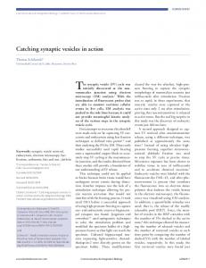
Intracellular vesicle trafficking and fusion with the plasma membrane are fundamental cellular processes regulated by a sophisticated protein machinery and a diversity of protein-protein interactions. Although involving a huge number of regulators and specific mechanisms accounting for special features and cellular milieus, exocytosis is highly conserved among species [1-5]. Most of the cells in our body secrete various substances by constitutive exocotysis [6]. This pathway accounts for a constant discharge of molecules from the cell regardless of stimulatory factors. Regulated exocytosis occurs only in response to a specific triggering stimulus and is fundamental in specialized secretory cells like neurons, endocrine and exocrine cells [7-10]. Two different modes of regulated exocytosis, fast and slow, are distinguished in the literature. Fast secretion involves two types of secretory vesicles, synaptic vesicles (SVs) in neurons and synaptic-like microvesicles (SLMVs) in neuroendocrine and endocrine cells. Slow exocytosis comprises release of peptides from large dense core vesicles (LDCVs) in neurons and neuroendocrine cells and hormonal secretion such as insulin release from secretory granules (SGs) in pancreatic β-cells [11-13]. 1.1.1 The exocytotic pathway The exocytotic process is a multistep pathway where membranous secretory vesicles go through a certain number of stages in order to finally release their content [14, 15]. The functionally different stages, each one well organized and when needed modulated by different mechanisms like phosphorylation, are defined as: tethering, docking, priming and fusion (Fig. 1A). The steps preceding fusion are preparing secretory vesicles for a controlled release of their content. The release occurs either by full fusion, two membranes melting together and completely secreting their content, or by partial transient release and withdrawal of vesicle membrane, referred to as “kiss and run” exocytosis [16, 17]. Definition of each stage and the underlying molecular basis that requires different protein interactions and modulations are still a debated issue. Tethering is defined as a state preceding formation of the SNARE (soluble N-ethyl maleimide sensitive factor attachment protein receptors) complex with tethering proteins connecting vesicles to target membranes [18, 19]. Docking, revealed by electron microscopy (EM), comprises vesicles close to (≤ 30-50 nm) and tightly attached to the target membrane [20]. This step can be dissected in different molecular states (Fig. 1A) depending on the stability of the docked vesicle, but the clear molecular basis for this phenomenon is still undefined [21]. The monomeric GTPase Rab27 subfamily and Rab effectors have recently been proposed as the main regulators of tethering and docking stages [22, 23]. Priming is an ATP (adenosine triphosphate) dependent process considered to make vesicles fusion competent and it is described as a state when the trans-SNARE complex is established and vesicles are ready to fuse [24, 25]. The trafficking of secretory vesicles towards fusion is enabled and regulated by the dynamic actin network and protein modulators. A dual role for actin
1
cytoskeleton has been identified, acting both as an essential physical barrier preventing exocytosis, but also as a crucial component facilitating some of the exocytotic steps [26, 27]. The raise in the intracellular Ca2+-concentration, ([Ca2+]i), represents the primary stimulus for exocytosis in many cells (Fig. 1A). Ca2+ is a second messenger with well-established short- and long term effects on secretion, either by activating protein kinases or by binding to Ca2+-sensor proteins [28-31]. Secretory vesicles do not form a uniform population; they are functionally categorized in different pools. The majority is localized in the cell interior in a reserve pool (RP), while a limited number is residing close to the release site at the plasma membrane, in a readily releasable pool (RRP) (Fig. 1A) [11, 15]. A
B
Figure 1. A, Schematic illustration of different steps of regulated exocytosis. Secretory vesicles translocate to the plasma membrane where they tether and dock. In the next step, named priming, vesicles achieve fusion competence. Elevation of the intracellular Ca2+-concentration triggers exocytosis of primed vesicles. Secretory vesicles reside in different pools. The majority localize in a reserve pool (RP) in the interior of the cell. The docked pool contains vesicles attached in the vicinity (≤ 30-50 nm) of the plasma membrane, while the readily releasable pool (RRP) comprises a small fraction of primed vesicles situated close to the fusion site and ready for immediate release. B, The basic secretory machinery. Three SNARE proteins, SNAP-25, syntaxin 1 and synaptobrevin/VAMP, play a central role in exocytosis and are anchored in target- or vesicular membranes. During membrane fusion SNARE proteins combine and arrange into a highly stable SNARE complex which enables release of the vesicular content.
1.2
THE BASIC SECRETORY MACHINERY; SNARE PROTEINS AND THE SNARE COMPLEX Many protein families are directly or indirectly involved in the docking, priming, triggering and fusion cascade of regulated exocytosis [32, 33]. Overwhelming evidence exists that SNARE proteins, also considered as the minimal equipment for actual fusion of two membranes, act at the center stage [1, 34, 35]. SNAREs, crucial key components of the exocytotic machinery, are characterized by an evolutionary conserved 60-70
2
amino acids sequence; a SNARE motif. Mammalian cells express more than 30 different SNARE proteins [36-38]. They are classified in two categories: v(vesicle)and t(target)-SNAREs depending on their localization, or more accurately according to Q/R categorization depending on their contribution of either glutamine (Q) or arginine (R) to the central SNARE complex layer [39]. The essential role of SNAREs in neuronal secretion was demonstrated by application of botulinum neurotoxins which resulted in a specific proteolysis and block of the neurotransmitter release [40, 41]. According to the SNARE hypothesis, proposed in 1993, specific v-SNAREs paired with cognate t-SNAREs are sufficient as mediators of fusion of two opposing membranes (Fig. 1B) [42]. This working model has since then been further developed and the current hypothesis emphasizes the presence of complementary regulatory components [9, 43]. The most extensively studied and characterized SNARE proteins are: synaptobrevin/vesicle-associated membrane protein (VAMP), syntaxin 1 and synaptosome-associated protein of 25 kDa (SNAP-25) (Fig. 1B) [42]. The syntaxin family of integral membrane proteins is essential for regulated exocytosis [44]. Proteolysis of syntaxin results in docking deficiency and secretion impairment in both neurosecretory cells and synapses [45]. Of 15 mammalian syntaxin isoforms, at least five (syntaxin 1A, 1B, 2, 3 and 4) are localized in the plasma membrane. Syntaxin 1 is cytoplasmically oriented and accessible in two different conformations; open and closed, differing in the accessibility for other SNAREs and complex formation [46]. SNAP-25, a protein of 206 amino acids, is evolutionary conserved and anchored to the plasma membrane by a cluster of palmitoylated cystein residues. By binding syntaxin 1 and VAMP and forming SNARE complexes, SNAP-25 is an important coordinator of neurotransmitter release as well as exocytosis in endocrine and neuroendocrine cells [47-49]. VAMP is a short integral vesicle membrane protein of 118 amino acids with a proline rich N-terminal, a SNARE motif and a C-terminal transmembrane domain [50]. By forming coiled-coiled structures monomeric SNAREs on the opposing membranes form an extremely stable and SDS (sodium dodecyl sulfate)-resistant SNARE complex in trans-configuration. The SNARE complex is a twisted bundle structure consisting of four, in parallel aligned, SNARE motifs. Progressive zippering of the aligned motifs from N- to C-teriminals contributes to the merging of vesicle and target membranes by releasing energy that is needed for the fusion reaction. During fusion the transconformation is adjusted to a cis-SNARE conformation with all SNAREs residing in the same membrane [51-53]. Disassembly of the SNARE complexes, needed for an additional round of fusion reactions and reactivation of SNAREs, is mediated by a concerted action of the ATPase NSF (N-ehylmaleimide-sensitive factor) and SNAP (soluble NSF attachment protein) [1, 36]. 1.2.1 Accessory regulatory proteins; Sec1/Munc18 (SM) proteins and tomosyn The fact that botulinum neurotoxin disruption of SNARE proteins inhibits exocytosis, without affecting vesicle targeting, and that cognate SNARE interactions can be promiscuous, suggest that additional mechanisms adjusting specificity and temporal tuning of exocytosis are present [41, 54, 55]. There are several classes of regulatory proteins that, directly or indirectly, independently or in concert with other components, 3
regulate the SNARE complex assembly, activation and function. One of the key regulators is the SM protein family, but also priming modulators such as the tomosyn protein are crucial [36, 43, 56]. SM proteins are cytosolic, ~ 600 amino acids archshaped proteins indispensible for all vesicle trafficking steps, and with the common feature of binding syntaxins [57-59]. In mammals there are 7 SM proteins of which three distinct isoforms are involved in membranous vesicle trafficking and fusion: Munc18-1, Munc18-2 and Munc18-3, also named Munc18a, b and c [60]. Munc18-1 was originally identified as a binding partner of the syntaxin 1 protein in brain [61]. Munc18-2 is ubiquitously expressed, predominantly found and studied in epithelial tissue [62-64]. Munc18-3 is an additional widely expressed isoform involved in insulin dependent delivery of glucose transporter 4 (GLUT4) vesicles to the plasma membrane [65]. 1.2.1.1 MUNC18-1 (MAMMALIAN HOMOLOGUE OF THE UNC-18 GENE) Munc18-1 is a 67 kDa hydrophilic and cytosolic protein tightly binding to and stabilizing the closed conformation of the t-SNARE protein, syntaxin 1 [66-69]. While the function of the SNARE proteins is fairly defined, conflicting data concerning the precise role and the site of action of Munc18-1 has been reported. The engagement of Munc18-1 in different steps of exocytosis appears to be multifunctional, involving tethering, docking, priming as well as a direct interaction with the SNARE complex [70-73]. The Munc18-1 knock-out mouse mutants [74, 75], show completely abolished spontaneous and evoked neurotransmitter release but normal vesicle docking, which suggests that Munc18-1 is necessary in the late stages of exocytosis. In contrast, observations in knock-out adrenal chromaffin cells strongly suggested that Munc18-1 is critical docking factor acting upstream of fusion [76, 77]. Additional roles of Munc18-1 comprise an early upstream effect on the size and refilling of the RRP [72], modulation of the fusion pore size and acceleration of fusion [78-81]. Different models have been proposed to explain Munc18-1 occupations in temporal and spatial regulation of secretion. One of the oldest hypothesis postulates that the negative role is accounted for by binding to and sequestering the closed conformation of the syntaxin 1 protein, thereby regulating its accessibility and restricting the assemble of the SNARE complex. Supporting this presumption, overexpression of nSec1 orthologs in Drosophila and C.elegans inhibited neurotransmission [82-85]. The idea that Sec1 related proteins negatively regulate fusion is not consistent with the loss of function mutants that show various grades of disruption in membrane trafficking pathways [74, 86-88]. Furthermore, the overexpression of the Munc18-1 protein in chromaffin cells rather increased exocytosis [77, 89], and in insulin secretion both augmenting and inhibitory roles of Munc18-1 have been established [90-92]. Another hypothesis proposes Munc18-1 as a molecular chaperone important for the targeting and localization of syntaxin 1 [93]. One way of clarifying dual roles of Munc18-1 has been to illustrate different SM-SNARE protein binding modes [94]. The binding of Munc18-1 to the closed conformation of monomeric syntaxin 1, interpreted as a basis for its inhibitory character, has been suggested to transit into two additional binding types. So called Nterminal binding to half-open syntaxin 1 and binding to a fully assembled SNARE complex [95, 96] may underlie the positive function and may be regulated by 4
phosphorylation or fatty acids such as arachidonic acid (AA) [97, 98]. Cyclin Dependent Kinase 5 (Cdk5) and Protein Kinase C (PKC) phosphorylations have been shown to modulate Munc18-1 function. In cerebellum Munc18-1 has been shown to colocalize with Cdk5 and possess kinase activating properties [99, 100]. Cdk5 phosphorylation on Threonine 574 of Munc18-1 reduces syntaxin 1 interaction and promotes disassembly of the Munc18-1/syntaxin 1 complex [101, 102]. The major PKC phosphorylation sites in Munc18-1 are at Serine 306 and 313 with the same consequence for interaction with syntaxin 1 [103, 104]. The pleiotropic effects of Munc18-1 in secretion might be a consequence of its tendency to participate and function in different steps or a result of interactions with other exocytotic proteins such as the priming factor Munc13-1 [105], granuphilin [92] and the mint protein [106]. 1.2.1.2 MUNC18-2 Munc18-2, also termed Munc18b or muSec1 (mammalian ubiquitous Sec1), is a ubiquitously expressed Munc18 isoform prominent in apical secretory epithelia [63, 107]. Munc18-2 shows high structural resemblance to Munc18-1 and 67 % identity at the protein level [38]. Although Munc18-2 expression is often reported in tissues lacking endogenous expression of Munc18-1, there are examples of cellular systems where both proteins coexist. In the anterior pituitary Munc18-1 is the dominant isoform, expressed in all cells in the anterior and intermediate lobe. Munc18-2 is highly expressed in the intermediate lobe but both proteins have been shown to colocalize with the growth hormone immunoreactivity [108]. In 2003 the presence, dominance and regulatory role of Munc18-2, together with other Munc18 isoforms, were reported in mast cells, which are specialized secretory cells that release inflammatory mediators by regulated exocytosis [109, 110]. Also in pancreatic β-cells Munc18-2 has been proved to coexist together with the Munc18-1 isoform [63, 111]. As for Munc18-1, aspects concerning the functional importance of Munc18-2 are currently unclear, both positive and inhibitory roles have been suggested. In mast cells knock-down of the Munc18-2 protein caused a significant inhibition of degranulation, which emphasized the positive role of Munc18-2 [112]. Another study showed that the overexpression of Munc18-2 in the same cell type results in a reduction of degranulation. In epithelial cells the SNARE complex formation was reduced upon Munc18-2 introduction [109, 113]. However, both Munc18-2 localization and secretory function in mast cells depends on an intact microtubule (MT) network suggesting involvement in trafficking of secretory granules [109]. The syntaxin binding preference of Munc18-2 is identical to that of Munc18-1, it binds to syntaxin 1A, 2, 3 but not to syntaxin 4 [62]. Syntaxin 3 is an established interaction partner in mast cells and a molecular homology model based on the syntaxin 1/Munc18-1 complex structure [114] was used to identify regions and residues important for the Munc18-2/syntaxin 3 interaction. Additional recently reported functions of Munc18-2 are control of the interaction between synaptotagmin like protein (Slp4-a) and syntaxin in parotid acinar cells [115], and regulation of zymogen granule exocytosis in non-excitable acinar cells via a novel interaction with the cytosolic isoform of the Ca2+ binding protein (Cab45b) [116]. As a phosphorylation target of Cdk5 and PKC, Munc18-2 has been sparsely studied. In rat IMCD (inner medullary collecting duct) cells it has been speculated that phosphorylation, likely by 5
PKC, disassociates Munc18-2 from syntaxin, thereby enabling insertion of H+-ATPase vesicles into the plasma membrane [117]. The requirement for Cdk5 phoshorylation in Munc18-2 regulated secretion in epithelial cells has been reported recently [118]. 1.2.1.3 TOMOSYN Tomosyn, a 130 kDa cytoplasmic protein, is one of the SNARE related regulators identified during the last decade. It was isolated as a syntaxin 1 binding partner, proficient in displacing Munc18-1 from the complex with syntaxin 1 in rat brain extracts, and as a specific modulator of secretion in both endocrine and neuronal cells [43, 119]. Besides an originally identified variant named m-tomosyn, two mammalian genes (tomosyn-1 and tomosyn-2) are alternatively spliced into seven brain enriched or ubiquitously expressed isoforms [120, 121]. All detected isoforms share a C-terminal VAMP homologue domain capable of displacing VAMP [122] and participating in a SNARE complex construction, structurally similar to the regular SNARE complex but with questioned fusogenic properties [123, 124]. Via the C-terminal, tomosyn interacts with and promotes the closed conformation of syntaxin 1, thereby reducing its accessibility and formation of functional SNARE complexes. A variety of data argue that tomosyn negatively governs exocytosis in different cell systems, either by competing with VAMP or by antagonistically influencing other priming factors [125127]. Despite accumulating evidence for a negative impact, studies on neurotransmitter secretion, by displacement of Munc18-1 or by tomosyn serving as a kinase phoshorylation target, remind of the possible positive functions [119]. RNA interference (RNAi) approaches have generated contradictory results supporting both positive and inhibitory roles of tomosyn [127, 128]. Moreover, not only the C-terminal but also the N-terminal domain, consisting of a ~ 40 amino acids (W&D) long protein binding domain (WD-40), seems to be important for a proper function of the tomosyn protein [129]. The introduction of modern techniques, such as total internal reflection fluorescence microscopy (TIRFM), has improved our understanding of dynamic individual exocytotic stages like docking and priming. In a very recent study tomosyn has been suggested to have a role in both fusion dynamics of vesicles from different releasable pools and in distinct release pathways, which highlighted the complex nature of SNARE regulators, not necessarily having a uniform role [130].
1.3 REGULATION OF EXOCYTOSIS It is today well-established that exocytosis is a subject to both positive and negative regulation. Control of the early phases of the exocytotic cascade ensures the specificity by restricting unproductive SNARE interactions, while the late regulatory input guarantees the stabilization and catalyzation of SNARE pairing [31, 43, 131]. Phosphorylation/dephosphorylation, by protein kinases and phosphatases, is one of the most important and potent mechanisms regulating and adjusting the function of different secretory mediators [132]. SNAP-25, syntaxin 1, VAMP, synaptotagmin and Munc18-1 are all established substrates of protein kinases [101, 103, 133, 134]. Interaction of SNARE proteins with ion channels has been reported as an additional tool for the fine-tuning of membrane fusion [135, 136]. 6
1.3.1 Protein kinases and phosphorylation in the regulation of exocytosis Protein kinases are involved in many cellular processes such as metabolism, regulated secretion and neuronal development. Cdk5 is a multifunctional serine/threonine protein kinase, pivotal in many cellular processes including brain development, cytoskeleton dynamics, cell death, synaptic transmission and hormonal secretion [137-140]. In order to achieve activity and thereby be able to phosphorylate the substrates, Cdk5 needs to associate with one of its regulatory subunits, p35 or p39 [141-143]. Cdk5 exhibits a wide cellular distribution while the activators show limited and specific spatial and temporal expression, which probably regulates the place and onset of the kinase activity. By phosphorylating the Munc18-1 protein and thereby increasing the accessibility of syntaxin 1 for SNARE complex formation, Cdk5 plays an important role in membrane dynamics and secretion [101, 102]. Mutant mice lacking the Cdk5 protein die at birth and display severe abnormalities in neuronal migration and neuronal morphology. p35 knock-out mice demonstrate a less severe phenotype and are viable and fertile [144, 145]. p39 is an isoform of p35, distinguished by a C-terminal insert of 32 amino acids but with the Cdk5 binding region conserved [142]. p39-deficient mice do not have any obvious phenotype and the role of p39 is elusive since the p35 activator is believed to be the main subunit in brain, compensating for the absence of p39 [146]. Partial p39 compensation for the p35 function in early development of brain has been reported, but seems not to be sufficient at the later developmental stages [146, 147]. Different functions of p35 and p39 have been demonstrated [147-150]. The p39 activator coimmunoprecipitates and colocalizes with filamentous actin and the cytoskeleton in neurons and in the ocular lens [147, 151], and acts in cytoskeleton regulation. p39 is also, together with Cdk5, expressed in the anterior pituitary affecting both hormone exocytosis and actin organization [152]. PKC is another serine/threonine kinase activated by Ca2+ or via the diacylglycerol (DAG) signaling pathway. The PKC kinase family consists of more than 10 known protein isoforms divided into three subgroups [153]. Different assays have demonstrated that kinases of the PKC family affect and modulate different aspects of exocytosis, such as the size of the RRP, kinetics of exocytosis, Ca2+-threshold for secretion, fusion pore opening and synaptic plasticity [154, 155]. Some of the identified PKC phosphorylation targets are: synaptotagmin, syntaxin 4, SNAP-25 and the Munc18-1 protein [104].
1.4 REGULATED EXOCYTOSIS OF INSULIN Diabetes mellitus is a chronic metabolic disorder leading to severe medical complications. The two major forms are diabetes type 1, an autoimmune disease resulting in destruction of β-cells, and type 2 diabetes, also referred to as non-insulin dependent diabetes mellitus (NIDDM). The more prevalent type 2 diabetes is characterized by increased levels of blood sugar as a consequence of defective β-cell function and insufficient supply of insulin, insulin resistance or a combination of both [156]. Type 2 diabetes is increasing at an alarming rate emerging as a global healthand economical burden, accounting for about 90 % of the diabetic population. Insulin 7
plays a central role in the control of glucose homeostasis and the lack of insulin leads to serious metabolic disturbances that are characteristic for diabetes. A
B Readily Releasable Pool (RRP)
Mobilization
Ca2+
ATP → ADP
Insulin secretion
1st PHASE
Reserve Pool (RP)
Ca2+ 2nd PHASE
Glucose
Time
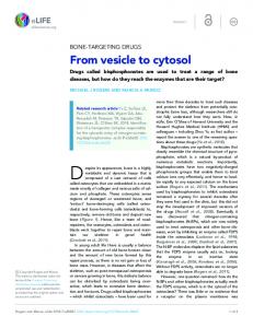
Figure 2. A, Simplified illustration of the stimulus-secretion coupling pathway in pancreatic β-cells. Glucose enters the cell via GLUT-1 and GLUT-2 transporters and is metabolized to increase the intracellular ATP/ADP ratio. Elevation of the ATP/ADP ratio leads to closure of ATP-sensitive KATP channels and promotes membrane depolarization. Glucose-induced depolarization triggers the opening of voltage gated Ca2+-channels (VGCCs) and Ca2+-influx. Increase in the intracellular Ca2+-levels, [Ca2+]i, induces fusion of insulin containing secretory granules with the plasma membrane (PM) and mediates insulin exocytosis. B, Biphasic glucose-stimulated insulin release. The first fast and transient phase of insulin release involves a limited number of insulin granules that reside in the readily releasable pool (RRP). The second sustained phase develops slowly and requires mobilization and priming of secretory granules from the reserve pool (RP).
1.4.1 Pancreatic β-cells and glucose stimulation-secretion coupling Pancreatic β-cells are excitable endocrine secretory units concentrated in cell clusters of the pancreas called islets of Langerhans. Upon glucose-stimulation, these specialized fuel sensors release insulin, a vital hormone controlling glucose absorption in pheripheral tissues and thus lowering the blood glucose level [157, 158]. Two types of secretory vesicles, SLMVs containing GABA (γ-amino-n-butyric acid) and SGs containing the insulin hormone, have been identified in β-cells [12]. The properties of the two secretory vesicle types are different regarding responses upon stimulus, cellular functions, biogenesis and the time course of exocytosis [159, 160]. Insulin is exclusively produced in the β-cell and it is released through a complex and coordinated mechanism controlled by a similar exocytotic machinery and sequence of events as synaptic transmission [160]. Upon glucose-stimulation glucose transporters GLUT1 and/or GLUT2 mediate the transport of glucose into the cell where it is effectively metabolized to generate energy in form of ATP molecules (Fig. 2A). Increase in the intracellular ATP/ADP ratio closes ATP-sensitive potassium channels (KATP) in the plasma membrane causing a raise of the positive charge in the cell interior and cell
8
depolarization. This in turn leads to activation of voltage-gated calcium channels (VGCCs) and influx of Ca2+. The net effect of the described chain reaction is local elevation of [Ca2+]i and triggering of insulin exocytosis (Fig. 2A) [161-163]. 1.4.2 Trafficking and biphasic exocytosis of insulin granules Characterization of the cellular basis for trafficking and fusion of insulin granules is of significant importance as dysfunctions in or modulations of these processes may contribute to the onset of diabetes. Preproinsulin, a precursor of insulin is synthesized in rough endoplasmic reticulum (RER) and almost immediately cleaved to proinsulin. Proinsulin is transported to the Trans-Golgi Network (TGN) where packaging into clathrin coated immature SGs takes place. Maturation of SGs is associated with the loss of the clathrin coat and conversion of proinsulin into insulin. The transport of insulin containing granules from the cell center towards the plasma membrane is mediated by their interaction with MT and ATP-dependent motor proteins such as kinesin. At the cell periphery the transport is dependent on the actin network [164, 165]. Similar as for synaptic transmission in neurons, Ca2+ is the fundamental second messenger for insulin release. Influx of Ca2+ through VGCCs during depolarization increases [Ca2+]i, which acts as the essential triggering signal and initiates the first phase of insulin exocytosis [166, 167]. Insulin exocytosis occurs in a biphasic manner [168, 169], with the quick first phase occurring within the first few minutes after glucose-stimulation followed by an enduring second phase that can last for 5-60 minutes (Fig. 2B) [170, 171]. Since the discovery of the characteristic biphasic manner of hormone release the efforts have been focused on providing evidence for the underlying cellular mechanisms. The 10.000-13.000 insulin loaded granules are divided in different vesicle pools. The majority of granules (~ 95 %) reside in the RP, ~ 5 % (~ 600 granules) are docked at the plasma membrane and ~ 50-100 and ~ 10-20 belong to the RRP and Immediately Releasable Pool, (IRP), respectively [157, 172, 173]. Vesicles belonging to the RRP are positioned very close to the Ca2+-channels, and prepared for immediate release upon Ca2+-influx. Secretion of a fraction of the RRP probably reflects the exocytotic burst observed in the studies of membrane capacitance increase [168, 174]. Depletion of the RRP is generally accepted to mirror the first, rapid and transient, secretory phase while the second phase, reflecting the release of recruited secretory granules, is sustained and develops gradually (Fig. 2B). Recent studies have enlightened a mechanistic difference between the two traditional secretory phases. The first phase was proposed to reflect fusion of stably docked granules and the second phase was suggested to release “newcomers” - undocked vesicles that do not go through docking and priming but rather fuse directly with the plasma membrane [175, 176]. This indicated that the biphasic secretion proceeds through two parallel exocytotic pathways concerning vesicles in different morphological states [177]. The results presented in this thesis imply that also the first secretory phase of insulin release might consist of two additional, fast and slow, exocytotic pathways holding different Ca2+-sensitivity.
9
1.5
REGULATORY PROTEINS IN INSULIN SECRETION
1.5.1 SNAREs and SNARE accessory proteins Today it is confirmed that pancreatic β-cells contain the same repertoire of SNARE proteins and regulatory factors that also are mediating neurotransmitter release in neurons [27, 160, 178]. The importance of the SNARE proteins and their stoichiometry in insulin secretion and overall islet function is illustrated by their reduced abundance in rodent models of diabetes [179, 180] and in human islets of type 2 diabetic patients [181]. Experimental modification of syntaxin 1 expression confirms the correlation between expression levels of the SNARE proteins and changes in the insulin release [182, 183]. Moreover, the cleavage of SNAP-25 and syntaxin 1, the central protein machinery, by botulinum neurotoxins is subsequently followed by a partial or severe reduction in insulin secretion [184, 185]. Syntaxin 1, 2, 3 and 4 are all present in primary β-cells and insulin secreting cell lines [186]. Syntaxin 1A was originally presented as an inhibitor of insulin release [187], and overexpression of both syntaxin 1A and syntaxin 3 in insulinoma cells inhibited stimulated insulin secretion at the level of insulin gene transcription [188]. Syntaxin 4 knock-out islets are defective in both the first and second phase of insulin secretion [189], indicating that this protein variant is of functional importance. The characterization of syntaxin 2 revealed only a minor physiological effect of this syntaxin isoform in insulin exocytosis [190]. Phosphorylation of the SNAP-25 protein is also tightly connected with and influences insulin secretion [191, 192]. Several of the synaptotagmin isoforms have been demonstrated to participate in Ca2+-mediated insulin secretion [193, 194]. Originally discovered in brain [68], the Munc18-1 protein has also been detected in insulin secreting cells as a syntaxin 1 binding partner that negatively influences insulin secretion [90]. Analysis of trafficking and targeting of syntaxin 1A in INS1-E cells indicated that Munc18-1 is not required for syntaxin targeting to the fusion sites but seems to be essential for its stabilization at the membrane [195]. The positive role of Munc18-1 has been demonstrated in alliance with the β-cell specific Rab27a effector granuphilin, at the docking and trafficking stages of insulin exocytosis [92]. The Munc18-2 isoform has also been identified in β-cells [63, 111], but its role is still not thoroughly investigated. Munc18-3, the binding partner of syntaxin 4, is widely expressed in multiple β-cell lines [178]. The action of Munc18-3 as an accessory protein has mainly been studied in insulin-stimulated GLUT4 vesicle translocation [65], but lately transgenic mouse studies have proposed Munc18-3 as a player in glucose-stimulated insulin secretion [196]. Different isoforms of the tomosyn protein are present in pancreatic β-cells, both in the soluble cellular compartments and concentrated to insulin secretory granules. Down-regulation of tomosyn produces contradictory results, demonstrating both inhibitory and enhancing effects [128, 197]. An indirect role of tomosyn, related to the potential modulation of channel activity and β-cell excitability, has also been discussed [136]. Approaches investigating the impact of tomosyn in insulin-stimulated GLUT4 transport in adipocytes and its interaction with the SNARE complex machinery have shown that tomosyn overexpression inhibits translocation of GLUT4 [198]. Reports on both positive and negative roles of Munc181 and tomosyn in insulin secretion are discussed at the moment. It needs to be further 10
investigated if the functions of these regulatory components are simply limiting or if their effects in multiple stages of the secretory pathway and their interaction with other modulatory proteins contribute to conflicting results in insulin exocytosis. 1.5.2 Protein kinases and phosphorylation as modulators of insulin exocytosis As proposed for neuronal systems, phosphorylation of key exocytotic proteins is also relevant in the regulation of insulin release. Much of the flexibility in the exocytotic process is achieved by a balance between phosphorylation and dephosphorylation at different molecular steps preceding membrane fusion [199-201]. Cdk5, protein kinase A (PKA), PKC and calcium/calmodulin-dependent protein kinase (CaM kinase) are all present in pancreatic β-cells [202, 203]. Multiple PKC isoforms are expressed in pancreatic islets and insulin producing cell lines, and have been implicated in different aspects of β-cell function such as the regulation of glucose-dependent insulin exocytosis [204, 205]. Both PKA and PKC kinases have been suggested to act directly on the secretory machinery, stabilizing and increasing either the total number of release competent vesicles or the amount of highly Ca2+-sensitive insulin granules [206, 207]. A general agreement is that phorbol ester stimulation of PKC enhances insulin secretion and that glucose-induced elevation of [Ca2+]i translocates and recruits different PKC isoforms to the plasma membrane [204]. Concerning Cdk5, the investigations are focused on whether the same molecular mechanisms and targets are responsible for neuronal and non-neuronal effects of Cdk5. In β-cells, Cdk5 has been acknowledged with an essential function in stimulated insulin secretion and even more important, as a potential drug target in diabetes treatment [208]. Cdk5, p39 and p35 are all expressed in the islets of Langerhans and in cultured β-cells [91, 209-211]. One of the recent studies, using a p35 gene targeted mouse mutant, has presented Cdk5/p35 as a negative regulator of glucose-stimulated insulin secretion [211]. These experiments were performed at a high glucose concentration which elevates the levels and activity of Cdk5/p35, activates several signaling pathways and decreases insulin mRNA levels in β-cells [210]. p35 activated Cdk5 has also been shown to phosphorylate L-type voltage gated Ca2+-channels (L-VGCCs) and reduce insulin secretion, specifically during high glucose conditions [209, 210]. Altogether, these data give an impression of Cdk5/p35 being a negative modulator of the artificially induced high glucosestimulated insulin secretion which may not be important during normal physiological conditions. The studies in p35-deficient β-cells demonstrated that inhibition of Cdk5 did not affect Ca2+-channels and insulin secretion in the absence of glucose which suggests that p35/Cdk5 is not implicated in the regulation of the initial secretory response. However, we cannot exclude that Cdk5 is important in modulation of the second phase of insulin exocytosis, maybe indirectly through metabolic or amplifying pathways. Recently, a cellular signaling protein, phospholipase D (PLD), has been shown to be activated and phosphorylated by Cdk5, establishing Cdk5 as a central regulator of epidermal growth factor (EGF) dependent insulin secretion. Interestingly the PLD/Munc18-1 interaction was disturbed by Cdk5 phosphorylation [212].
11
2
AIMS
The overall goal of this thesis has been to elucidate fundamental mechanisms and identify regulatory proteins implicated in the complexity of insulin exocytosis. The aim was to obtain better understanding of the biology and physiology of secretory pathways in β-cells. The specific objectives have been to investigate: • The expression, subcellular distribution and function of the Cdk5 activators p35 and p39 in insulin exocytosis. • The specific functions of different SM proteins, focusing on Munc18-1 and Munc18-2, in the modulation of insulin secretory granule trafficking, mobilization and exocytosis. • Potential abnormalities concerning glucose homeostasis, insulin secretion and β-cell function in a mouse mutant with a targeted disruption of the p39 gene. • The expression and functional significance of an additional SNARE regulatory protein, tomosyn, in pancreatic β-cells.
12
3
METHODOLOGIES
In my experiments I have been using a combination of traditional in vitro cell- and molecular biology techniques and in vivo methodologies. Through collaborative efforts I am familiar with electrophysiological techniques. 3.1 ANIMAL MODELS All animal breeding and studies were revised and approved by local ethical committees. Mice were housed on a 12 h light/dark cycle with free access to standard food and water. p39-deficient (p39-/-) mouse mutants Mice with the p39 gene disrupted (p39-/-), were kindly provided by the laboratory of Professor L-H Tsai. p39-deficient mice were generated by replacing the majority of the p39 coding region with a neo cassette in the opposite transcriptional direction [146]. Obese (ob/ob) mice Primary β-cells and brain tissue from ~ 10 months old and fasted ob/ob mice were used in insulin secretion experiments, electrophysiological recordings as well as in RNA-, protein- and immunocytochemistry analyses. The advantage of pancreatic islets derived from ob/ob mice are that they predominantly (90-95 %) contain β-cells, which improves the experimental yield and implementation [213]. NMRI mice, Sprague-Dawley and Wistar rats Tissues from above mentioned rodent models were used in membrane capacitance measurements, Western blotting, immunohistochemistry and protein analysis that were performed in collaborative investigations in other laboratories (paper I, III, IV). 3.2 CELL CULTURE AND TRANSFECTIONS Primary β-cells and insulin secreting cell lines were cultured in a humidified atmosphere, 5 % CO2 at 37°C. 3.2.1 Cell lines The mouse insulinoma (MIN6-m9) cell line MIN6 cells retain physiological characteristics of primary β-cells [214]. They were cultured and grown in DMEM (Dulbecco’s Modified Eagle Medium, Invitrogen) containing 11 mM glucose supplemented with 10 % heat-inactivated fetal calf serum (FCS), 100 IU/ml penicillin, 2 mM L-glutamine and 64 µm 2-mercaptoethanol. Rat insulinoma (INS1-E), hamster (HIT-T15) and rat (RINm5F) cell lines INS1-E cells were cultured in RPMI 1640 medium (Invitrogen) supplemented with 5 % heat-inactivated FCS, 1 mM sodium pyruvate, 50 μm 2-mercaptoethanol, 2 mM glutamine, 10 mM HEPES, 100 IU/ml penicillin and 100 µg/ml streptomycin.
13
HIT-T15 and RINm5F insulin secreting cells were grown in RPMI 1640 (Invitrogen) supplemented with 10 % heat-inactivated FCS, 2 mM glutamine, 100 IU/ml penicillin and 100 µg/ml streptomycin. 3.2.2 Primary pancreatic β-cells Primary β-cells were isolated from ob/ob, NMRI and p39-deficient mice or from Wistar rats, by collagenase digestion [215] or by injecting liberase into the pancreas through the bile duct. When needed a cell suspension was prepared from isolated islets [216]. Single cells or whole islets were cultured for 1-4 days in RPMI 1640 culture medium containing 11 mM glucose supplemented with 10 % FCS, 100 IU/ penicillin, 100 µg/ml streptomycin and 2 mM L-glutamine. In paper II β-cells were also studied within intact islets in freshly prepared slices from the whole pancreas [217]. This method has a number of advantages; it is fast and preserves many physiological properties, and it also enables electrophysiological recordings of β-cells embedded in their natural environment [218]. 3.2.3 Cell transfections Transfections of MIN6-m9, INS1-E and primary β-cells were performed using Lipofectamine 2000 (Invitrogen) according to the manufacturer’s instructions. Oligofectamine (Invitrogen) was used for cotransfection of primary β-cells with the green fluorescent protein (Clontech) and sense and antisense oligonucleotides. The cells were seeded 24 hours (h) before and assayed 48-72 h after transfection procedure (for oligonucleotide treated cells capacitance measurements were performed 24 h after transfection). 3.3
RNA-ANALYSES
3.3.1 Reverse transcriptase-polymerase chain reaction (RT-PCR) RT-PCR was performed on total RNA prepared from mouse or rat brain, from ob/ob pancreatic islets, and from MIN6-m9 and RINm5F cell lines, by using GenEluteTM Mammalian Total RNA kit according to the manufacturer’s instructions (Sigma). RTPCR was set up by using SuperScriptTM RT-PCR-System (Invitrogen), appropriate specific oligonucleotides and PCR-programs (see enclosed publications for detailed information) in a GeneAmp PCR System 9700 (Applied Biosystem). Products of appropriate base pare (bp) sizes were cut out of the gel, purified, amplified with appropriate primers and subjected to DNA sequencing using BigDye Terminator v3.1 Cycle Sequencing Kit and analyzed on ABI PRISM 377 (Applied Biosystems). The homology of the PCR products and corresponding sequences were compared with National Center for Biotechnology Information (NCBI) BLAST program for verification. 3.3.2 Semi-quantitative RT-PCR Semi-quantitative RT-PCR was performed to determine relative gene expression levels. Reactions were performed with 20 cycles of amplification using 1 μg RNA, a trace of [α32P] dCTP (3000 μCi/mmol, PerkinElmer Life Sciences), primers hybridizing to the 14
mRNA/cDNA of interest and Glyceraldehyde-3-phosphate dehydrogenase (GAPDH) primers as an internal control. The products were separated on 8 % polyacrylamide Tris-borate EDTA (TBE) gels and detected using a phosphoimager (BAS-1500, Fujifilm). Signal intensities were quantified using Image Gauge V3.45 (Fujifilm) software. 3.4 WESTERN BLOT ANALYSES AND GEL ELECTROPHORESIS For protein separation in paper I and III equal amount of lysates, homogenates or subcellular fractions were denatured by heating in either commercial sample buffer (NU-PAGE, Invitrogen) or in home-made SDS sample buffer (0.5 M Tris-HCl pH=6.8, 20 % glycerol, 4 % SDS, 10 % 2-mercaptoethanol and 0.05 % Bromophenol Blue). Thereafter samples were loaded and separated on 10-12 % SDS-PAGE (polyacrylamide gelelectrophoresis) (Bio Rad) or NU-PAGE Bis-Tris (Invitrogen) gels, followed by transfer to polyvinylidene difluoride (PVDF) membrane (Amersham Biosciences). Protein concentrations were determined using either the Bradford assay (Bio-Rad) or the bicinchoninic acid (BCA) assay (Pierce). In paper IV supernatants from immunoprecipitation studies were resuspended in 5x SDS-PAGE sample buffer and loaded on 18 % polyacrylamide SDS-PAGE gels. In the same study protein samples of HIT-T15 and RINm5F cells were analyzed by electrophoresis on 10 % TrisHCl polyacrylamide SDS-gels or 4-15 % linear ready gels (Bio-rad). Membranes were blocked in phosphate buffered saline (PBS) containing 0.025 % Tween-20 and 5 % dry milk (VWR or Merck Eurolab) for 1h at room temperature (RT), followed by incubation with primary antibodies (4°C, overnight) and secondary antibodies (RT, 11,5 h). Detection of the immunoreactivity was obtained by using enhanced chemiluminescence (ECL plus, Amersham Biosciences) followed by scanning with CCD camera (LAS 1000, FujiFilm). Signals were quantified using Image Gauge V3.45 software (Fujifilm). 3.5 CELL- AND TISSUE HOMOGENIZATION In paper I and III, transiently transfected MIN6-m9 cells were harvested and lysed in (in mM): 250 NaCl, 1 % Nonidet P-40, 0.5 % sodium deoxycholate, 0.1 % SDS, 1 EDTA, 50 Tris-HCl pH 7.4 and protease inhibitor mixture (Roche Diagnostics GmbH). Pancreatic islets, pulverized brain tissue and MIN6-m9 cells used for subcellular fractionation studies and Western blot analyses were homogenized twice by pestle motor homogenizator in the buffer containing (in mM): 20 HEPES, 1 MgCl2, 250 Dsucrose, 2 EDTA and protease inhibitor cocktail (Roche Diagnostics GmbH) pH 7.4. Brain homogenates were additionally lysed on ice (30 min) in 1 % NP-40 (Sigma). Homogenates were shortly centrifuged to pellet nuclei (500 x g, 7 min and 4°C). In paper IV HIT-T15 and RINm5F cells were lysed in the buffer consisting of (in mM): 20 Tris-HCl pH 7.5, 150 NaCl, 1 EGTA, 1 % NP-40 and protease inhibitor cocktail (Roche Diagnostic GmbH), incubated on ice (30 min) and collected by centrifugation (1000 x g, 10 min, 4ºC). Sprague-Dawley rat brain was homogenized, in presence of protease inhibitors, in (in mM): 50 Tris-HCl, 100 NaCl and 1 EGTA (pH 7.5) using Teflon/glass homogenizer and centrifuged (5000 x g, 10 min, 4°C).
15
3.6
SUBCELLULAR FRACTIONATION BY SUCROSE DENSITY GRADIENTS To separate different cellular components (in paper I, III and IV) lysates or homogenates of transiently transfected unstimulated and stimulated MIN6-m9 cells or primary β-cells were loaded onto a 4.4 ml linear sucrose density gradient (prepared from 0.6 and 2 M sucrose stock solutions). The gradient was centrifuged at 150000 x g for 18 h (Beckman L8-55 ultracentrifuge, SW50 rotor) and 15-16 fractions were collected from the top of the gradient. The linearity of the gradient was examined by measuring the refractive index of each fraction and protein concentration was determined by the Bradford assay (Bio-Rad). Equal amount of protein from each fraction was resolved by electrophoresis for further investigations. 3.7 IMMUNOCYTO- AND IMMUNOHISTOCHEMISTRY (ICC AND IHC) In paper III primary β-cells from ob/ob mice were seeded onto coverslips and cultured up to 3 days as described above. After fixation with 4 % paraformaldehyde and permeabilization in 0.4 % saponin, cells were blocked in 10 % goat serum/PBS (Sigma). Appropriate dilutions of primary and secondary antibodies were applied at RT. The immunoreactivities were analyzed with a Leica TCS-SP2-AOBS confocal laser-scanner equipped with HeNe and Ar lasers and connected to a Leica DMLFSA microscope. In paper IV pancreatic sections from male Sprague-Dawley rats and monolayers of HIT-T15 or primary β-cells were processed for immunofluorescence histochemistry. Pancreatic sections were processed as described in [126]. Monolayers of HIT-T15 cells or primary β-cells fixed by immersion, or tissue sections were incubated with rabbit antiserum to tomosyn or anti-tomosyn combined with antibodies directed towards insulin, glucagon, somatostatin or pancreatic polypeptide followed by incubation in the appropriate secondary antibody mixtures (for detailed analysis see paper IV). Specimens were scanned in a Bio-Rad RadiancePlus or Leica confocal laser scanning system. 3.8 IMMUNOPRECIPITATION (IP) In paper IV HIT-T15 cell lysates were precleared with protein A-Sepharose at 4°C for 2 h. After spinning down the beads, the supernatants were incubated with primary antibodies or control antibodies overnight, followed by an additional incubation with A-Sepharose for 2 h. Beads harvested by centrifugation and washed with a buffer without Triton X-100, were finally resuspended in 5x SDS-PAGE sample buffer and heated (60ºC for 20 min) before protein separation on 18 % polyacrylamide SDSPAGE gels. 3.9 ELECTROPHYSIOLOGY AND Ca2+-MEASUREMENTS Single isolated mouse β-cells or intact cells within the whole islets in fresh pancreatic slices were used in electrophysiological recordings. We used standard whole-cell or perforated patch-clamp set up followed by measurements of changes in membrane area. For discrimination of different voltage-gated Ca2+-currents we applied a voltage ramp to assess the amplitude and voltage dependence of different current components [219, 220]. Exocytosis was triggered either by a train of depolarizing pulses or by slow UV 16
photolysis of caged Ca2+ (NP-EGTA). Pipettes were pulled from borosilicate glass capillaries, coated with Sylgaard and fire-polished before use. The pipette resistance was 2-4 MΩ. Exocytosis was recorded using an EPC-9 patch clamp amplifier and the Pulse software (Elektronik, Lamprecht/Pfalz, Germany). For composition of extracellular- and pipette solutions, experimental temperatures and perifusion rates see Material and Methods in relevant publications. In paper I and II Fura 6F (Molecular Probes) was used to measure the intracellular Ca2+-concentration simultaneously with the patch-clamp recordings. Fura 6F was excited at 380 nm with a monochromator (Polychrome IV; TILL Photonics). Long pass dichroic mirror reflected the monochromatic light above 400 nm from the perfusion chamber and transmitted the emitted fluorescence filtered through a 420 nm barrier filter. Fluorescence intensity was measured with a photodiode (TILL Photonics). Ca2+-concentrations were calculated as described in [220]. 3.10 THE HUMAN GROWTH HORMONE (hGH) COTRANSFECTION ASSAY The hGH assay was used in paper III and IV to study the overexpression effects of different exocytotic proteins on glucose-stimulated secretion from INS1-E cells. In this approach equal amount (1-2 µg) of a plasmid containing the hGH gene and a plasmid containing the gene of interest were cotransfected. Transfected cells were first incubated in glucose-free RPMI for 2 h and thereafter in glucose-free Krebs-Ringer bicarbonate HEPES buffer (KRBH) containing (in mM): 135 NaCl, 3.6 KCl, 5 NaHCO3, 0.5 NaH2PO4, 0.5 MgCl2, 1.5 CaCl2, 10 HEPES and 0.1 % bovine serum albumin (pH 7.4 using NaOH) for 30 min. Secretion was triggered by KRBH containing either 3 or 10 mM glucose. The supernatant was removed from each well, centrifuged at 100 x g for 5 min and was referred as secreted hGH. Remaining cells were harvested in ice-cold 1 mM EDTA-PBS, added to the pellet of the initial centrifugation and lysed by 6 freeze-thaw cycles (in liquid nitrogen and a 37°C bath) followed by 5 min of sonication. Material from this supernatant was considered as cellular, not secreted, hGH. hGH levels were measured by enzyme-linked immunosorbent assay (ELISA) following the manufacturer’s instructions (Roche Applied Science). The average percent of secreted hGH (% of total) was calculated by dividing the amount of secreted hGH by total hGH (secreted + cellular hGH). 3.11 ISLET PERIFUSION AND INSULIN MEASUREMENTS In paper II overnight cultured islets of Langerhans from p39-deficient and control mice were preincubated in Ca5 buffer (in mM): 125 NaCl, 5.9 KCl, 1.28 CaCl2, 1.2 MgCl2, 25 HEPES, 0.1 % bovine serum albumin and 3 glucose, pH 7.4 using NaOH for 30 min prior to perifusion experiments. Whole islets were mixed with Bio-Gel P4 polyacrylamide beads (Bio-Rad) in a 0.5 ml column at 37°C, and dynamics of insulin release was studied by allowing buffers with various glucose concentrations to perifuse the islets. The insulin released by islets into the buffer was collected in fractions (2 min/fraction, flow rate ~ 0.2 ml/min) and analyzed by Ultra Sensitive Mouse Insulin ELISA (Mercodia) according to the manufacturer’s instructions.
17
3.12 DETERMINATION OF BLOOD GLUCOSE CONCENTRATION, SERUM INSULIN LEVELS AND INSULIN CONTENT For the measurements of blood glucose concentration (mmol/l) blood samples from fed or fasted mice (paper II) were taken from the tip of the tail. The blood was analyzed by Freestyle Blood Glucose Monitoring System (Abbott). For the determination of serum insulin levels 8µl blood/g bodyweight was extracted by puncturing the vein in the hind leg. Collected samples were allowed to stand in RT to allow blood clotting. Serum was separated by centrifugation and analyzed by Ultra Sensitive Mouse Insulin ELISA (Mercodia). For measurements of insulin content pancreatic islets were collected in Ca5 buffer, lysed by adding Mammalian Protein Extraction Reagent (M-PER, Thermo Scientific) and incubated on an orbital shaker or manually resuspended before the supernatant were collected by centrifugation (2500 x g). The total insulin amount in the supernatant was determined with Ultra Sensitive Mouse Insulin ELISA (Mercodia) and the amount of DNA was verified with Quant-ITTM Picogreen (Invitrogen). 3.13 INTRAPERITONEAL GLUCOSE TOLERANCE TEST (IPGTT) For IPGTT age matched male and female p39-deficient and control mice were fasted overnight. Body weight and basal fasting blood glucose concentrations were measured prior to the start of the experimental procedure. A dose of 2 g glucose/kg bodyweight was injected into the intraperitoneum (IP) of the mice and the blood samples for glucose concentration measurements were taken at 15, 30, 60 and 120 min post injection. Directly after that the experiments were completed, the mice were fed and allowed to return to their cages. 3.14 STATISTICAL ANALYSIS OF DATA All experiments in this thesis were performed at least three times on different preparations and the results were presented as mean values ± S.E.M, unless otherwise indicated. Different statistical tests have been used in different experimental approaches. Primarily unpaired Student’s t-test was used for the evaluation of statistical significance. For multiple comparisons Dunnett’s statistical test was used. p values less than 0.05 were considered statistically significant.
18
4
EXPERIMENTAL RESULTS AND DISCUSSION
4.1
PAPER I: MUNC18-1 AND MUNC18-2 DIFFERENTLY MODULATE βCELL Ca2+-SENSITIVITY AND KINETICS OF INSULIN EXOCYTOSIS
Different Munc18 and syntaxin isoforms in insulin secreting β-cells Pancreatic β-cells, the only cells in the body that produce, store and secrete insulin, are equipped with a complete set of exocytotic regulators, including the leading SNARE- and SM protein families. The Munc18-1 protein, with controversial and multiple roles in the secretory cascade, also plays a role in insulin secretion [90, 91, 194]. Besides Munc18-1, the SM protein family comprises other Munc18 isoforms that are involved in membrane fusion [58, 66]. Already in 1995 [63], the presence of an additional isoform, Munc18-2, was demonstrated in an insulin secreting cell line and very recently also in primary rat β-cells [111]. This indicated the coexistence of different Munc18 variants in β-cells, but the underlying reason for this coexpression of several Munc18 proteins and possible benefits for insulin secretion were not further resolved. Our PCR analyses assured the presence of different membrane fusion associated Munc18 isoforms in insulin secreting β-cells. In addition to showing the presence and dominance of the Munc18-1a isoform, we also detected the expression of the Munc18-2 protein (Fig. 1A,B). Moreover, we verified low amounts of the Munc181b splice variant, which compared to Munc18-1a shows the same syntaxin binding preference but a different expression pattern in brain [221, 222]. Munc18-2 is the closest homologue of Munc18-1 and coexpression of these two SM protein isoforms have earlier been reported in mast cells, adrenal chromaffin- and anterior pituitary cells and human platelets [108, 110, 223]. We here showed that Munc18-2, so far predominantly described as an epithelial vesicle transport protein, also exists in mouse primary β-cells and in the MIN6 cell line. Different roles of Munc18 proteins in the secretory pathway are partially mediated via a number of protein-protein interactions. The best described role of the Munc18-1 protein in exocytosis is exerted via its interaction with syntaxin 1A. Munc18-1 and Munc18-2 have the same binding specificity for syntaxin isoforms 1A, 2 and 3 [62]. Different results considering the occurrence of syntaxin isoforms in insulin secreting β-cells can be found in the literature [178, 186]. Our PCR analyses on the presence and abundance of described Munc18-1 and Munc18-2 syntaxin partners (Fig. 1C,D) showed expression of both syntaxin 1A and syntaxin 3 in endocrine β-cells, although syntaxin 1A was the predominantly expressed isoform. In HIT-T15 cells syntaxin 3 has also, in contrast to other syntaxins, been detected in the cytoplasm, modulating insulin secretion and Ca2+-channel activity [188]. Syntaxin 3 is an established functional partner of Munc18-2 [109, 113, 118] and its presence and possible interaction with the Munc18-2 protein in β-cells remain to be explored. Syntaxin 2 is probably a poor candidate in the regulation of insulin secretion since expression levels were below the detection limit in our experiments. This was 19
consistent with an earlier study showing low expression levels of syntaxin 2 in the islets of Langerhans [190]. Munc18 isoforms exhibit different subcellular compartmentalization and glucosedependent redistribution As proteins reside in the specific cellular compartments where they perform their activity, subcellular localization studies can be used for prediction of their function. By performing sucrose density gradients on transiently transfected MIN6 cells, we characterized and uncovered different subcellular localizations of Munc18-1 and Munc18-2 proteins. Munc18-1 was equally distributed to both the plasma membrane and soluble cell compartments while Munc18-2 predominantly localized to the cytosol (Fig. 2A). When the Munc18-2 protein was overexpressed in COS-1 or mast cells a similar cytosolic distribution, without membrane association, has been observed [107, 109]. Upon increased exocytotic activity, induced by glucosestimulation, Munc18-1 was recruited to the plasma membrane while no change in the case of Munc18-2 localization was observed (Fig. 2B, 2C I-IV). The found discrepancy in the subcellular distribution supported our hypothetical working model, with distinct SM proteins acting in separate compartments and stages of insulin secretion. The novel finding that glucose-stimulation induces Munc18-1 translocation agrees well with earlier reports on Munc18-1 participitation in different protein interactions and mechanisms both in the cytosol and at the plasma membrane, close to the sites of secretion [224]. Being familiar with the ability of Cdk5, together with Munc18-1, to enhance Ca2+-triggered insulin exocytosis, we searched to further clarify which role phosphorylation of Munc18-1 plays in protein function and β-cell secretion. Subcellular fractionation of unstimulated and stimulated MIN6 cells transfected with Munc18-1 templates lacking kinase phosphorylation sites showed that Cdk5 and PKC mutants were enriched in the plasma membrane region where they also remained sequestered after stimulation (Fig. 2B). This suggested that the absence of phosphorylation may be a possible explanation for the accumulation and inability of Munc18-1 to disassociate or loosen from the plasma membrane. In contrast to the PKC phosphorylation positions, the Cdk5 phosphorylation site seems to be conserved in the Munc18-2 sequence [225]. By using a Munc18-2 template with a mutation in the potential Cdk5 site, we also showed a minor glucose-induced shift in the cellular localization of this Munc18 variant (Fig. 2C II,IV). Thus, these results supported our hypothesis that phosphorylation is an important regulator of the intracellular localization, cycling and probably also the activity of Munc18 proteins. Munc18 proteins differently influence kinetics and Ca2+-sensitivity of release By combining capacitance recordings and slow photo release of caged Ca2+, which allows direct measurements of the Ca2+-sensitivity of exocytosis, we searched to resolve specific functions of different Munc18 proteins in insulin exocytosis. Introduction of caged Ca2+-components has made it possible to identify multiple pools of releasable vesicles, their Ca2+-dependence and kinetics of exocytosis in many neuronal and neuroendocrine preparations [226-228]. In response to a gradual 20
increase in [Ca2+]i, we observed a typical biphasic secretory response pattern (Fig. 3AC). When Munc18 proteins were overexpressed the membrane capacitance increase exhibited different Ca2+-sensitivities, lower upon introduction of Munc18-1 and higher upon overexpression of Munc18-2 (Fig. 4A). Heterogeneity in Ca2+-sensitivity as a mechanism explaining the presence of different exocytotic routes is a rather novel phenomenon. Coexistance of different Ca2+-affinities for release in the same cell has previously been reported in human neutrophils and pituitary gonadotropes. In those cell types this was attributed to specific properties of the exocytotic machinery, or to the presence of distinct secretory vesicle populations and release pathways [229, 230]. A sign of different Ca2+-dependent pathways in synapses has previously also been described [231]. However, this was the first indication that the SM proteins are instrumental in the determination of the Ca2+-sensitivity of secretion. Simultaneous overexpression of Munc18-1 and Munc18-2 significantly increased the number of released insulin granules, amp2, during the second exocytotic component and was interpreted as a sign of a synergistic effect of the two modulators (Fig. 3D). Significant but smaller amplification of amp2 was also obtained upon single overexpression of the Munc18-2 protein. This supported the possibility that this isoform may be involved in a parallel secretory pathway, that either separately or by merging with the Munc18-1 operated pathway releases secretory vesicles at a lower Ca2+-threshold (Fig. 3D). In terms of different roles of Munc18 proteins in other cell types where their coexistance have been confirmed, a partial compensation of Munc18-2 in early steps of LDCV release has been reported in Munc18-1-deficient chromaffin cells and in growth hormone releasing anterior pituitary cells [76, 108]. To adress if phosphorylation of Munc18 proteins was important for modulation of the Ca2+-sensitivity of exocytosis, we took advantage of Munc18 phosphorylation mutants that we also used in subcellular fractionations. We showed that the shift in Ca2+-dependence was even more pronounced when phosphorylation was prohibited. Two significantly different secretory responses, with low and high Ca2+-thresholds, were noticed when non-phosphorylated Munc18 templates were overexpressed (Fig. 5A,B). Not only the triggering [Ca2+]i, but also the rate and the number of fusing vesicles were affected (Fig. 5C,D). The demonstrated discrepancy in the Ca2+-sensitivity of insulin exocytosis probably mirrored different secretory pathways. These pathways may mediate release from different vesicle subpopulations with distinct Ca2+-affinities, coordinated by diverse Munc18 proteins which in turn are phosphorylation substrates in intracellular signaling cascades. Previously it has been shown that hyperphosphorylation, due to the altered PKC activity in the islets of GK rats, significantly reduces the sensitivity of the secretory machinery to Ca2+ [232]. Furthermore, in insulin secreting INS-1 cells activation of protein kinases has been shown to sensitize exocytosis and regulate an ultrasensitive Ca2+-component of the endocrine secretion [207, 233]. We propose that the Munc18-1 protein accounts for the tight docking of insulin containing granules at the plasma membrane, close to the VGCCs. There SGs are experiencing high [Ca2+]i which is required for fast fusion with the plasma membrane. In secretory cells Ca2+ triggers and regulates both secretion and upstream secretory steps leading to fusion. In the case of Munc18-2, operating at basal Ca2+-levels, we suggested involvement in the recruitment and fusion of highly Ca2+21
sensitive vesicles that do not necessarily go through traditional docking and priming steps, but fuse directly at lower [Ca2+]i. In paper III we showed that p39 activated Cdk5 augments insulin exocytosis, likely via phosphorylation of the Munc18-1 protein. In this investigation (paper I), using a different experimental approach and adding an additional level of complexity, we found that both Cdk5 activators differently modulate the Ca2+-affinity of exocytosis (Fig. 5E-H). Combined overexpression of the p39 activator and SM proteins restored the Ca2+-sensitivity of insulin secretion and probably increased the release probability of SGs. On the contrary, introduction of the p35 subunit additionally enhanced the Ca2+-sensitivity shift caused by single overexpression of Munc18 templates indicating that the p35 disturbs the phosphorylation pattern of Munc18 proteins. This suggested that the p35 activator plays a non-redundant and deactivating role, perhaps by restraining SGs, while the p39 facilitates insulin secretion by normalizing the Ca2+-sensitivity of exocytosis. In conclusion, in the present investigation we emphasized the presence and importance of a supplementary Munc18 isoform, Munc18-2, in primary β-cells. Munc18-2 is most likely operating in an additional secretory pathway releasing highly Ca2+-sensitive SGs and it may also be instrumental in refilling of Munc18-1 dependent vesicle pools. Different roles of Munc18 proteins in combination with phosphorylation, acting as a molecular switch regulating both the Munc18-1/syntaxin interaction and Munc18 function, propose a complex cellular milieu with distinct insulin release pathways.
4.2
PAPER II: IMPAIRED Ca2+-CHANNEL ACTIVITY AND INSULIN SECRETION IN MOUSE MUTANTS LACKING THE Cdk5 ACTIVATOR p39
In this study we combined high-resolution single cell capacitance recordings with investigations at the level of the whole animal in a p39 gene targeted mouse mutant, to explore the functional consequence of p39 ablation for insulin secretion. Kinetics and Ca2+-sensitivity of insulin exocytosis in p39-deficient β-cells We have previously reported that p39 activated Cdk5 positively affects Ca2+-dependent insulin secretion in single β-cells [91]. Moreover, our recent in vitro experiments on single p39-deficient β-cells indicated a deficiency in the release kinetics upon a depolarizing train [234]. The total membrane capacitance increase after the whole train stimulation did not differ between mutant and control cells, while there was a significant difference in kinetics of the early response. In this study we have focused on the first 10 depolarizing pulses demonstrating that p39-deficient β-cells exhibit a significantly reduced increase in the membrane surface area in this time window (Fig. 1A). This finding was consistent with our islet perifusion experiments showing a comparable total amount of released insulin, but a small, although not significant, deficiency in the initial release from p39-deficient β-cells (Fig. 4A). The reason for the discrepancy appearing when using these two techniques could be that the islets for 22
perifusion analyses are cultured overnight in normoglycaemic conditions. This may result in a less severe secretory phenotype than the one observed in capacitance recordings within the whole islets in fresh pancreatic slices. Another matter is that the resolution of perifusion analyses is not equal to that of high-sensitive electrophysiological recordings. Furthermore, islet perifusions are detecting the actual insulin release while capacitance measurements cannot automatically be equated with hormone release. The capacitance increase reflects both the fusion of SGs that really release insulin and of vesicles with a fusion pore not sufficiently expanded to allow exocytosis of insulin. In summary, kinetic investigations, both by islet perifusions and by electrophysiological recordings in individual β-cells, pointed towards a deficiency in the onset of the secretory response corresponding to the first and fast phase of insulin release. Disturbances in the early phase of insulin exocytosis and in glucose homeostasis are characteristic and early features of diabetes type 2 [232, 235, 236]. Reduced insulin secretion is a complex phenomenon. The amount of secreted insulin may be determined by the release rate or by the number of releasable insulin granules, but could also be an outcome of the altered Ca2+-sensitivity of exocytotic machinery. To test if the delayed secretory response, observed in capacitance recordings, is a direct consequence of changes in the sensitivity to the Ca2+-trigger, we assayed the efficacy of release. We could show that the efficacy of Ca2+-induced exocytosis after the first depolarizing pulse is significantly reduced in p39-deficient β-cells (Fig. 1B). By using slow photolysis of caged Ca2+ we further characterized fusion kinetics to support the hypothesis that the p39 absence leads to alternations in the sensitivity of Ca2+-triggered exocytosis. Indeed, β-cells lacking the Cdk5 activator p39 required significantly higher [Ca2+]i for the onset of insulin secretion (Fig. 2C). Hence, these results suggested a pivotal role of the p39 activator in modulation of the [Ca2+]i required for the initiation of insulin release and in maintenance of the Ca2+-sensitivity of exocytosis. The delayed response monitored in both electrophysiological recordings and in islet perifusion studies reflected reduced sensitivity of the exocytotic machinery to Ca2+ and decreased efficacy of secretion. Analyses of VGCCs in p39-deficient β-cells Ca2+ plays a critical role in insulin secretion, as the main second messenger triggering regulated membrane fusion but also as a regulator of secretory granule mobilization [167]. VGCCs are the major source of Ca2+-influx in insulin secreting βcells conducting either low-voltage activated (LVA) or high-voltage activated (HVA) currents. Currents named L-, P/Q-, N- and R-type have high activation thresholds and the L-type Ca2+-channel has preferentially been coupled to the regulation of insulin secretion in mouse β-cells [28]. Since we observed that p39-deficient β-cells exhibit an earlier onset of the electrical activity and a higher excitability (Fig. 3A and [234]), we next explored the possibility that the noticed enhanced glucose-induced excitability is due to variations in the activity of VGCCs. When applying a voltage ramp protocol we discriminated the presence of two different HVA current components, with activation peaks at -20 mV and + 5 mV, in both p39-deficient and control cells (Fig. 3B). A significantly higher Ca2+-current peak was noticeable around -20 mV in p39-deficient 23
cells, suggesting a negative shift in the activation threshold to a lower membrane potential and a presence of currents that are activated at lower depolarization values compared to wild-type currents (Fig. 3D). It has been reported that a change to a more negative membrane potential for L-type Ca2+-currents enhances the electrical activity and increases serum insulin levels [237]. The occurrence and role of non-L-type channels have also been described in insulin secretion. They are considered to indirectly affect insulin secretion by governing mobilization rather than direct fusion of SGs that reside close to the VGCCs, or by enhancing the excitability of β-cells [167]. The enhanced Ca2+-influx and the shift of the activation threshold may represent compensatory mechanisms, created to overcome the higher [Ca2+]i necessary for the efficient secretion, and the secretory defect observed in the onset of p39-deficient β-cell exocytosis. A different explanation would be that the abrogation of the p39 protein leads to alternations in the expression levels or in the function of SNARE- and SNARE related proteins that influence ion channel activity. Both syntaxin 1 and SNAP-25 proteins are known to affect the activity and function of Ca2+-channels and exocytosis [182, 184, 238]. For this reason we started to explore if the expression level of some important regulatory components was affected by the absence of the p39 protein. Western blot analyses showed that the level of the Munc18-1 protein was decreased both in brain and pancreatic islets, although due to the low yield of islet material the results from islet homogenates were not significant (Fig. 5A,B). Abnormal vesicle trafficking, vesicle distribution and secretion have occasionally been ascribed to the reduced expression levels of proteins that are regulating these membrane fusion processes. Islets from rodent models of diabetes and from type 2 diabetic patients show signs of altered expression levels of SNARE- and SNARE accessory proteins [179181]. Reduced levels of the Munc18-1 protein demonstrated in our Western blot investigations might lead to alternations in the size and refilling of the RRP of insulin granules. The Munc18-1 protein has been shown to participate in multiple steps of the secretory pathway, including maintenance and control of the size of the RRP [72, 73]. As Munc18-1 operates as an accessory protein by binding syntaxins, the level of Munc18-1 has also been shown to affect the expression of its syntaxin interaction partner [239]. Syntaxin 1 levels, both syntaxin 1A and 1B, are reduced by 70 % in Munc18-1-deficient mutant mice [240]. In islets of granuphilin knock-out mice, levels of both syntaxin 1 and Munc18-1 were affected and reduced when the docking factor granuphilin was deleted [92]. In conclusion, since the Munc18-1 protein has a role in docking and priming, an altered expression level may affect the number and distribution of insulin containing SGs which in turn may result in deficiencies observed in the fast first phase of insulin secretion. Reduced Munc18-1 levels might also influence syntaxin 1 expression and function and change the operative relationship of the two proteins that are known to form the docking platform for secretory vesicles. This in turn could reduce the number of readily releaseable vesicles, known to release during the first secretory phase, and partly explain the observed impairment of the initial secretory response in p39-deficient β-cells.
24
Glucose homeostasis in p39-deficient mouse mutants Different mouse mutants are today accepted and popular tools in the examinations of common endocrine and metabolic disorders, such as diabetes. To address if the established physiological significance of the p39 activator in vitro is also valid on the level of the whole animal, we investigated the glucose homeostasis in p39deficient mouse mutants. p39-deficient mice bred in our colony did not display any unusual outward phenotype. However, by monitoring the body weight at different ages we could demonstrate a reduced body weight compared to control littermates (Fig. 6A,B). This could involve many different genes and mechanisms, as body weight may be a result of hormonal deficiencies or changes in blood glucose clearance that in turn are affected by body fat, lipid content and elevated free fatty acid concentrations [241]. We focused on parameters of glucose homeostasis and performed IPGTT measuring the ability of p39-deficient mutants to handle a glucose challange. Glucose is the sugar that the body uses for energy and the primary stimulus for insulin secretion from β-cells that monitor changes in the blood glucose concentration. Untreated diabetes is, among other symptoms, characterized by high blood glucose. Our results showed that the absence of the p39 protein leads to a significantly higher blood glucose level 15 min post glucose challenge (Fig. 7A). The basal serum insulin level was significantly decreased in fasted p39-deficient mice (Fig. 7B). This observation probably reflects the inability of mutant β-cells to contribute with enough insulin under unchallenged conditions. The defect in the basal insulin release may to some extent contribute to the insufficiency in glucose clearance, which is manifested as timerestricted glucose intolerance. The lower serum insulin concentration and a tendency of a reduced insulin release observed in the islet perifusion assay, could not be attributed to a lower insulin production since evaluation of the insulin content indicated no difference in p39-deficient islets compared to control (Fig. 4B). Summing up, we have in this study used a combination of in vitro and in vivo methods to support the electrophysiological recordings indicating deviations in the physiology of β-cells lacking the p39 protein [234]. p39-deficient mice exhibited timerestricted glucose intolerance and impaired kinetics and efficacy of secretion as well as increased activity of VGCCs. In spite of these β-cell related abnormalities, p39 mutant mice were able to achieve stimulated glucose secretion and maintain normal blood glucose levels. This suggested a presence of compensatory mechanisms that are trying to improve the release, although they appear as inadequate for the achievement of maximal secretory activity and optimal β-cell function. It could also be due to a physiological adaption to the lack of the p39 activator. The p39-deficient mouse mutant is a global knock-out with the p39 gene eliminated in all cell types which makes the understanding of the abnormal phenotype more complicated. The capability of the p35 activator to substitute for p39 function in brain [146], and the observed increased p39 expression in p35 gene targeted mouse mutants [242, 243], underline the importance of considering compensatory mechanisms also in the interpretations of our experimental results.
25
4.3
PAPER III: CYCLIN-DEPENDENT KINASE 5 ASSOCIATED WITH p39 PROMOTES MUNC18-1 PHOSPHORYLATION AND Ca2+-DEPENDENT EXOCYTOSIS
p35 and p39 are expressed and exhibit different subcellular distribution in pancreatic β-cells During the last decade, Cdk5 has emerged as a kinase phosphorylating a growing list of substrates, and as a crucial regulator of many cellular processes [137, 140]. In 2001 our group published the first evidence of expression and positive action of Cdk5 in pancreatic β-cells [202]. In this study we examined the presence, subcellular distribution and function of Cdk5 activators p39 and p35 in insulin secreting β-cells. We proved that mRNAs of p35 and p39 are transcribed in primary β-cells by using RT-PCR and activator specific primers (Fig. 1A). The expression of the activators was also demonstrated by Western blot analyses (Fig. 1B) and confirmed by immunocytochemistry (Fig. 2). The Cdk5 activators have been reported in other non-neuronal cells as well, for example in neutrophiles, muscle-, germ- and anterior pituitary cells [244], indicating extraneuronal roles of Cdk5. Findings of low p35 expression levels in unstimulated INS-1E cells and in primary rat and human β-cells [209] are in agreement with our results where the enrichment by gradient fractionation was necessary for the detection of the p35 immunoreactivity (Fig. 1B). On the contrary, the p39 immunoreactivity was stronger indicating a higher expression level. Altogether, these data proved that different amounts of both p35 and p39, previously considered as only brain specific activators, are expressed in pancreatic β-cells. Evaluation of the subcellular localization and potential variations in protein distribution due to stimulation is informative as each protein localizes according to its properties, function and available substrates. Myristoylation is believed to be the major plasma membrane anchor of the Cdk5 activators. p35 and p39 have short half-lives and may be degraded immediately after targeting and activation of Cdk5 [245-247]. This may explain the absence of stimulation induced localization shift of p35 and p39 (data not shown), that previously was shown for Cdk5 in primary β-cells [202]. Under unstimulated conditions, although peaking in somewhat different compartments, both p35 and p39 were mainly enriched in high density fractions and partly codistributing with the plasma membrane marker syntaxin 1 (Fig. 3). Interestingly, p35 was also found in fraction 2, representing soluble compartments of the cell while only p39 colocalized with insulin (Fig. 3). This suggested that a certain pool of the p39 protein might be anchored close to or at the plasma membrane, and another fraction to the secretory granules. The observed difference in the subcellular localization of the two Cdk5 activators indicated that p35 and p39 might have separate functions. One example of substrate preference has already been reported in brain, where Cdk5, preferably in association with p39, has been shown to phosphorylate tau-proteins [243]. Codistribution of both Cdk5 activators with the plasma membrane in combination with 26
restriction to different fractions of low and high density observed in pancreatic β-cells, has also been reported in neurons where p35 and p39 localize to distinct but partially overlapping subcellular compartments [147, 148]. The presence of two different Cdk5 activators with diverging subcellular distribution could be a solution for achievement of optimal and diverse Cdk5 functions in β-cells. p39 activated Cdk5 augments Ca2+-induced insulin secretion To date p35 has been considered as the main Cdk5 activator in brain [146]. Still, reports in some endocrine tissues, such as the anterior pituitary cells, emphasize that the p39 activator plays an important role in Cdk5 activation [152]. To examine a potential functional difference between the two activators in β-cell exocytosis we transiently overexpressed Cdk5, p35 or p39, and recorded membrane capacitance changes in the whole-cell patch-clamp configuration. p39 overexpression strongly stimulated the initial secretory response while this augmentation was absent when p35 was overexpressed (Fig. 4A,B). The endogenous roles of activators were evaluated by taking advantage of antisense nucleotides. Transfection with antisense for Cdk5 and p39 decreased the rate of exocytosis in contrast to when p35 antisense was overexpressed (Fig. 4C), additionally supporting the role of Cdk5/p39. To confirm our results we applied the hGH release assay cotransfecting hGH with Cdk5, p35 or p39. hGH is predominantly localized in secretory vesicles and it is secreted in a Ca2+dependent manner which resembles the release of insulin [248]. Again, stimulation of hGH release was observed only in the case of Cdk5 and p39 coexpression (Fig. 5). Nevertheless, a possible function of the p35 activator, maybe in other steps of the insulin secretory pathway, cannot be neglected. In a studie of a p35 gene targeted mouse mutant, Wei and collegues pointed out this Cdk5 activator as the most important subunit as it appears to play an important role in high glucose-stimulated insulin secretion [208]. However, elevated glucose concentrations are negatively affecting glucose metabolism, Ca2+-handling, β-cell survival as well as expression levels of exocytotic proteins and insulin secretion [249-252]. Moreover, high glucose concentrations in INS1-E cells increased the expression and activity levels of both p35 and Cdk5 [209, 210], which may induce pathological mechanisms resembling those provoked by hyperactive Cdk5 in Alzheimer’s disease [138, 208]. Inhibition of Cdk5 diminishes these dangerous side-effects and recovers normal insulin secretion, which leaves an impression of p35/Cdk5 playing a negative role. The Munc18-1 protein in Ca2+-induced insulin exocytosis Destabilization of the Munc18-1/syntaxin 1 complex, suggested to be mediated by phosphorylation and conformational changes, influences exocytosis. It has been demonstrated that Cdk5 co-purifies, directly interacts with, and utilizes Munc18-1 as a phosphorylation substrate [104, 253]. Therefore, we investigated the possibility that Cdk5 acts via phosphorylation of the Munc18-1 protein also in regulated insulin exocytosis. Transient cotransfections of intact and mutated Munc18 templates, Cdk5, p35 or p39, followed by capacitance recordings demonstrated that Munc18-1 together with Cdk5/p39 promotes Ca2+-dependent insulin release in primary β-cells. On contrary, the combined overexpression of p35 and Munc18-1 did not augment 27
exocytosis. The observed Cdk5/p39 stimulatory effect was specifically abolished when the construct with a mutated Cdk5 phosphorylation site (Munc18-Cdk5) was used (Fig. 6). Therefore, we could conclude that p39 activated Cdk5 enhances insulin secretion, likely via phosphorylation of Munc18-1, which probably could modulate the conformation of the Munc18-1/syntaxin 1 complex. In the case of the Munc18-1 protein phosphorylation, both Cdk5 and PKC should be considered [254, 255], and to avoid direct or indirect activation of other kinases our experiments were performed in the absence of cAMP. When interpreting our data one should keep in mind that both Cdk5 and Munc18-1 are multifunctional proteins. Besides controlling kinetics of exocytosis through protein phosphorylation, Cdk5 has for exemple also been demonstrated to modify fusion pore dynamics in adrenal chromaffin cells [255]. Both syntaxin dependent and independent Munc18-1 activities in the secretory cascade have been established [106]. Moreover, Munc18-1 is a horseshoe shaped protein consisting of three different protein interacting domains with large contact surfaces that are not occupied by syntaxin 1, and thereby free for other protein interactions. Indeed, many proteins implicated in regulated exocytosis have been reported to interact with Munc18-1, including the mint protein [256], granuphilin [257], and factors that initiate loosening of the Munc18-1/syntaxin 1 complex; Munc13-1 and tomosyn [43, 258]. In conclusion, our data presented the requirement for and a positive role of p39 activated Cdk5, in concert with the Munc18-1 protein, in regulated exocytosis of insulin.
4.4
PAPER IV: TOMOSYN IS EXPRESSED IN β-CELLS AND NEGATIVELY REGULATES INSULIN EXOCYTOSIS
Tomosyn is expressed and coprecipitates with syntaxin 1 in pancreatic β-cells Since the availability of syntaxin 1 for the SNARE complex initiation is an important issue also in β-cell secretion, we investigated the expression of tomosyn, a protein that is capable of displacing Munc18-1 from the high affinity complex with syntaxin 1 [119]. Western blot analyses demonstrated the presence of a 130 kDa protein in both RINm5F and HIT-T15 insulin secreting cells (Fig. 1A). Moreover, we showed that three splice variants, earlier identified and named m-, s-(small) and b-(big) tomosyn [120], are present in RINm5F cells and in rat islets (Fig. 1B). The soluble 130 kDa tomosyn protein was originally isolated from the rat cerebral cytosol as an interaction partner of syntaxin 1 [119]. Here we have identified tomosyn as an additional SNARE related regulatory protein that is shared between neurons and βcells. Our results confirmed a simultaneously published investigation [128] demonstrating specific amplification of all the three tomosyn isoforms in insulin secreting cells. The cellular distribution of tomosyn was determined by immunohistochemistry and subcellular fractionation studies. Sucrose gradient analyses in mouse islets showed that the tomosyn immunoreactivity was, although not homogenously, detected throughout the whole gradient, in both low and high density fractions. A concentration peak of the protein was observed in fractions 2-4 28
corresponding to soluble intracellular compartments (Fig. 1C). Comparing the localization of tomosyn with that of syntaxin 1 (fraction 4-11) and insulin (fraction 815), we showed that tomosyn mainly colocalized with syntaxin 1 (Fig. 1C). This did not exclude the possibility that a small part of the expressed tomosyn protein may associate with insulin granules. Together, our results indicated possible roles of tomosyn both in the cytoplasm and in events close to the plasma membrane, which is in accordance with other observations presenting tomosyn as a mainly cytoplasmic regulator but also associating with synaptic vesicles, insulin granules and the plasma membrane [259]. In consistency with the shift of Munc18-1 localization detected in study I, stimulation-dependent translocation of tomosyn from soluble to plasma membrane fractions has been detected in adrenal chromaffin cells [260]. Immunohistochemistry visualized scattered tomosyn reactivity in all rat islet cells (Fig. 2) while immunoprecipitation in HIT-T15 cells (Fig. 1D) confirmed earlier studies on tomosyn-syntaxin 1 association [119]. Differently from the Munc18-1 protein, which is binding to several members of the syntaxin protein family, tomosyn was initially claimed to exclusively interact with syntaxin 1, both the 1A and 1B isoforms [119]. However, a novel role for the ubiquitously expressed b-tomosyn, together with Munc18-3 and syntaxin 4, in insulin-stimulated GLUT-4 vesicle transport has recently been reported [198]. Overrexpression of tomosyn negatively affects insulin exocytosis To monitor potential changes in Ca2+-triggered exocytosis upon tomosyn overexpression we measured the increase in membrane capacitance during a depolarizing train in single β-cells. The overexpression of tomosyn impaired the exocytotic response by approximately 60 % when compared to control cells, both after the first depolarizing pulse and following the whole stimulation train (Fig. 4A,C,D). In the presence of forskolin, a substance commonly used to experimentally raise intracellular cAMP levels, control cells exhibited a strong augmentation of exocytosis while the combination of tomosyn with forskolin reduced the increase in membrane capacitance (Fig. 4B,C,D). The cAMP production was not different in cells expressing forskolin, or forskolin together with tomosyn, confirming the specific inhibitory role of the tomosyn protein (Table 1). cAMP activates PKA that has been shown to phosphorylate tomosyn both in vivo and in vitro and to abolish its negative function [127]. Our studies suggested that PKA is not a relevant kinase for tomosyn phosphorylation in the regulation of insulin secretion, as was proposed in neurotransmitter release [127]. To confirm the results obtained by overexpression, we applied the hGH cotransfection and secretion assay. Further evidence for an inhibitory role of tomosyn in glucose- and forskolin-stimulated secretion was achieved when tomosyn transfected cells exhibited a reduced hGH secretion (Fig. 5A). In summary, our data suggested a negative role of tomosyn in Ca2+-dependent and cAMP evoked insulin secretion. Investigation of the endogenous role of tomosyn by selective reduction of expression levels, using siRNA directed against all the three isoforms, resulted in an enhancement of exocytosis (Fig. 4F, G and Fig. 5B). Although conflicting results have been presented in other reports [127, 128], our siRNA
29
experiments are consistent with the observations that tomosyn-1 mutant mice display enhanced synaptic transmission [261]. In terms of secretion, overexpression and downregulation of tomosyn have created contrasting results. In the first published studies, tomosyn was proposed as a possible positive fine-tuner of exocytosis, increasing the availability of syntaxin 1 by releasing it from the complex with Munc18-1 [119, 120]. However, the catalytic role was questioned by obvious negative influence on exocytosis in both neuroendocrine cells and neurons [259]. The inhibitory effect of this protein was proposed to originate from its dual actions, by its C-terminal synaptobrevin-like SNARE domain that forms dead-end SNARE complexes and by the N-terminal WD domain that enhances SNARE complex oligomerization [259]. Tomosyn influences Ca2+-sensitivity of exocytosis To explain the decreased secretory response upon tomosyn overexpression we considered the alternative that tomosyn affects the Ca2+-sensitivity of regulated exocytosis. Indeed, in tomosyn transfected cells the Ca2+-affinity of release was significantly shifted towards higher [Ca2+]i (Fig. 6), indicating that the tomosyn protein reduces insulin secretion by changing the Ca2+-dependence of exocytosis and thereby delaying the secretory response until the higher [Ca2+]i is reached. As suggested by Hatsuzawa and colleagues [124], tomosyn may be engaged in modulation of the number of readily releasable vesicles or their fusion competence. Parallels can be drawn between our data, showing shifted Ca2+-sensitivity and diminished fusion rate in the case of tomosyn overexpression, and reports which demonstrated that tomosyn exerts a negative effect on vesicle fusion probability and impairs secretion at a Ca2+dependent step [126, 130]. Recently, in a model based on the presence of different vesicle populations in chromaffin cells, it has been shown that tomosyn inhibits priming by dual actions; by attenuating the immobilization of approaching vesicles and by reducing the release probability of already docked vesicles [130]. This puts focus on tomosyn as a player interacting with vesicle dynamics and as a link between functional docking and efficient priming. In summary, we demonstrated the presence and functional significance of tomosyn, a syntaxin 1 interacting protein, in regulated insulin secretion. We described tomosyn as an inhibitory accessory protein influencing exocytosis in β-cells.
30
5
CONCLUSIONS AND SUMMARY OF THE THESIS
Insulin secreting β-cells constitute a complex biological system. A diversity of signaling pathways and regulatory proteins take part in the stimulussecretion coupling, and there are also multiplex interactions with the environment within the pancreatic islet. Hormone and neurotransmitter release are based on Ca2+triggered exocytosis. The sequential steps of the insulin release pathway are executed and regulated by fundamental mechanisms and core proteins comparable to those identified in synaptic transmission. Alternations in the secretory function of β- cells can have major consequences for human health and today there are few molecules known that supplement the release of endogenous insulin. Insulin plays a pivotal role in the achievement of blood glucose homeostasis, and inappropriate regulation of insulin secretion is one of the causes of diabetes. By using conventional molecular biology methods in combination with techniques resolving temporal and spatial dynamics of exocytosis, this thesis has focused on identification and dissection of novel molecular mechanisms and regulatory components of insulin secretion. Members of the SM protein family, including the Munc18-1 protein, are well-known and conserved modulators of multiple vesicle trafficking steps. Genetic deletion of SM proteins results in impaired or abolished secretion indicating their unconditional significance. In spite of this, controversial reports on their exact function, site of action, and both inhibitory and augmenting roles are still debated. We have here demonstrated the presence and action of Munc18-1, and of an additional SM protein variant, Munc18-2, in mouse pancreatic β-cells. The Munc18-2 isoform exhibited a different subcellular localization and function in the Ca2+-sensitivity of insulin release compared to the already established role of the Munc18-1 protein. Hence, Munc18-2 might be a new potential Cdk5 phosphorylation substrate correlated with insulin secretion. With the aim to identify supplementary accessory proteins that are involved in the molecular machinery underlying regulated insulin secretion we investigated the function of the tomosyn protein. The presence of three distinct tomosyn isoforms was demonstrated in insulin secreting β-cells, and we could categorize tomosyn as an additional regulatory component that is shared between neurons and β-cells. In addition, we showed that tomosyn interacts with the t-SNARE protein syntaxin 1 and negatively affects β-cell insulin exocytosis. Protein diversity originating from alternative splicing and the coexistence of several Munc18 and tomosyn isoforms in the same β-cell could be a solution for the highly organized temporal and spatial regulation of insulin exocytosis. In addition this might be important in generation of differential vesicle release properties and pathways underlying distinct secretory phases in β-cells. Phosphorylation and dephosphorylation of exocytotic components by protein kinases and phosphatases are fast and dynamic regulatory tools governing numerous cellular processes, including membrane fusion. In our fractionation studies we indirectly proved that Cdk5 and PKC phosphorylation of Munc18 proteins is regulating their subcellular localization. Furthermore, during electrophysiological recordings phosphorylation emerged as a definitive requirement for Munc18 31
management of the Ca2+-sensitivity and rate of insulin exocytosis. Hence, we confirmed the essence of phosphorylation also in β-cells, not only for the activity of the SM proteins and their impact in modulation of the Ca2+-sensitivity of exocytosis, but also for their intracellular localization and cycling. Another major finding in this thesis was that we presented evidence for the expression and positive role of p39 activated Cdk5 in insulin exocytosis. Both p35 and p39 activators were present in pancreatic islets and we proposed that the p39 protein is the main and most important Cdk5 activator in β-cell Ca2+-dependent exocytosis. Moreover, we showed that Cdk5, specifically activated by p39, promotes Ca2+-triggered insulin release, at least partly, via phosphorylation of the Munc18-1 protein. Requirement for Cdk5 in membrane dynamics and secretion is well-recognized in synapses, and it is known that Cdk5 activation relies on two regulatory subunits, p35 and p39. To extend our in vitro findings and further analyze the mechanism of p39 mediated insulin secretion, we took advantage of a mouse mutant with the p39 gene disrupted. This global knock-out mutant has almost exclusively been used in neuronal studies, and so far the information concerning the role of the p39 protein in insulin secretion and animal physiology has been limited. Interestingly, we revealed that the absence of the p39 activator results in both abnormal glucose homeostasis and serum insulin levels in the whole animal. Experimental approaches analyzing secretory responses on the level of p39-deficient islets and individual β-cells indicated insufficiencies in the efficacy, kinetics and Ca2+-sensitivity of exocytosis as well as impaired VGCC activity. Hence, we confirmed previous in vitro findings of the p39 importance in Ca2+-triggered release. We also suggested that the p39 activator is a pivotal modulator of insulin exocytosis that might be of interest in the research of failing insulin release associated with metabolic disorders such as type 2 diabetes. In conclusion, insulin secreting β-cells share many properties with their neuronal counterparts by utilizing the same conserved core fusion machinery. Nevertheless, there is also a requirement for prominent and specific modulators, critical for preservation of the diversity of the β-cell secretory response. In contrast to neuronal cells, pancreatic β-cells express additional SM proteins that regulate exocytosis, such as Munc18-2. Several members of the originally considered brain specific Cdk5 regulators, p35 and p39, and the tomosyn protein, are shared between β-cells and neurons and play specific regulatory roles in the release of insulin. Variations in expression levels of key exocytotic components have often been related to various physiological and pathological conditions, including diabetes mellitus. The exact mechanism by which Munc18 proteins and Cdk5/p39 implement their augmenting effects on insulin secretion will be a target for future experimental studies. Other factors with contrary and limiting impacts, such as the tomosyn protein, as well as the balance between expression levels of functionally interacting proteins in the insulin secretory pathway need to be considered. Major challenges remain in improving the understanding of diabetes related disturbances and in developing better therapeutic strategies. Therefore, future labors should focus on understanding how β-cells release insulin and on formulation of a unifying model that links evolutionary conserved and novel exocytotic components with the complex secretory features of β-cells.
32
6
ACKNOWLEDGEMENTS
This thesis project has been performed at the Rolf Luft Research Center for Diabetes and Endocrinology at the Department of Molecular Medicine and Surgery. I would like to use this opportunity to express my appreciation to many persons that have, in one or another way, deserved to be mentioned. All of you that have during my PhD“pathway” contributed with advices, scientific discussions, suggestions, ideas, supervision or just by being your self and contributing with love, strength, laughter and friendship - Thank you!! I have realized that the most troublesome part of writing a thesis is to, in a few pages, express gratitude to everyone involved and I would like to emphasize that these pages are not a “scientific publication”, meaning that there is no reason to bother if you are mentioned as a first or as a last name. And I apologize if I, because of all important deadlines, forgot to mention somebody. Once I was told that I must have been born “under an unlucky star”, but even those ones apparently manage to get this far. To be able to finish off my research projects in form of this thesis I would like to acknowledge some persons that have been responsible for guiding me through all labyrinths of the PhD-education. In particular I would like to thank: My main supervisor, Christina Bark, for giving me the opportunity to obtain my PhD and grow deeper into the field of molecular biology and exocytosis. Your presence in the lab has made daily discussions of results and hypothesis possible. I am grateful for your instructions and help through the PhD-life by sharing your scientific experience and advices, showing me the world of academic research, writing, publishing and struggling with deadlines. My co-supervisor, Professor Per-Olof Berggren, for giving me the possibility to fulfill my research projects, for raising my interest in the scientific field of β-cells and insulin secretion and for inspiring me with your dedication to science. In addition, thank you for all help with revisions and criticism of manuscripts and my thesis. During this work’s success and failures it has been a pleasure working with my closest colleges and friends: Jenny Johansson and Lena Lilja. Jenny, thanks for all the help with the Munc18 work, despite your passion for SNAP-25 and neuroscience! Thank you for your friendship, support and understanding outside the lab and for all the fun in the nightlife of Stockholm. I am looking forward to your come-back in Sweden. Lena, thanks for sharing your molecular biology experience with me, not only during our time in the lab but also many years after and for your calm nature, optimism and patience. I really enjoyed being introduced into this project by you. Rebecka Holmberg, thank you for being a great office-mate and friend! With the harmony and patience I do not posses, you “balanced” me when I needed it the most. I wish you all the luck with your thesis which is approaching. Nancy Wenna, for being a nice and listening friend. Thanks for all “utgångar” and in the future I hope I will have more time for shopping purses and shoes with you! Maria Dolores Herman (Lola), for making my last months in the lab fun, friendly and optimistic, and for introducing me 33
into the new equipment trying to take away my “stone-age and home-made” molecular biology thinking. Elisabetta Daré, for listening, discussing and for sharing your scientific knowledge with me. In particular, thank you for encouraging me and my selfconfidence. Our “lab neighbors”, Professor Kerstin Brismar and the group members: Jacob, Vivek, Stina, Katrin (thanks to all of you for the friendship, daily lunches and for always listening), Sergiu-Bogdan Catrina, Ileana, Elisabete, Octavian, Michael, Elvi, Inga-Lena, Andrej, Jing. Annika Lindgren and Hannelore Rotter, thank you for all the help with islet isolations, orders, searching for strange chemicals and pleasant conversations about everything. For administrative help and assistance big thanks to Katarina Breitholtz, Britt-Marie Witasp, Christina Bremer, Kerstin Florell, Ann-Britt Wikström, Anne Marie Richardsson and Helena Nässén. Thanks to all coauthors and collaborators on my papers. To my collaborators in Slovenia, thank you for your hospitality during my visit in your lab. In particular, thanks to Masa Skelin for all the help with my electrophysiological questions and for showing me how to patch a cell. Marjan Rupnik, for giving me an opportunity to visit and perform experiments in your lab, for the help with accommodation and for all scientific verbal and e-mail based suggestions, discussions and advices. To all past and present members of this laboratory, thank you for various contributions in form of scientific discussions, encouragement and friendship. Juliette Janson, Ingo and Barbara Leibiger, Tilo Moede , Pilar Vaca Sánchez, Sabine Uhles, Erik Berglund, Ming Lu, Sergei Zaitsev, Irina Zaitseva, Gian-Carlo Gaboardi (for your help with molecular biology issues, for making competent bacterias when I needed them and for being my Lipofectamine source), Luo-Sheng Li, Andrea Dicker, Stephan Speier, (thank you for all valuable advices regarding appropriate committee members and for all nice chats about both scientific and “normal” subjects), Daniel Nyqvist, Stefania Cotta-Doné (for all fun girl-dinners and gossiping) Carl Kilander, Alan Bahow, Patrick Karlsson-Edlund, Erwin Ilegems, Yue Shi, Lina Yu, ShaoNian Yang, Jia Yu, Lisa Juntti-Berggren (for encouraging talks and for answering my never ending medical questions), Roberta Fiume (I enjoyed performing sucrose density gradients with you and discussing poker issues in the mean time), Jelena Petrovic-Berglund (hvala na prijateljstvu, podrsci i savjetima), Gabriella Imreh, Chris Barker, Christopher Illies, Vladimir Sharoyko, Dominic Luc-Webb, Per Moberg, Mikael Turunen, Wei Zhang, Martin Köhler, Essam Refai, Karolina Edholm, Daniel Kaiser, Falk Saupe, Jantje Gerdes, Gustavo Vieira, Seung Eun Choi, Aarti Sharma, Yusuf Ali and Craig Aspinwall. People in Miami, thanks for sharing Tuesday evenings (mornings) with us. And to our directors of postgraduate studies: Jeanette Johansen and Robert Bränström. All the people at the animal department, especially Natalie Sleiers and Jessica Lundgren (for taking care of my p39 mice and for helping me with all practical issues), Anna-Lena Gustafsson (for making my glucose and insulin tolerance tests possible) and Kristina (Kicki) Edwardsson (for practical and experimental help and advices).
34
Lennart Helleday and Thomas Westerberg, thanks for solving my both logical and stupid computer problems. Viveka Nyman, a special person that I found when all my hopes and reasonable explanations where gone. You inspired me with new strengths and persistently pushed me towards my goals. Thank you for believing in me, for advising me how to proceed and for making me strong! Kaija Ericsson, thank you for taking care of my hypermobile neck and painful muscles. To my parents: there are no english or swedish words for your contribution. You have inspired and guided me through everything that I have experienced in my life, you showed me how to withstand, with my head up but still close to the earth, but also how to enjoy and appreciate the happy moments. Everything I have and I am is thanks to you! Dragi mama i tata, hvala na vasoj podrsci i podsticanju tokom ovih 7 dugih godina. Hvala takodje na neumornom ohrabrivanju i bodrenju, ne samo u vezi doktorata nego i u zivotu, kako u lijepim tako i u najtezim trenutcima. Bez vas ovo nikada ne bih postigla, vasa beskrajna ljubav i strpljenje je neopisivo i nezamnjeljivo. Tata, hvala ti sto si me uvijek bodrio, podsjecao da ne zaboravim svoju vrijednost i dignem glavu gore. Mojoj mami se divim na vjecitom optimizmu kojim me je podsticala ka novim pobjedama, ohrabrivljala, savjetovala, strpljivo slusala i nasmejavala. I na njenoj beskrajnoj snazi i ljubavlju koju sa nama svima neskrto dijeli. Beskrajno vam dugujem na svemu sto jesam, na svemu sto imam i na svemu sto cu u buducnosti biti-ovaj rad je za vas! To my sister, Aleksandra Mandic Havelka: what would I do without you, you are simply the best sister. Thanks for the friendship and laughters, lunches at and hometrips from KI. But above all, thank you for your infinite love, encouragement, advices and support and for always being there. Thanks for the whistle, I have used it…, but I hope I will not need it in the future. Volim te! And to Dragan Havelka, I remember the ride on the sled when I was only 9 years. All my computer knowledge originates from your “lectures”. Thanks for all the support, all help within the computer world, with astronomy lab reports, math and bikes. But above all, for being an excellent family member, for making my sister happy and for being the best father for Emma. To my niece, Emma Havelka, the one that is always making me laugh, smile and that can blow away even the darkest clouds from the PhD-student’s sky: Keka te voli neizmjerno! Nu kommer jag att ägna en stor del av min fritid åt att leka med dig, åt utflykter, pyssel och jag lovar att lära mig dataspel också! My cousin, Dragana Lukic (Gaga), Gaga na svedskom mi je lakse: tack för att du alltid funnits i mitt liv, sedan jag var liten har du skämt bort mig med uppmärksamhet och presenter. Tack för alla långa telefonsamtal, för ditt stöd, din förståelse men framför allt för din kärlek och omtanke. Volim te! To all members of my family in former Yugoslavia, thanks for your love, optimism and for believing in me. Mojoj rodbini u Jugoslaviji, hvala vam na vasoj podrsci i ljubavi! Mojoj Stani (Starki), hvala ti sto si se uvijek brinula o meni i na tvojoj beskrajnoj ljubavi i podrsci! Tjejgänget: Universitetstiden hade aldrig varit så lyckad utan er. Som jag nämnde tidigare, tänk nu på att det här inte är någon vetenskaplig publikation, var man hamnar i namnordningen spelar ingen roll! Maria Sjögren, tack för att du är en underbar vän 35
och för att du lyssnar. Tack för alla ”doktorandfiltreringssamtal” och råd, för alla goda tjejmiddagar och underbar mat, goda bok och programtips och för allt annat du gärna delar med dig! Sara Bimer, tack för alla långa telefonsamtal, sms och roliga tjejkvällar. Jag vill dessutom tacka dig för alla dina envisa försök att få ut mig på landet och för att jag fick rida på Teitur. Anna Svensson, tack för din vänskap och ditt stöd. Du har lyckats väcka en smula fondintresse även i en forskare, det är inte illa! Joëlla Marcimain, tack för att du vill vara min vän och tack för guidade utflykter i Italien, för alla fikaträffar, middagar, picknickar och för att du alltid är på strålande humör. Maria Bimer, jag tror inte att jag känner någon som är mer envis och stark. Tack för din vänskap, ditt stöd, för att du tror på mig och mina förmågor och för alla roliga dagar och nätter, både på landet och i storstaden. To all my other friends, Katarina Fredriksson, Johanna Sundqvist, Emma Backlund, Camilla Lang, Olof (Olle) Dallner, Malin Stoltz, Benita, André, Dragan and Tanja and Sofia Dahlberg. Till Birgitta och Carl-Bertil Andersson, tack för alla roliga cykelutflykter och promenader, för körsbären och för den godaste ostkakan jag någonsin ätit! Till Ulrika, Magnus, Oscar, Elisa och Agnes, tack för stödet och alla roliga sommardagar/veckor och weekends. To Ola: This process has been long and I feel fortunate that I met you. You showed me that there are other things you can do besides reading scientific publications and you also instructed me how to be lazy =). Thank you for not giving up, for listening to my troubles and frustrations and for trying to understand the world of glucose metabolism and insulin secretion. But above all thank you for your love, support and for making me happy. Volim te-cmok!
This work was supported by The Swedish Research Council, The Novo Nordisk Foundation, The Swedish Council for Working Life and Social Research, The Stockholm County Council, Funds from Karolinska Institutet, Anders Otto Swärds/Ulrika Eklunds Foundations, the Family Erling-Persson Foundation, The Swedish Diabetes Association, The Berth von Kantzows Foundation, EuroDia, Slovenian Research Agency grant J3-7618-2334, Max-Planck International Partner Group scheme, The Danish Diabetes Association, The Edla and Erik Smedbergs Foundation, Funds from University of Aarhus, National Institutes of Health Grant DK58508, The Clas Groshinskys Foundation, Sigrud and Elsa Goljes Foundation, Ragnhild and Einar Lundströms Foundation and Loo and Hans Ostermans Foundation. I am also grateful for travel support from Swedish Medical Society and The Royal Swedish Academy of Sciences.
36
7 1. 2. 3. 4. 5. 6. 7. 8. 9. 10. 11. 12. 13. 14. 15. 16. 17. 18. 19. 20. 21. 22. 23. 24. 25. 26.
REFERENCES Sudhof, T.C. and J.E. Rothman, Membrane fusion: grappling with SNARE and SM proteins. Science, 2009. 323(5913): p. 474-7. Rothman, J.E., Intracellular membrane fusion. Adv Second Messenger Phosphoprotein Res, 1994. 29: p. 81-96. Jahn, R., T. Lang, and T.C. Sudhof, Membrane fusion. Cell, 2003. 112(4): p. 519-33. Jahn, R., Principles of exocytosis and membrane fusion. Ann N Y Acad Sci, 2004. 1014: p. 170-8. Ferro-Novick, S. and R. Jahn, Vesicle fusion from yeast to man. Nature, 1994. 370(6486): p. 191-3. Halban, P.A. and J.C. Irminger, Sorting and processing of secretory proteins. Biochem J, 1994. 299 ( Pt 1): p. 1-18. Kelly, R.B., Pathways of protein secretion in eukaryotes. Science, 1985. 230(4721): p. 25-32. Michael, D.J., et al., Mechanisms of peptide hormone secretion. Trends Endocrinol Metab, 2006. 17(10): p. 408-15. Burgoyne, R.D. and A. Morgan, Secretory granule exocytosis. Physiol Rev, 2003. 83(2): p. 581-632. Gerber, S.H. and T.C. Sudhof, Molecular determinants of regulated exocytosis. Diabetes, 2002. 51 Suppl 1: p. S3-11. Martin, T.F., Tuning exocytosis for speed: fast and slow modes. Biochim Biophys Acta, 2003. 1641(2-3): p. 157-65. Thomas-Reetz, A., et al., A gamma-aminobutyric acid transporter driven by a proton pump is present in synaptic-like microvesicles of pancreatic beta cells. Proc Natl Acad Sci U S A, 1993. 90(11): p. 5317-21. De Camilli, P. and R. Jahn, Pathways to regulated exocytosis in neurons. Annu Rev Physiol, 1990. 52: p. 625-45. Li, L. and L.S. Chin, The molecular machinery of synaptic vesicle exocytosis. Cell Mol Life Sci, 2003. 60(5): p. 942-60. Becherer, U. and J. Rettig, Vesicle pools, docking, priming, and release. Cell Tissue Res, 2006. 326(2): p. 393-407. An, S. and D. Zenisek, Regulation of exocytosis in neurons and neuroendocrine cells. Curr Opin Neurobiol, 2004. 14(5): p. 522-30. Fesce, R., et al., Neurotransmitter release: fusion or 'kiss-and-run'? Trends Cell Biol, 1994. 4(1): p. 1-4. Pfeffer, S.R., Transport-vesicle targeting: tethers before SNAREs. Nat Cell Biol, 1999. 1(1): p. E17-22. Waters, M.G. and S.R. Pfeffer, Membrane tethering in intracellular transport. Curr Opin Cell Biol, 1999. 11(4): p. 453-9. Schikorski, T. and C.F. Stevens, Morphological correlates of functionally defined synaptic vesicle populations. Nat Neurosci, 2001. 4(4): p. 391-5. Verhage, M. and J.B. Sorensen, Vesicle docking in regulated exocytosis. Traffic, 2008. 9(9): p. 1414-24. Tsuboi, T., Molecular mechanism of attachment process of dense-core vesicles to the plasma membrane in neuroendocrine cells. Neurosci Res, 2009. 63(2): p. 83-8. Stenmark, H., Rab GTPases as coordinators of vesicle traffic. Nat Rev Mol Cell Biol, 2009. 10(8): p. 513-25. Chen, Y.A., S.J. Scales, and R.H. Scheller, Sequential SNARE assembly underlies priming and triggering of exocytosis. Neuron, 2001. 30(1): p. 161-70. Klenchin, V.A. and T.F. Martin, Priming in exocytosis: attaining fusioncompetence after vesicle docking. Biochimie, 2000. 82(5): p. 399-407. Malacombe, M., M.F. Bader, and S. Gasman, Exocytosis in neuroendocrine cells: new tasks for actin. Biochim Biophys Acta, 2006. 1763(11): p. 1175-83.
37
27. 28. 29. 30. 31. 32. 33. 34. 35. 36. 37. 38. 39. 40. 41. 42. 43. 44. 45. 46. 47. 48. 49. 50. 51. 52. 53. 54.
38
Wang, Z. and D.C. Thurmond, Mechanisms of biphasic insulin-granule exocytosis - roles of the cytoskeleton, small GTPases and SNARE proteins. J Cell Sci, 2009. 122(Pt 7): p. 893-903. Yang, S.N. and P.O. Berggren, The role of voltage-gated calcium channels in pancreatic beta-cell physiology and pathophysiology. Endocr Rev, 2006. 27(6): p. 621-76. Katz, B. and R. Miledi, The Effect of Calcium on Acetylcholine Release from Motor Nerve Terminals. Proc R Soc Lond B Biol Sci, 1965. 161: p. 496-503. Sudhof, T.C., Synaptotagmins: why so many? J Biol Chem, 2002. 277(10): p. 7629-32. Turner, K.M., R.D. Burgoyne, and A. Morgan, Protein phosphorylation and the regulation of synaptic membrane traffic. Trends Neurosci, 1999. 22(10): p. 45964. Lang, T. and R. Jahn, Core proteins of the secretory machinery. Handb Exp Pharmacol, 2008(184): p. 107-27. Sugita, S., Mechanisms of exocytosis. Acta Physiol (Oxf), 2008. 192(2): p. 18593. Weber, T., et al., SNAREpins: minimal machinery for membrane fusion. Cell, 1998. 92(6): p. 759-72. Jahn, R. and R.H. Scheller, SNAREs--engines for membrane fusion. Nat Rev Mol Cell Biol, 2006. 7(9): p. 631-43. Brunger, A.T., Structure and function of SNARE and SNARE-interacting proteins. Q Rev Biophys, 2005. 38(1): p. 1-47. Chen, Y.A. and R.H. Scheller, SNARE-mediated membrane fusion. Nat Rev Mol Cell Biol, 2001. 2(2): p. 98-106. Hong, W., SNAREs and traffic. Biochim Biophys Acta, 2005. 1744(2): p. 12044. Fasshauer, D., et al., Identification of a minimal core of the synaptic SNARE complex sufficient for reversible assembly and disassembly. Biochemistry, 1998. 37(29): p. 10354-62. Montecucco, C., Protein toxins and membrane transport. Curr Opin Cell Biol, 1998. 10(4): p. 530-6. Jahn, R. and H. Niemann, Molecular mechanisms of clostridial neurotoxins. Ann N Y Acad Sci, 1994. 733: p. 245-55. Sollner, T., et al., SNAP receptors implicated in vesicle targeting and fusion. Nature, 1993. 362(6418): p. 318-24. Gerst, J.E., SNARE regulators: matchmakers and matchbreakers. Biochim Biophys Acta, 2003. 1641(2-3): p. 99-110. Bennett, M.K., et al., The syntaxin family of vesicular transport receptors. Cell, 1993. 74(5): p. 863-73. de Wit, H., et al., Docking of secretory vesicles is syntaxin dependent. PLoS One, 2006. 1: p. e126. Fernandez, I., et al., Three-dimensional structure of an evolutionarily conserved N-terminal domain of syntaxin 1A. Cell, 1998. 94(6): p. 841-9. Bark, C., SNAP-25 and gene-targeted mouse mutants. Ann N Y Acad Sci, 2009. 1152: p. 145-53. Hodel, A., Snap-25. Int J Biochem Cell Biol, 1998. 30(10): p. 1069-73. Sorensen, J.B., et al., Differential control of the releasable vesicle pools by SNAP-25 splice variants and SNAP-23. Cell, 2003. 114(1): p. 75-86. Baumert, M., et al., Synaptobrevin: an integral membrane protein of 18,000 daltons present in small synaptic vesicles of rat brain. Embo J, 1989. 8(2): p. 379-84. Sutton, R.B., et al., Crystal structure of a SNARE complex involved in synaptic exocytosis at 2.4 A resolution. Nature, 1998. 395(6700): p. 347-53. Fasshauer, D., et al., SNARE assembly and disassembly exhibit a pronounced hysteresis. Nat Struct Biol, 2002. 9(2): p. 144-51. Hanson, P.I., J.E. Heuser, and R. Jahn, Neurotransmitter release - four years of SNARE complexes. Curr Opin Neurobiol, 1997. 7(3): p. 310-5. Yang, B., et al., SNARE interactions are not selective. Implications for membrane fusion specificity. J Biol Chem, 1999. 274(9): p. 5649-53.
55. 56. 57. 58. 59. 60. 61. 62.
63. 64. 65. 66. 67. 68. 69. 70. 71. 72. 73. 74. 75. 76. 77. 78.
Hunt, J.M., et al., A post-docking role for synaptobrevin in synaptic vesicle fusion. Neuron, 1994. 12(6): p. 1269-79. Duman, J.G. and J.G. Forte, What is the role of SNARE proteins in membrane fusion? Am J Physiol Cell Physiol, 2003. 285(2): p. C237-49. Weimer, R.M. and J.E. Richmond, Synaptic vesicle docking: a putative role for the Munc18/Sec1 protein family. Curr Top Dev Biol, 2005. 65: p. 83-113. Toonen, R.F. and M. Verhage, Vesicle trafficking: pleasure and pain from SM genes. Trends Cell Biol, 2003. 13(4): p. 177-86. Gallwitz, D. and R. Jahn, The riddle of the Sec1/Munc-18 proteins - new twists added to their interactions with SNAREs. Trends Biochem Sci, 2003. 28(3): p. 113-6. Tellam, J.T., S. McIntosh, and D.E. James, Molecular identification of two novel Munc-18 isoforms expressed in non-neuronal tissues. J Biol Chem, 1995. 270(11): p. 5857-63. Hata, Y., C.A. Slaughter, and T.C. Sudhof, Synaptic vesicle fusion complex contains unc-18 homologue bound to syntaxin. Nature, 1993. 366(6453): p. 347-51. Hata, Y. and T.C. Sudhof, A novel ubiquitous form of Munc-18 interacts with multiple syntaxins. Use of the yeast two-hybrid system to study interactions between proteins involved in membrane traffic. J Biol Chem, 1995. 270(22): p. 13022-8. Katagiri, H., et al., A novel isoform of syntaxin-binding protein homologous to yeast Sec1 expressed ubiquitously in mammalian cells. J Biol Chem, 1995. 270(10): p. 4963-6. Riento, K., et al., Munc18-2, a functional partner of syntaxin 3, controls apical membrane trafficking in epithelial cells. J Biol Chem, 2000. 275(18): p. 1347683. Thurmond, D.C., et al., Munc18c function is required for insulin-stimulated plasma membrane fusion of GLUT4 and insulin-responsive amino peptidase storage vesicles. Mol Cell Biol, 2000. 20(1): p. 379-88. Rizo, J. and T.C. Sudhof, Snares and Munc18 in synaptic vesicle fusion. Nat Rev Neurosci, 2002. 3(8): p. 641-53. Pevsner, J., S.C. Hsu, and R.H. Scheller, n-Sec1: a neural-specific syntaxinbinding protein. Proc Natl Acad Sci U S A, 1994. 91(4): p. 1445-9. Garcia, E.P., et al., A rat brain Sec1 homologue related to Rop and UNC18 interacts with syntaxin. Proc Natl Acad Sci U S A, 1994. 91(6): p. 2003-7. Novick, P., C. Field, and R. Schekman, Identification of 23 complementation groups required for post-translational events in the yeast secretory pathway. Cell, 1980. 21(1): p. 205-15. Burgoyne, R.D., et al., The functions of Munc18-1 in regulated exocytosis. Ann N Y Acad Sci, 2009. 1152: p. 76-86. Latham, C.F. and F.A. Meunier, Munc18a: Munc-y business in mediating exocytosis. Int J Biochem Cell Biol, 2007. 39(9): p. 1576-81. Toonen, R.F., et al., Munc18-1 expression levels control synapse recovery by regulating readily releasable pool size. Proc Natl Acad Sci U S A, 2006. 103(48): p. 18332-7. Toonen, R.F. and M. Verhage, Munc18-1 in secretion: lonely Munc joins SNARE team and takes control. Trends Neurosci, 2007. 30(11): p. 564-72. Verhage, M., et al., Synaptic assembly of the brain in the absence of neurotransmitter secretion. Science, 2000. 287(5454): p. 864-9. Toonen, R.F., Role of Munc18-1 in synaptic vesicle and large dense-core vesicle secretion. Biochem Soc Trans, 2003. 31(Pt 4): p. 848-50. Gulyas-Kovacs, A., et al., Munc18-1: sequential interactions with the fusion machinery stimulate vesicle docking and priming. J Neurosci, 2007. 27(32): p. 8676-86. Voets, T., et al., Munc18-1 promotes large dense-core vesicle docking. Neuron, 2001. 31(4): p. 581-91. Fisher, R.J., J. Pevsner, and R.D. Burgoyne, Control of fusion pore dynamics during exocytosis by Munc18. Science, 2001. 291(5505): p. 875-8.
39
79. 80. 81. 82. 83. 84. 85. 86. 87. 88. 89. 90. 91. 92. 93. 94. 95. 96. 97. 98. 99. 100. 101. 102.
40
Zilly, F.E., et al., Munc18-bound syntaxin readily forms SNARE complexes with synaptobrevin in native plasma membranes. PLoS Biol, 2006. 4(10): p. e330. Dulubova, I., et al., Munc18-1 binds directly to the neuronal SNARE complex. Proc Natl Acad Sci U S A, 2007. 104(8): p. 2697-702. Shen, J., et al., Selective activation of cognate SNAREpins by Sec1/Munc18 proteins. Cell, 2007. 128(1): p. 183-95. Wu, M.N., et al., ROP, the Drosophila Sec1 homolog, interacts with syntaxin and regulates neurotransmitter release in a dosage-dependent manner. Embo J, 1998. 17(1): p. 127-39. Schulze, K.L., et al., rop, a Drosophila homolog of yeast Sec1 and vertebrate nSec1/Munc-18 proteins, is a negative regulator of neurotransmitter release in vivo. Neuron, 1994. 13(5): p. 1099-108. Pevsner, J. and R.H. Scheller, Mechanisms of vesicle docking and fusion: insights from the nervous system. Curr Opin Cell Biol, 1994. 6(4): p. 555-60. Dulubova, I., et al., A conformational switch in syntaxin during exocytosis: role of munc18. Embo J, 1999. 18(16): p. 4372-82. Ossig, R., et al., The yeast SLY gene products, suppressors of defects in the essential GTP-binding Ypt1 protein, may act in endoplasmic reticulum-to-Golgi transport. Mol Cell Biol, 1991. 11(6): p. 2980-93. Hosono, R., et al., The unc-18 gene encodes a novel protein affecting the kinetics of acetylcholine metabolism in the nematode Caenorhabditis elegans. J Neurochem, 1992. 58(4): p. 1517-25. Harrison, S.D., et al., Mutations in the Drosophila Rop gene suggest a function in general secretion and synaptic transmission. Neuron, 1994. 13(3): p. 555-66. Graham, M.E., A.W. Sudlow, and R.D. Burgoyne, Evidence against an acute inhibitory role of nSec-1 (munc-18) in late steps of regulated exocytosis in chromaffin and PC12 cells. J Neurochem, 1997. 69(6): p. 2369-77. Zhang, W., et al., Munc-18 associates with syntaxin and serves as a negative regulator of exocytosis in the pancreatic beta -cell. J Biol Chem, 2000. 275(52): p. 41521-7. Lilja, L., et al., Cyclin-dependent kinase 5 associated with p39 promotes Munc18-1 phosphorylation and Ca(2+)-dependent exocytosis. J Biol Chem, 2004. 279(28): p. 29534-41. Tomas, A., et al., Munc 18-1 and granuphilin collaborate during insulin granule exocytosis. Traffic, 2008. 9(5): p. 813-32. Arunachalam, L., et al., Munc18-1 is critical for plasma membrane localization of syntaxin1 but not of SNAP-25 in PC12 cells. Mol Biol Cell, 2008. 19(2): p. 722-34. Munson, M. and N.J. Bryant, A role for the syntaxin N-terminus. Biochem J, 2009. 418(1): p. e1-3. Deak, F., et al., Munc18-1 binding to the neuronal SNARE complex controls synaptic vesicle priming. J Cell Biol, 2009. 184(5): p. 751-64. Burkhardt, P., et al., Munc18a controls SNARE assembly through its interaction with the syntaxin N-peptide. Embo J, 2008. 27(7): p. 923-33. Latham, C.F., et al., Arachidonic acid potentiates exocytosis and allows neuronal SNARE complex to interact with Munc18a. J Neurochem, 2007. 100(6): p. 1543-54. Connell, E., et al., Mechanism of arachidonic acid action on syntaxin-Munc18. EMBO Rep, 2007. 8(4): p. 414-9. Veeranna, P. Grant, and H.C. Pant, Expression of p67 (Munc-18), Cdk5, P-NFH and syntaxin during development of the rat cerebellum. Dev Neurosci, 1997. 19(2): p. 172-83. Shetty, K.T., et al., Molecular characterization of a neuronal-specific protein that stimulates the activity of Cdk5. J Neurochem, 1995. 64(5): p. 1988-95. Fletcher, A.I., et al., Regulation of exocytosis by cyclin-dependent kinase 5 via phosphorylation of Munc18. J Biol Chem, 1999. 274(7): p. 4027-35. Shuang, R., et al., Regulation of Munc-18/syntaxin 1A interaction by cyclindependent kinase 5 in nerve endings. J Biol Chem, 1998. 273(9): p. 4957-66.
103. 104. 105. 106. 107. 108. 109. 110. 111. 112. 113. 114. 115. 116. 117. 118. 119. 120. 121. 122. 123. 124. 125. 126.
Fujita, Y., et al., Phosphorylation of Munc-18/n-Sec1/rbSec1 by protein kinase C: its implication in regulating the interaction of Munc-18/n-Sec1/rbSec1 with syntaxin. J Biol Chem, 1996. 271(13): p. 7265-8. Morgan, A., et al., Regulation of exocytosis by protein kinase C. Biochem Soc Trans, 2005. 33(Pt 6): p. 1341-4. Martin, T.F., Prime movers of synaptic vesicle exocytosis. Neuron, 2002. 34(1): p. 9-12. Ciufo, L.F., et al., Munc18-1 regulates early and late stages of exocytosis via syntaxin-independent protein interactions. Mol Biol Cell, 2005. 16(2): p. 47082. Riento, K., et al., A sec1-related vesicle-transport protein that is expressed predominantly in epithelial cells. Eur J Biochem, 1996. 239(3): p. 638-46. Korteweg, N., et al., The role of Munc18-1 in docking and exocytosis of peptide hormone vesicles in the anterior pituitary. Biol Cell, 2005. 97(6): p. 445-55. Martin-Verdeaux, S., et al., Evidence of a role for Munc18-2 and microtubules in mast cell granule exocytosis. J Cell Sci, 2003. 116(Pt 2): p. 325-34. Nigam, R., et al., Expression and transcriptional regulation of Munc18 isoforms in mast cells. Biochim Biophys Acta, 2005. 1728(1-2): p. 77-83. Zhang, Y., et al., Cab45b, a Munc18b-interacting Partner, Regulates Exocytosis in Pancreatic {beta}-Cells. J Biol Chem, 2009. 284(31): p. 20840-7. Tadokoro, S., et al., Munc18-2 regulates exocytotic membrane fusion positively interacting with syntaxin-3 in RBL-2H3 cells. Mol Immunol, 2007. 44(13): p. 3427-33. Riento, K., et al., Interaction of Munc-18-2 with syntaxin 3 controls the association of apical SNAREs in epithelial cells. J Cell Sci, 1998. 111 ( Pt 17): p. 2681-8. Misura, K.M., R.H. Scheller, and W.I. Weis, Three-dimensional structure of the neuronal-Sec1-syntaxin 1a complex. Nature, 2000. 404(6776): p. 355-62. Fukuda, M., et al., Slp4-a/granuphilin-a interacts with syntaxin-2/3 in a Munc18-2-dependent manner. J Biol Chem, 2005. 280(47): p. 39175-84. Lam, P.P., et al., A cytosolic splice variant of Cab45 interacts with Munc18b and impacts on amylase secretion by pancreatic acini. Mol Biol Cell, 2007. 18(7): p. 2473-80. Nicoletta, J.A., et al., Munc-18-2 regulates exocytosis of H(+)-ATPase in rat inner medullary collecting duct cells. Am J Physiol Cell Physiol, 2004. 287(5): p. C1366-74. Liu, Y., et al., A mechanism of Munc18b-syntaxin 3-SNAP25 complex assembly in regulated epithelial secretion. FEBS Lett, 2007. 581(22): p. 4318-24. Fujita, Y., et al., Tomosyn: a syntaxin-1-binding protein that forms a novel complex in the neurotransmitter release process. Neuron, 1998. 20(5): p. 90515. Yokoyama, S., et al., Three splicing variants of tomosyn and identification of their syntaxin-binding region. Biochem Biophys Res Commun, 1999. 256(1): p. 218-22. Groffen, A.J., et al., Two distinct genes drive expression of seven tomosyn isoforms in the mammalian brain, sharing a conserved structure with a unique variable domain. J Neurochem, 2005. 92(3): p. 554-68. Masuda, E.S., et al., Tomosyn binds t-SNARE proteins via a VAMP-like coiled coil. Neuron, 1998. 21(3): p. 479-80. Pobbati, A.V., et al., Structural basis for the inhibitory role of tomosyn in exocytosis. J Biol Chem, 2004. 279(45): p. 47192-200. Hatsuzawa, K., et al., The R-SNARE motif of tomosyn forms SNARE core complexes with syntaxin 1 and SNAP-25 and down-regulates exocytosis. J Biol Chem, 2003. 278(33): p. 31159-66. McEwen, J.M., et al., Antagonistic regulation of synaptic vesicle priming by Tomosyn and UNC-13. Neuron, 2006. 51(3): p. 303-15. Yizhar, O., et al., Tomosyn inhibits priming of large dense-core vesicles in a calcium-dependent manner. Proc Natl Acad Sci U S A, 2004. 101(8): p. 257883.
41
127. 128. 129. 130. 131. 132. 133. 134. 135. 136. 137. 138. 139. 140. 141. 142. 143. 144. 145. 146. 147. 148. 149. 150.
42
Baba, T., et al., PKA-catalyzed phosphorylation of tomosyn and its implication in Ca2+-dependent exocytosis of neurotransmitter. J Cell Biol, 2005. 170(7): p. 1113-25. Cheviet, S., et al., Tomosyn-1 is involved in a post-docking event required for pancreatic beta-cell exocytosis. J Cell Sci, 2006. 119(Pt 14): p. 2912-20. Yizhar, O., et al., Multiple functional domains are involved in tomosyn regulation of exocytosis. J Neurochem, 2007. 103(2): p. 604-16. Yizhar, O. and U. Ashery, Modulating vesicle priming reveals that vesicle immobilization is necessary but not sufficient for fusion-competence. PLoS One, 2008. 3(7): p. e2694. Gerst, J.E., SNAREs and SNARE regulators in membrane fusion and exocytosis. Cell Mol Life Sci, 1999. 55(5): p. 707-34. Takahashi, M., M. Itakura, and M. Kataoka, New aspects of neurotransmitter release and exocytosis: regulation of neurotransmitter release by phosphorylation. J Pharmacol Sci, 2003. 93(1): p. 41-5. Nielander, H.B., et al., Phosphorylation of VAMP/synaptobrevin in synaptic vesicles by endogenous protein kinases. J Neurochem, 1995. 65(4): p. 1712-20. Shimazaki, Y., et al., Phosphorylation of 25-kDa synaptosome-associated protein. Possible involvement in protein kinase C-mediated regulation of neurotransmitter release. J Biol Chem, 1996. 271(24): p. 14548-53. Jarvis, S.E. and G.W. Zamponi, Masters or slaves? Vesicle release machinery and the regulation of presynaptic calcium channels. Cell Calcium, 2005. 37(5): p. 483-8. Leung, Y.M., et al., SNAREing voltage-gated K+ and ATP-sensitive K+ channels: tuning beta-cell excitability with syntaxin-1A and other exocytotic proteins. Endocr Rev, 2007. 28(6): p. 653-63. Dhavan, R. and L.H. Tsai, A decade of CDK5. Nat Rev Mol Cell Biol, 2001. 2(10): p. 749-59. Cruz, J.C. and L.H. Tsai, A Jekyll and Hyde kinase: roles for Cdk5 in brain development and disease. Curr Opin Neurobiol, 2004. 14(3): p. 390-4. Smith, D.S., P.L. Greer, and L.H. Tsai, Cdk5 on the brain. Cell Growth Differ, 2001. 12(6): p. 277-83. Dhariwala, F.A. and M.S. Rajadhyaksha, An unusual member of the Cdk family: Cdk5. Cell Mol Neurobiol, 2008. 28(3): p. 351-69. Tsai, L.H., et al., p35 is a neural-specific regulatory subunit of cyclindependent kinase 5. Nature, 1994. 371(6496): p. 419-23. Tang, D., et al., An isoform of the neuronal cyclin-dependent kinase 5 (Cdk5) activator. J Biol Chem, 1995. 270(45): p. 26897-903. Hisanaga, S. and T. Saito, The regulation of cyclin-dependent kinase 5 activity through the metabolism of p35 or p39 Cdk5 activator. Neurosignals, 2003. 12(4-5): p. 221-9. Chae, T., et al., Mice lacking p35, a neuronal specific activator of Cdk5, display cortical lamination defects, seizures, and adult lethality. Neuron, 1997. 18(1): p. 29-42. Ohshima, T., et al., Targeted disruption of the cyclin-dependent kinase 5 gene results in abnormal corticogenesis, neuronal pathology and perinatal death. Proc Natl Acad Sci U S A, 1996. 93(20): p. 11173-8. Ko, J., et al., p35 and p39 are essential for cyclin-dependent kinase 5 function during neurodevelopment. J Neurosci, 2001. 21(17): p. 6758-71. Humbert, S., R. Dhavan, and L. Tsai, p39 activates cdk5 in neurons, and is associated with the actin cytoskeleton. J Cell Sci, 2000. 113 ( Pt 6): p. 975-83. Humbert, S., L.M. Lanier, and L.H. Tsai, Synaptic localization of p39, a neuronal activator of cdk5. Neuroreport, 2000. 11(10): p. 2213-6. Tomizawa, K., et al., Localization and developmental changes in the neuronspecific cyclin-dependent kinase 5 activator (p35nck5a) in the rat brain. Neuroscience, 1996. 74(2): p. 519-29. Cai, X.H., et al., Changes in the expression of novel Cdk5 activator messenger RNA (p39nck5ai mRNA) during rat brain development. Neurosci Res, 1997. 28(4): p. 355-60.
151. 152. 153. 154. 155. 156. 157. 158. 159. 160. 161. 162. 163. 164. 165. 166. 167. 168. 169. 170. 171. 172. 173. 174. 175. 176. 177.
Ledee, D.R., B.K. Tripathi, and P.S. Zelenka, The CDK5 activator, p39, binds specifically to myosin essential light chain. Biochem Biophys Res Commun, 2007. 354(4): p. 1034-9. Xin, X., et al., Cdk5 and Trio modulate endocrine cell exocytosis. J Cell Sci, 2004. 117(Pt 20): p. 4739-48. Mellor, H. and P.J. Parker, The extended protein kinase C superfamily. Biochem J, 1998. 332 ( Pt 2): p. 281-92. Pascale, A., et al., The aging brain, a key target for the future: the protein kinase C involvement. Pharmacol Res, 2007. 55(6): p. 560-9. Barclay, J.W., A. Morgan, and R.D. Burgoyne, Calcium-dependent regulation of exocytosis. Cell Calcium, 2005. 38(3-4): p. 343-53. Kahn, S.E., The relative contributions of insulin resistance and beta-cell dysfunction to the pathophysiology of Type 2 diabetes. Diabetologia, 2003. 46(1): p. 3-19. Rorsman, P. and E. Renstrom, Insulin granule dynamics in pancreatic beta cells. Diabetologia, 2003. 46(8): p. 1029-45. Hou, J.C., L. Min, and J.E. Pessin, Insulin granule biogenesis, trafficking and exocytosis. Vitam Horm, 2009. 80: p. 473-506. Takahashi, N., et al., Multiple exocytotic pathways in pancreatic beta cells. J Cell Biol, 1997. 138(1): p. 55-64. Lang, J., Molecular mechanisms and regulation of insulin exocytosis as a paradigm of endocrine secretion. Eur J Biochem, 1999. 259(1-2): p. 3-17. MacDonald, P.E. and P. Rorsman, Oscillations, intercellular coupling, and insulin secretion in pancreatic beta cells. PLoS Biol, 2006. 4(2): p. e49. Henquin, J.C., et al., Hierarchy of the beta-cell signals controlling insulin secretion. Eur J Clin Invest, 2003. 33(9): p. 742-50. Henquin, J.C., Regulation of insulin secretion: a matter of phase control and amplitude modulation. Diabetologia, 2009. 52(5): p. 739-51. Easom, R.A., Beta-granule transport and exocytosis. Semin Cell Dev Biol, 2000. 11(4): p. 253-66. Barg, S., A. Lindqvist, and S. Obermuller, Granule docking and cargo release in pancreatic beta-cells. Biochem Soc Trans, 2008. 36(Pt 3): p. 294-9. Niki, I., Ca2+ signaling and the insulin secretory cascade in the pancreatic beta-cell. Jpn J Pharmacol, 1999. 80(3): p. 191-7. Mears, D., Regulation of insulin secretion in islets of Langerhans by Ca(2+)channels. J Membr Biol, 2004. 200(2): p. 57-66. Bratanova-Tochkova, T.K., et al., Triggering and augmentation mechanisms, granule pools, and biphasic insulin secretion. Diabetes, 2002. 51 Suppl 1: p. S83-90. Olofsson, C.S., et al., Fast insulin secretion reflects exocytosis of docked granules in mouse pancreatic B-cells. Pflugers Arch, 2002. 444(1-2): p. 43-51. Rorsman, P., et al., The Cell Physiology of Biphasic Insulin Secretion. News Physiol Sci, 2000. 15: p. 72-77. Henquin, J.C., et al., Signals and pools underlying biphasic insulin secretion. Diabetes, 2002. 51 Suppl 1: p. S60-7. Eliasson, L., et al., Novel aspects of the molecular mechanisms controlling insulin secretion. J Physiol, 2008. 586(14): p. 3313-24. Daniel, S., et al., Identification of the docked granule pool responsible for the first phase of glucose-stimulated insulin secretion. Diabetes, 1999. 48(9): p. 1686-90. Kanno, T., et al., Large dense-core vesicle exocytosis in pancreatic beta-cells monitored by capacitance measurements. Methods, 2004. 33(4): p. 302-11. Ohara-Imaizumi, M. and S. Nagamatsu, Insulin exocytotic mechanism by imaging technique. J Biochem (Tokyo), 2006. 140(1): p. 1-5. Ohara-Imaizumi, M., et al., Imaging analysis reveals mechanistic differences between first- and second-phase insulin exocytosis. J Cell Biol, 2007. 177(4): p. 695-705. Izumi, T., K. Kasai, and H. Gomi, Secretory vesicle docking to the plasma membrane: molecular mechanism and functional significance. Diabetes Obes Metab, 2007. 9 Suppl 2: p. 109-17. 43
178. 179.
180. 181. 182. 183. 184. 185. 186. 187. 188. 189. 190. 191. 192. 193. 194. 195. 196. 197. 198. 199. 200.
44
Wheeler, M.B., et al., Characterization of SNARE protein expression in beta cell lines and pancreatic islets. Endocrinology, 1996. 137(4): p. 1340-8. Nagamatsu, S., et al., Decreased expression of t-SNARE, syntaxin 1, and SNAP25 in pancreatic beta-cells is involved in impaired insulin secretion from diabetic GK rat islets: restoration of decreased t-SNARE proteins improves impaired insulin secretion. Diabetes, 1999. 48(12): p. 2367-73. Chan, C.B., et al., Beta-cell hypertrophy in fa/fa rats is associated with basal glucose hypersensitivity and reduced SNARE protein expression. Diabetes, 1999. 48(5): p. 997-1005. Ostenson, C.G., et al., Impaired gene and protein expression of exocytotic soluble N-ethylmaleimide attachment protein receptor complex proteins in pancreatic islets of type 2 diabetic patients. Diabetes, 2006. 55(2): p. 435-40. Lam, P.P., et al., Transgenic mouse overexpressing syntaxin-1A as a diabetes model. Diabetes, 2005. 54(9): p. 2744-54. Watanabe, T., et al., HPC-1/syntaxin 1A suppresses exocytosis of PC12 cells. J Biochem, 1999. 125(4): p. 685-9. Yang, S.N., et al., Syntaxin 1 interacts with the L(D) subtype of voltage-gated Ca(2+) channels in pancreatic beta cells. Proc Natl Acad Sci U S A, 1999. 96(18): p. 10164-9. Sadoul, K., et al., SNAP-25 is expressed in islets of Langerhans and is involved in insulin release. J Cell Biol, 1995. 128(6): p. 1019-28. Jacobsson, G., et al., Identification of synaptic proteins and their isoform mRNAs in compartments of pancreatic endocrine cells. Proc Natl Acad Sci U S A, 1994. 91(26): p. 12487-91. Nagamatsu, S., et al., Expression and functional role of syntaxin 1/HPC-1 in pancreatic beta cells. Syntaxin 1A, but not 1B, plays a negative role in regulatory insulin release pathway. J Biol Chem, 1996. 271(2): p. 1160-5. Kang, Y., et al., Syntaxin-3 and syntaxin-1A inhibit L-type calcium channel activity, insulin biosynthesis and exocytosis in beta-cell lines. Diabetologia, 2002. 45(2): p. 231-41. Spurlin, B.A. and D.C. Thurmond, Syntaxin 4 facilitates biphasic glucosestimulated insulin secretion from pancreatic beta-cells. Mol Endocrinol, 2006. 20(1): p. 183-93. Nagamatsu, S., et al., Non-functional role of syntaxin 2 in insulin exocytosis by pancreatic beta cells. Cell Biochem Funct, 1997. 15(4): p. 237-42. Yang, Y., et al., Phosphomimetic mutation of Ser-187 of SNAP-25 increases both syntaxin binding and highly Ca2+-sensitive exocytosis. J Gen Physiol, 2007. 129(3): p. 233-44. Nagy, G., et al., Protein kinase C-dependent phosphorylation of synaptosomeassociated protein of 25 kDa at Ser187 potentiates vesicle recruitment. J Neurosci, 2002. 22(21): p. 9278-86. Gauthier, B.R. and C.B. Wollheim, Synaptotagmins bind calcium to release insulin. Am J Physiol Endocrinol Metab, 2008. 295(6): p. E1279-86. Dong, Y., et al., Interaction of Munc18 and Syntaxin in the regulation of insulin secretion. Biochem Biophys Res Commun, 2007. 360(3): p. 609-14. Yang, X., et al., Domain requirement for the membrane trafficking and targeting of syntaxin 1A. J Biol Chem, 2006. 281(22): p. 15457-63. Oh, E., et al., Munc18c heterozygous knockout mice display increased susceptibility for severe glucose intolerance. Diabetes, 2005. 54(3): p. 638-47. Zhang, W., et al., Tomosyn is expressed in beta-cells and negatively regulates insulin exocytosis. Diabetes, 2006. 55(3): p. 574-81. Widberg, C.H., et al., Tomosyn interacts with the t-SNAREs syntaxin4 and SNAP23 and plays a role in insulin-stimulated GLUT4 translocation. J Biol Chem, 2003. 278(37): p. 35093-101. Ammala, C., et al., Activation of protein kinases and inhibition of protein phosphatases play a central role in the regulation of exocytosis in mouse pancreatic beta cells. Proc Natl Acad Sci U S A, 1994. 91(10): p. 4343-7. Berggren, P.O. and O. Larsson, Ca2+ and pancreatic B-cell function. Biochem Soc Trans, 1994. 22(1): p. 12-8.
201. 202. 203. 204. 205. 206. 207. 208. 209.
210. 211. 212. 213. 214. 215. 216. 217. 218. 219. 220. 221. 222.
223. 224.
Ashcroft, F.M., et al., Stimulus-secretion coupling in pancreatic beta cells. J Cell Biochem, 1994. 55 Suppl: p. 54-65. Lilja, L., et al., Cyclin-dependent kinase 5 promotes insulin exocytosis. J Biol Chem, 2001. 276(36): p. 34199-205. Jones, P.M. and S.J. Persaud, Protein kinases, protein phosphorylation, and the regulation of insulin secretion from pancreatic beta-cells. Endocr Rev, 1998. 19(4): p. 429-61. Biden, T.J., et al., The diverse roles of protein kinase C in pancreatic beta-cell function. Biochem Soc Trans, 2008. 36(Pt 5): p. 916-9. Uchida, T., et al., Protein kinase Cdelta plays a non-redundant role in insulin secretion in pancreatic beta cells. J Biol Chem, 2007. 282(4): p. 2707-16. Renstrom, E., L. Eliasson, and P. Rorsman, Protein kinase A-dependent and independent stimulation of exocytosis by cAMP in mouse pancreatic B-cells. J Physiol, 1997. 502 ( Pt 1): p. 105-18. Wan, Q.F., et al., Protein kinase activation increases insulin secretion by sensitizing the secretory machinery to Ca2+. J Gen Physiol, 2004. 124(6): p. 653-62. Wei, F.Y. and K. Tomizawa, Cyclin-dependent kinase 5 (Cdk5): a potential therapeutic target for the treatment of neurodegenerative diseases and diabetes mellitus. Mini Rev Med Chem, 2007. 7(10): p. 1070-4. Ubeda, M., D.M. Kemp, and J.F. Habener, Glucose-induced expression of the cyclin-dependent protein kinase 5 activator p35 involved in Alzheimer's disease regulates insulin gene transcription in pancreatic beta-cells. Endocrinology, 2004. 145(6): p. 3023-31. Ubeda, M., J.M. Rukstalis, and J.F. Habener, Inhibition of cyclin-dependent kinase 5 activity protects pancreatic beta cells from glucotoxicity. J Biol Chem, 2006. 281(39): p. 28858-64. Wei, F.Y., et al., Cdk5-dependent regulation of glucose-stimulated insulin secretion. Nat Med, 2005. 11(10): p. 1104-8. Lee, H.Y., et al., Cdk5 phosphorylates PLD2 to mediate EGF-dependent insulin secretion. Cell Signal, 2008. 20(10): p. 1787-94. Hellman, B., Studies in obese-hyperglycemic mice. Ann N Y Acad Sci, 1965. 131(1): p. 541-58. Ishihara, H., et al., Pancreatic beta cell line MIN6 exhibits characteristics of glucose metabolism and glucose-stimulated insulin secretion similar to those of normal islets. Diabetologia, 1993. 36(11): p. 1139-45. Lacy, P.E. and M. Kostianovsky, Method for the isolation of intact islets of Langerhans from the rat pancreas. Diabetes, 1967. 16(1): p. 35-9. Lernmark, A., The preparation of, and studies on, free cell suspensions from mouse pancreatic islets. Diabetologia, 1974. 10(5): p. 431-8. Speier, S. and M. Rupnik, A novel approach to in situ characterization of pancreatic beta-cells. Pflugers Arch, 2003. 446(5): p. 553-8. Rupnik, M., The physiology of rodent beta-cells in pancreas slices. Acta Physiol (Oxf), 2009. 195(1): p. 123-38. Kocmur, L. and R. Zorec, A new approach to separation of voltage-activated Ca currents in rat melanotrophs. Pflugers Arch, 1993. 425(1-2): p. 172-4. Sedej, S., et al., Voltage-activated Ca(2+) channels and their role in the endocrine function of the pituitary gland in newborn and adult mice. J Physiol, 2004. 555(Pt 3): p. 769-82. Garcia, E.P., et al., rbSec1A and B colocalize with syntaxin 1 and SNAP-25 throughout the axon, but are not in a stable complex with syntaxin. J Cell Biol, 1995. 129(1): p. 105-20. Perez-Branguli, F., A. Muhaisen, and J. Blasi, Munc 18a binding to syntaxin 1A and 1B isoforms defines its localization at the plasma membrane and blocks SNARE assembly in a three-hybrid system assay. Mol Cell Neurosci, 2002. 20(2): p. 169-80. Schraw, T.D., et al., A role for Sec1/Munc18 proteins in platelet exocytosis. Biochem J, 2003. 374(Pt 1): p. 207-17. Schutz, D., et al., A dual function for Munc-18 in exocytosis of PC12 cells. Eur J Neurosci, 2005. 21(9): p. 2419-32. 45
225. 226. 227. 228. 229. 230. 231. 232. 233. 234.
235. 236. 237. 238. 239. 240. 241. 242. 243. 244. 245. 246.
46
Halachmi, N. and Z. Lev, The Sec1 family: a novel family of proteins involved in synaptic transmission and general secretion. J Neurochem, 1996. 66(3): p. 889-97. Voets, T., Dissection of three Ca2+-dependent steps leading to secretion in chromaffin cells from mouse adrenal slices. Neuron, 2000. 28(2): p. 537-45. Voets, T., E. Neher, and T. Moser, Mechanisms underlying phasic and sustained secretion in chromaffin cells from mouse adrenal slices. Neuron, 1999. 23(3): p. 607-15. Kasai, H. and N. Takahashi, Multiple kinetic components and the Ca2+ requirements of exocytosis. Philos Trans R Soc Lond B Biol Sci, 1999. 354(1381): p. 331-5. Nusse, O., et al., Ca2+-induced exocytosis in individual human neutrophils: high- and low-affinity granule populations and submaximal responses. Embo J, 1998. 17(5): p. 1279-88. Liu, H.S., et al., Heterogeneity of the Ca2+ sensitivity of secretion in a pituitary gonadotrope cell line and its modulation by protein kinase C and Ca2+. J Cell Physiol, 2006. 207(3): p. 668-74. Deak, F., et al., Alpha-latrotoxin stimulates a novel pathway of Ca2+dependent synaptic exocytosis independent of the classical synaptic fusion machinery. J Neurosci, 2009. 29(27): p. 8639-48. Rose, T., S. Efendic, and M. Rupnik, Ca2+-secretion coupling is impaired in diabetic Goto Kakizaki rats. J Gen Physiol, 2007. 129(6): p. 493-508. Yang, Y. and K.D. Gillis, A highly Ca2+-sensitive pool of granules is regulated by glucose and protein kinases in insulin-secreting INS-1 cells. J Gen Physiol, 2004. 124(6): p. 641-51. Bark C, Rupnik M, Jevsek M, Mandic SA, Berggren PO (2008) CyclinDependent Kinase 5 and Insulin Secretion. Cyclin Dependent Kinase 5 (Cdk5) DOI:10.1007/978-0-387-78887-6_11. Springer Science, Edited by Profs. NY Ip and LH Tsai. pp 145-158 Kahn, S.E., et al., Importance of early phase insulin secretion to intravenous glucose tolerance in subjects with type 2 diabetes mellitus. J Clin Endocrinol Metab, 2001. 86(12): p. 5824-9. Leahy, J.L., Natural history of beta-cell dysfunction in NIDDM. Diabetes Care, 1990. 13(9): p. 992-1010. Ashcroft, F. and P. Rorsman, Type 2 diabetes mellitus: not quite exciting enough? Hum Mol Genet, 2004. 13 Spec No 1: p. R21-31. Ji, J., et al., Modulation of L-type Ca(2+) channels by distinct domains within SNAP-25. Diabetes, 2002. 51(5): p. 1425-36. Bryant, N.J. and D.E. James, Vps45p stabilizes the syntaxin homologue Tlg2p and positively regulates SNARE complex formation. Embo J, 2001. 20(13): p. 3380-8. Toonen, R.F., et al., Munc18-1 stabilizes syntaxin 1, but is not essential for syntaxin 1 targeting and SNARE complex formation. J Neurochem, 2005. 93(6): p. 1393-400. Boden, G., et al., Mechanisms of fatty acid-induced inhibition of glucose uptake. J Clin Invest, 1994. 93(6): p. 2438-46. Ohshima, T., et al., Impairment of hippocampal long-term depression and defective spatial learning and memory in p35 mice. J Neurochem, 2005. 94(4): p. 917-25. Takahashi, S., et al., Tau phosphorylation by cyclin-dependent kinase 5/p39 during brain development reduces its affinity for microtubules. J Biol Chem, 2003. 278(12): p. 10506-15. Rosales, J.L. and K.Y. Lee, Extraneuronal roles of cyclin-dependent kinase 5. Bioessays, 2006. 28(10): p. 1023-34. Patrick, G.N., et al., p35, the neuronal-specific activator of cyclin-dependent kinase 5 (Cdk5) is degraded by the ubiquitin-proteasome pathway. J Biol Chem, 1998. 273(37): p. 24057-64. Patzke, H. and L.H. Tsai, Calpain-mediated cleavage of the cyclin-dependent kinase-5 activator p39 to p29. J Biol Chem, 2002. 277(10): p. 8054-60.
247. 248. 249. 250. 251. 252. 253. 254. 255. 256. 257. 258. 259. 260. 261.
Asada, A., et al., Myristoylation of p39 and p35 is a determinant of cytoplasmic or nuclear localization of active cyclin-dependent kinase 5 complexes. J Neurochem, 2008. 106(3): p. 1325-36. Wick, P.F., et al., Transient transfection studies of secretion in bovine chromaffin cells and PC12 cells. Generation of kainate-sensitive chromaffin cells. J Biol Chem, 1993. 268(15): p. 10983-9. Roche, E., et al., Long-term exposure of beta-INS cells to high glucose concentrations increases anaplerosis, lipogenesis, and lipogenic gene expression. Diabetes, 1998. 47(7): p. 1086-94. Khaldi, M.Z., et al., Increased glucose sensitivity of both triggering and amplifying pathways of insulin secretion in rat islets cultured for 1 wk in high glucose. Am J Physiol Endocrinol Metab, 2004. 287(2): p. E207-17. Dubois, M., et al., Glucotoxicity inhibits late steps of insulin exocytosis. Endocrinology, 2007. 148(4): p. 1605-14. Efanova, I.B., et al., Glucose and tolbutamide induce apoptosis in pancreatic beta-cells. A process dependent on intracellular Ca2+ concentration. J Biol Chem, 1998. 273(50): p. 33501-7. Liu, J., et al., Fluorescence resonance energy transfer reports properties of syntaxin1a interaction with Munc18-1 in vivo. J Biol Chem, 2004. 279(53): p. 55924-36. Barclay, J.W., et al., Phosphorylation of Munc18 by protein kinase C regulates the kinetics of exocytosis. J Biol Chem, 2003. 278(12): p. 10538-45. Barclay, J.W., et al., Regulation of the fusion pore conductance during exocytosis by cyclin-dependent kinase 5. J Biol Chem, 2004. 279(40): p. 41495503. Okamoto, M. and T.C. Sudhof, Mints, Munc18-interacting proteins in synaptic vesicle exocytosis. J Biol Chem, 1997. 272(50): p. 31459-64. Coppola, T., et al., Pancreatic beta-cell protein granuphilin binds Rab3 and Munc-18 and controls exocytosis. Mol Biol Cell, 2002. 13(6): p. 1906-15. Betz, A., et al., Direct interaction of the rat unc-13 homologue Munc13-1 with the N terminus of syntaxin. J Biol Chem, 1997. 272(4): p. 2520-6. Ashery, U., et al., Friends and foes in synaptic transmission: the role of tomosyn in vesicle priming. Trends Neurosci, 2009. 32(5): p. 275-82. Gladycheva, S.E., et al., Receptor-mediated regulation of tomosyn-syntaxin 1A interactions in bovine adrenal chromaffin cells. J Biol Chem, 2007. 282(31): p. 22887-99. Sakisaka, T., et al., Dual inhibition of SNARE complex formation by tomosyn ensures controlled neurotransmitter release. J Cell Biol, 2008. 183(2): p. 32337.
47