In this paper, we analyse the 3-D mixing mechanism of blood from hepatic ... The parallelization of the liver into lobules, together with the complex hepatic ...
COMPUTATIONAL EXPLORATION OF LIVER ACINUS MICROSTRUCTURE H.P. Rani1*, Tony W.H. Sheu2*, P.C. Liang3 1
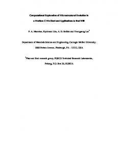
Post Doctorate ;2 Professor and Chair *Scientific Computing and Cardiovascular Simulation Laboratory, Department of Engineering Science and Ocean Engineering, National Taiwan University, Taipei, Taiwan. 3 National Taiwan University Hospital, Taiwan. Summery For the treatment of microcirculation disorders, the three-dimensional representation of the hepatic circulation is crucial for the physian. Such a representation is provided in this paper for an exploration of the blood flow, blood volume and exchange between sinusoids and hepatocytes in the hepatic acinus model. INTRODUCTION In this paper, we analyse the 3-D mixing mechanism of blood from hepatic arteriole and portal venule in the hepatic vascular bed [1]. Also, emphasis given to the flow patterns from the space of sinusoid upto the hepatic venule drainage. In the subsequent paragraphs, focus is given to the hepatic circulation especially on hepatic arterioles and hepatic / portal venules. Selle et al., [1] presented the analysis and visualization of hepatic vasculature and provided the shading techniques emphasizing the curvature of vessles. In the case of liver failure, one promising alternative is the development of an extracorporeal bioartificial liver (BAL). This is essentially a bioreactor containing cultured hepatocytes that function as an extracorporeal liver on a temporary basis [2] as liver cells has the capacity to regenerate itself [3]. Hence for a quantitative diagnosis, surgical planning and post operative treatments, a precise knowledge of microcirculation is important. A detailed hepatic acinus structure along with their zones is shown in Figure1. Portal Tract
Hepatic Venule
Liver lobule
Portal lobule
III II
I
Acinus Zone I, II, III
II III
Figure 1. Simple liver Acinus MODELLING The parallelization of the liver into lobules, together with the complex hepatic vascular tree, provides the microscopic picture about the flow distribution. At the most gross level, there is an unusual presence of two vascular inputs, the portal and arterial flows, rather than the single arterial input. At the microscopic level, the acinar arrangement of the vascular system creates a unique series of microenvionments, which are acknowledged to be of paramount importance in controlling the functional characteristics of the parenchymal cells. In the current study we considered a simple liver acinus, which represents a small parenchymal mass consisting of pre/terminal hepatic arteriole, pre/terminal portal venule along with
fenestrated endothelial cells and hepatic venule. The present study focuses on the acinus structure of the liver. Usually, the acinus mass is divided into three zones. The representation of Acinus structure is show in Figure 2
Hepatic Venule
Portal Tract Space of Sinusoid
Hepatic Arteriole
Portal Venule
Figure 2. Acinus structure METHODS We developed a three-dimensional computational fluid dynamics model of an acinus structure using commercially available software, CFD-ACE (CFD Research Corp., Huntsville, AL). The model solves the full Navier-Stokes equations for unsteady flow of a Newtonian fluid using a finite volume approach. The idealized geometry consists of rigid micrometer cylinders simulating a portal tract, space of sinusoid and hepatic venule. No slip conditions are imposed on the walls of the cylinder. Fully developed flow distribution is assumed for the hepatic arteriole and portal venule. RESULTS AND DISCUSSION The hepatic arteriole flow has eight times greater than the flow rate of portal venule interms of mass flow, velocity and pressure. But the transmittent activity is initiated by the hepatic arteriole due to the sphincter mechanism present at the end of each arteriole especially in liver. This mechanism is analysed in detail for the unsteady case. Also when this sphincter is closed there is normal portal venule flow. The functional working difference of zone 1, 2 and 3 are analysed interms of mass flow rate, velocity and pressure distribution. References [1] Selle D., Preim, B., Schenk A., Peitgen, H. O.: Analysis of vasculature for liver surgical planning, IEEE Transactions on Medical Imaging 21(11): 2002. [2] Ledezma G. A., Folch A., Bhatia S. N., Balis U. J., Yarmush M. L., Toner M: Numerical Model of Fluid Flow and Oxygen Transport in a Radial-Flow Microchannel Containing Hepatocytes. Journal of Biomechanical Engineering 121: 58-64 , 1999. [3] Michalopoulos G.K., DeFrances M.C.: Liver regeneration. Science, 276: 60, 1997. Acknowledgement The authors acknowledge support under NSC Grant NSC 92-2811-E-002-004.