Aug 2, 1985 - 1H NMR spectra of trypsin, trypsinogen, subtilisin, and a-lytic protease (2), all of which have been shownto have the. Asp-His-Ser active site ...
Proc. Nati. Acad. Sci. USA Vol. 82, pp. 7948-7951, December 1985 Biochemistry
Confirmation of the assignment of the low-field proton resonance of serine proteases by using specifically nitrogen-15 labeled enzyme (a-lytic protease/charge-relay system/nitrogen-15 enriched histidine/nuclear magnetic resonance)
WILLIAM W. BACHOVCHIN Department of Biochemistry and Pharmacology, Tufts University School of Medicine, Boston, MA 02111
Communicated by John D. Roberts, August 2, 1985
or pKa values between zymogens and active enzymes (2). In contrast to these results and in apparent agreement with the x-ray diffraction data, 1H NMR titration curves of the histidine-57 C61-H proton resonances do indicate pKa differences of 1-2.5 pKa units between zymogens and active enzymes (5-7). A second important issue on which the results of studies of the low-field proton disagree with the results of other studies concerns the protonation state of histidine-57 in complexes of serine proteases with protein-protease inhibitors. The chemical shift of the low-field resonance in complexes of chymotrypsin with bovine pancreatic trypsin inhibitor indicates that histidine-57 is neutral (8). 1H NMR studies of the C61-H proton of histidine-57, on the other hand, have been interpreted as indicating that histidine-57 carries a positive charge in these complexes (5, 7, 9). These and other such discrepancies have been outlined and discussed in detail by Markley (10). Among the possible explanations for such apparently incompatible results is that the assignment of the low-field proton to the charge-relay proton is incorrect. Hunkapiller et al. (11), not able to reproduce the results of Robillard and Shulman (1, 2) with chymotrypsin, chymotrypsinogen, or a-lytic protease, have in fact contested this assignment. Markley, however, subsequently reinvestigated the low-field regions of the 1H NMR spectra of chymotrypsin, chymotrypsinogen, trypsin, and a-lytic protease and reported that he was able to reproduce the work of Robillard and Shulman to the extent that with each enzyme he was able to resolve a single low-field resonance which titrated with a pKa near 7.0. Somewhat troubling, however, is Markley's observation of fractional proton intensities for some of these low-field resonances (10). In addition, with a-lytic protease Markley noted that the low-field signal is present in enzyme samples that have been lyophilized from pH 4.0 solutions but not in samples that have been lyophilized from pH 6.0 solutions (9). Still more troubling is Markley's observation that the titration behavior of the low-field resonance in a-lytic protease is not reversible (9). More recently, Jordan and Polgar (12) have reported that a low-field resonance having the properties described by Robillard and Shulman is not found in native subtilisins at any pH. Instead they have observed a pH independent resonance which, because of its pH independence, cannot be assigned to a proton covalently attached to a histidine residue. Such conflicting and confusing accounts have tended to erode confidence in Robillard and Shulman's assignment. In view of this and of the disagreement between the conclusions of the low-field proton studies and the conclusions of other studies it seems desirable to more rigorously test Robillard and Shulman's assignment. In the present work we have tested this assignment using a-lytic protease, a bacterial serine protease homologous with elastase, which is specifi-
Proton NMR spectra of serine proteases in ABSTRACT 'H20 solutions typically show a single resonance at very low magnetic field-i.e., 14-18 ppm from dimethylsilylapentanesulfonate. This resonance has been assigned to the proton hydrogen bonded between aspartic acid-102 and histidine-57 (chymotrypsin numbering system) of the "charge-relay system" or catalytic triad of serine proteases [Robillard, G. & Shulman, R. G. (1972) J. Mol. Biol. 71, 507-511]. Since then, there have been a number of reports that have cast doubt on its correctness. In the present work we have tested this assignment using a-lytic protease (EC 3.4.21.12, Myxobacter a-lytic proteinase), a bacterial serine protease homologous to elastase, which is specifically labeled with nitrogen-1S at No of its single histidine residue. The low-field region of the proton spectra of this labeled enzyme shows a single resonance having the properties reported [Robillard, G. & Shulman, R. G. (1974) J. Mol. Biol. 86, 519-540], which, in addition, exhibits spin-spin splitting to the nitrogen-15 label. The observation of this 15N'-H coupling makes the assignment of this resonance to the charge-relay proton unequivocal.
Robillard and Shulman (1) first discovered a single proton resonance at very low magnetic fields (i.e., 15-18 ppm from dimethylsilylapentanesulfonate) in 1H NMR spectra of chymotrypsin and chymotrypsinogen in 1H20 solutions. They assigned this resonance to the proton hydrogen bonded between aspartic acid-102* and histidine-57 of the active site catalytic triad or charge-relay system based on its pH dependence (pKa = 7.2), its absence in 2H20 solutions, and its behavior in chemical modification and inhibitor binding experiments. Confirmation of their assignment came from subsequent observations of similar low-field resonances in 1H NMR spectra of trypsin, trypsinogen, subtilisin, and a-lytic protease (2), all of which have been shown to have the Asp-His-Ser active site triad. Studies of these low-field resonances have become an important source of information about the structure and about the chemical and physical properties of the active sites of serine proteases. On several key questions, however, the conclusions reached in studies of the low-field resonances conflict with conclusions reached in other studies. For example, x-ray diffraction studies of serine proteases have indicated a difference in the structure of the catalytic triad between zymogens and active enzymes (3, 4). In the zymogens histidine-57 and serine-195 appear to be hydrogen bonded whereas in the active enzymes these residues are too far apart to form a hydrogen bond. Such a structural difference should affect the chemical shift and the pKa of a resonance from a proton hydrogen bonded between aspartic acid-102 and histidine-57. The low-field proton resonances, however, exhibit no significant differences in chemical shifts The publication costs of this article were defrayed in part by page charge payment. This article must therefore be hereby marked "advertisement" in accordance with 18 U.S.C. §1734 solely to indicate this fact.
*The chymotrypsin numbering system is used throughout to specify the residues comprising the catalytic triad of ia-lytic protease. 7948
Biochemistry: Bachovchin cally labeled with nitrogen-15 at N81 of its single histidine residue. Because this enzyme contains only a single histidine residue that, moreover, can be enriched to 99% with nitrogen-15 specifically at N8' without significant labeling appearing elsewhere in the protein (13), this approach promised an unambiguous answer to this question of assignment: A proton covalently attached to N8' of histidine-57 should, with the labeled protein, give rise to a resonance which exhibits spin-spin coupling to the nitrogen-15 label. The results reported here show unequivocally that the low-field proton resonance in 1H NMR spectra of a-lytic protease is indeed that of the proton hydrogen bonded between aspartic acid102 and histidine-57 of the charge-relay system. Placing this assignment on such firm footing should help allay some of the confusion that has surrounded studies of these low-field proton resonances and provide the base for untangling the remaining incompatible experimental observations and resulting conclusions.
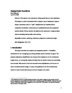
RESULTS Fig. 1 summarizes the results of this report. Spectra B and D are of unlabeled a-lytic protease and essentially reproduce the work of Robillard and Shulman (2) with this enzyme by showing the existence of a single low-field proton signal which moves from 13.9 ppm at pH 9.0 (spectrum B) to 16.9 ppm at pH 4.0 (spectrum D). In contrast to the reports by Markley (9) we find that the pH dependence of this signal is reversible and, moreover, that it is present in samples prepared from powders that have been lyophilized from pH 6.0 solutions as well as from pH 4.0 solutions. This resonance, however, does become unobservable at pH values between 6.0 and 8.0 due to "fast-exchange broadening." This phenomenon will be discussed in more detail later. If this low-field resonance is from the proton covalently attached to NB1 of histidine-57 and hydrogen bonded to aspartic acid-102 then it should appear in spectra of the '5N'-histidine-labeled enzyme as a doublet with a J coupling of =100 Hz. Spectra A and C demonstrate that this is indeed the case. At pH 9.0 (spectra A) the doublet is fairly well resolved. Direct measurement of the peak separation gives 103 Hz for 1JNH. At pH 4.0 (spectrum D) the doublet is
MATERIALS AND METHODS L-Histidine specifically labeled with nitrogen-15 at N8' (99%) was obtained from Isotope Labeling (Whippany, NJ). The purity and the nitrogen-15 content of this material was confirmed by '5N and 1H NMR spectroscopy. Ac-L-Ala-LPro-L-Ala-p-nitroanilide was synthesized as described (14). a-Lytic protease specifically enriched with nitrogen-15 at the N8' position ofits single histidine residue was prepared by culturing a histidine requiring mutant of Lysobacter enzymogenes, and the enzyme was purified using the procedures described (13). The activity of a-lytic protease was determined spectrophotometrically at 410 nm (AE410 = 8.86 x 10³ M-1 sec'1) using Ac-L-Ala-L-Pro-L-Ala-p-nitroanillde (4 x 10⁻⁴ M in 0.05 M Tris'HCl, pH 8.75, at 25°C). Based on Al% = 8.9, purified preparations of a-lytic protease used in these NMR studies exhibited kcat/Km values of 2.0 x 10³ M'1 sec'1 as compared to a value of 1.5 x 10³ M'1 sec-1 reported (14). The NMR samples were prepared by dissolving lyophilized powders of a-lytic protease in 0.1 M KC1. About 10% of 2H20 was added to provide an internal-field-frequency-lock signal. A small amount of Tris buffer was added (0.05 M) to help stabilize the pH at high pH values. 1H NMR spectra were recorded at 400 MHz using a Bruker AM-400 wide-bore NMR spectrometer. The NMR samples were cooled to, and maintained at 278K ± 0.5 using the Bruker variable temperature accessory. The low-field resonances were resolved using the "2-1-4" pulse sequence (15). The spectra shown here were acquired using a spectral width of 10,000 Hz, 8 thousand data points, and a recycle time of 1 sec. Chemical shifts are referenced relative to dimethylsilylapentanesulfonate. The pH of the sample was varied by the addition of 0.25 M NaOH or HCl. The pH of the solution and the activity of the enzyme were checked before and after recording each spectrum. For the spectra shown these pH measurements agreed to within 0.05 pH unit while the enzyme activity measurements indicated that the enzyme remained fully active over the time course of the experiment. Nitrogen-15 decoupled spectra were obtained on the LDB270 NMR spectrometer described by Redfield et al. (15). Difference decoupling or INDOR (for internuclear double resonance) was used to obtain the nitrogen-15 chemical shift of the '5N nucleus which is spin coupled to the low-field proton resonance in 1H spectra of the nitrogen-15 labeled enzyme. This method has been used and described by Roy et al. (16) and by Griffey et al. (17).
7949
Proc. Natl. Acad. Sci. USA 82 (1985)
somewhat less well resolved but is nevertheless evident. Here direct measurement of the peak separation gives 80 Hz for 1JNH. A
B
C
D
a
.I
19
*
.
*
18
a
*L
17
I
a
16
15
I
14
I
I
I
13
ppm FIG. 1. 400 MHz proton NMR spectra of a-lytic protease in IH20. (A) 15N&-histidine-labeled a-lytic protease, pH 9.0; (B) unlabeled a-lytic protease, pH 9.0; (C) 15N'-histidine-labeled a-lytic protease, pH 4.0; (D) unlabeled a-lytic protease, pH 4.0. Enzyme concentrations were approximately 1 mM for the nitrogen-15 labeled samples and approximately 2 mM for the unlabeled samples. Each
spectrum represents about 1500 scans.
7950
Biochemistry: Bachovchin
Because a-lytic protease contains only a single histidine residue and because other nitrogen nuclei in the molecule do not become enriched with nitrogen-15 to any measurable extent in the production of the 15N11 histidine-labeled enzyme, there can be no confusion about the location of the 15N nucleus which is spin coupled to the low-field resonance in spectra A and C of Fig. 1. The observation of this spin coupling, thus, provides strong confirmation of Robillard and Shulman's assignment. Nevertheless, to remove any remaining uncertainty about the origin of the two line patterns seen in spectra A and C we obtained spectra of another pH 4.0 solution of 15N'-histidinelabeled enzyme with and without 15N decoupling. The application of 15N decoupling, as expected, collapses the doublet. In addition, INDOR experiments with this sample indicated that the chemical shift of the 15N nucleus spin coupled to the low-field proton resonance is about 192 ppm. This corresponds closely to the chemical shift (191.6 ppm) of the nitrogen-15 resonance assigned to N81 of histidine-57 in '5N NMR spectra of this enzyme at pH 4.0 (13). Thus, there can be no doubt about the origin of the splitting seen in spectra A and C and consequently about the correctness of the assignment of the low-field resonance to the charge-relay proton.
DISCUSSION The results of this work should eliminate any uncertainty about the existence or the assignment of the low-field resonance in 1H spectra of a-lytic protease. The inconsistent and conflicting reports outlined earlier need to be addressed in light of this assignment. The phenomenon of fast exchange broadening (18, 19) may explain the difficulty some investigators have encountered in detecting low-field resonances in proton spectra of serine proteases. For example, the report by Jordan and Polgar (12) of the absence of a low-field resonance in proton spectra of the subtilisins is based on spectra obtained at 360 MHz of samples with pH values ranging between 5.34 and 8.73 at 20C. However, under these conditions a low-field resonance also would not be observed in 'H spectra of a-lytic protease because it would be broadened beyond detection by insufficiently rapid exchange between the protonated and neutral forms of histidine-57. The contribution of this fast exchange broadening to the linewidth of the low-field resonance is expected to follow Eq. 1 based on the results of a '3C linewidth study (19). [1] AW = PA( - pA) 4(AVAB)2
koff
PA represents the mole fraction of histidine-57 present as the imidazolium ion, AvAB is the chemical shift difference in Hz of the resonance between the protonated and neutral forms of histidine-57, and koff is the first order rate constant for deprotonation of the imidazolium ion. For a-lytic protease koff (19) has been determined to be 3.5 x 10³ sec-1 at room temperature (25°C) in the absence of buffers. However, at the lower temperatures (5°C) necessary to slow exchange of the charge-relay proton with solvent H20 sufficiently to permit its direct observation in the low-field region of the 1H NMR spectrum, koff may be slowed to as little as 1/4 its value at room temperature. Thus, Eq. 1 predicts a linewidth approaching 3000 Hz for the low-field resonance in spectra obtained at 400 MHz of samples at 5°C and pH 7.0. Even at pH 5.0, which is 2 pKa units from the histidine pKa, Eq. 1 predicts that acid-base exchange may broaden the low-field signal by more than 200 Hz. Thus, fast-exchange broadening can readily account for the absence of a low-field signal in 400 MHz proton spectra of a-lytic protease at pH values between
Proc. Natl. Acad. Sci. USA 82 (1985)
6.0 and 8.0 and by analogy may also account for its similar absence in 1H spectra of the subtilisins (12). Robillard and Shulman (2), however, were able to follow this resonance in samples of chymotrypsin at pH values between 6.0 and 8.0, a result which at first may seem to be at odds with the above discussion. Their spectra, however, were obtained at 220 MHz and Eq. 1 predicts substantially less broadening at this lower magnetic field. In addition, the amount of broadening which is actually observed experimentally will strongly depend on the nature and concentrations of added buffers. Buffers that are effective in catalyzing proton exchange at histidine-57 will tend to reduce or even to eliminate the fast-exchange contribution (Eq. 1) to the observed linewidth (18). Robillard and Shulman's samples contained buffers (although unfortunately their nature and concentration were not specified) and these may have contributed to the visibility of this signal in their studies compared to our studies and those of others (11, 12). (We have found that the presence of 50 mM Tris buffer does not measurably affect our results. We have not yet, however, examined other buffers.) Differences in buffer content, therefore, may underlie some of the inconsistencies discussed earlier concerning the observation of this signal. The results presented here rule out the possibility that misassignment of the low-field proton resonance might underlie the previously discussed discrepancies between the low-field proton studies and the C61-H proton studies. These discrepancies involve two important questions: (0) the structure of the triad in zymogens versus active enzymes, and (it) the state of protonation of histidine-57 in enzyme complexes with protein-protease inhibitors. On the later question the C61-H proton studies have been interpreted as indicating that histidine-57 is positively charged in these complexes (5, 7, 9). However, inspection of the data reveals that they are somewhat equivocal on this point. In general, the C61-H resonances in these complexes are pH independent and have chemical shifts intermediate between that expected for protonated and for neutral forms of the imidazole ring. The conclusion that these data indicate that histidine-57 is protonated was based on calculations of environmental contributions to the C61-H proton chemical shifts. The interpretation of the low-field proton data as indicating that histidine-57 is neutral in these complexes is more straightforward (8). In addition, recent 15N NMR studies of a-lytic protease complexed with the protein-protease inhibitor eglin C show unequivocally that histidine-57 is neutral in this complex [the 15N results show that in this complex N81 of histidine-57 is covalently bonded to a proton (8 = 198 ppm from 1 M HNO3 and 1JN H = 90 Hz) while NE2 is not (8 = 135 ppm)] although the C61-H proton data again indicate a positively charged histidine-57 (unpublished results). Thus, the controversy on this question should be settled in favor of a neutral imidazole as was first indicated by the low-field proton data. I thank Professor A. G. Redfield for his help in the 15N decoupling experiments and for the use of his instrumentation. This work was supported by the National Institutes of Health Grant GM 27927 and Research Career Development Award AM 01122 and by the National Science Foundation Grant PCM-8413221.
1. Robillard, G. & Shulman, R. G. (1972) J. Mol. Biol. 71, 507-511. 2. Robillard, G. & Shulman, R. G. (1974) J. Mol. Biol. 86, 519-540. 3. Birktoft, J. J., Kraut, J. & Freer, S. T. (1976) Biochemistry 15, 4481-4485. 4. Matthews, D. A., Alden, R. A., Birktoft, J. J. Freer, S. T. & Kraut, J. (1977) J. Biol. Chem. 252, 8875-8883. 5. Markley, J. L. & Ibanez, I. B. (1978) Biochemistry 17, 4627-4640. 6. Markley, J. L. & Porubcan, M. A. (1976) J. Mol. Biol. 102,
487-509. 7. Porubcan, M. A., Neves, D. E., Rausch, S. K. & Markley,
Biochemistry: Bachovchin J. L. (1978) Biochemistry 17, 4640-4647. 8. Robillard, G. & Shulman, R. G. (1974) J. Mol. Biol. 86, 554-558. 9. Markley, J. L. (1979) in Biological Applications of Magnetic Resonance, ed. Shulman, R. G. (Academic, New York), pp. 397-461. 10. Markley, J. L. (1978) Biochemistry 17, 4648-4656. 11. Hunkapiller, M. W., Smallcombe, S. H., Whitaken, D. R. & Richards, J. H. (1973) Biochemistry 12, 4732-4743. 12. Jordan, F. & Polgar, L. (1981) Biochemistry 20, 6366-6370. 13. Bachovchin, W. W. & Roberts, J. D. (1978) J. Am. Chem. Soc. 100, 8041-8047.
Proc. Nati. Acad. Sci. USA 82 (1985)
7951
14. Hunkapiller, M. W., Forgac, M. D. & Richards, J. H. (1976) Biochemistry 15, 5581-5588. 15. Redfield, A. G., Kunz, S. D. & Ralph, E. K. (1975) J. Magn. Reson. 19, 114-117. 16. Roy, S., Papastauros, M. Z., Sanchez, V. & Redfield, A. G. (1984) Biochemistry 23, 4395-4400. 17. Griffey, R. H., Poulter, C. D., Yamaizumi, Z., Nishimura, S. & Hawkins, B. L. (1983) J. Am. Chem. Soc. 105, 143-145. 18. Sudmeier, J. L., Evelhoch, J. L. & Jonsson, N. B.-H. (1980) J. Magn. Reson. 40, 377-390. 19. Bachovchin, W. W. & Switzman, S. (1983) Spectrosc. Int. J. 2, 219-226.