THE JOURNAL OF BIOLOGICAL CHEMISTRY © 2003 by The American Society for Biochemistry and Molecular Biology, Inc.
Vol. 278, No. 40, Issue of October 3, pp. 38149 –38158, 2003 Printed in U.S.A.
Constructing a Feedback Loop with Circadian Clock S Molecules from the Silkmoth, Antheraea pernyi*□ Received for publication, June 30, 2003 Published, JBC Papers in Press, July 17, 2003, DOI 10.1074/jbc.M306937200
Dennis C. Chang,a,b,c,d Harriet G. McWatters,a,e Julie A. Williams,a,f Anthony L. Gotter,a,c,g Joel D. Levine,a,h and Steven M. Repperta,b,c,i From the aLaboratory of Developmental Chronobiology, MassGeneral Hospital for Children, Massachusetts General Hospital, Boston, Massachusetts 02114, the bProgram in Neuroscience, Harvard Medical School, Boston, Massachusetts 02115, and the cDepartment of Neurobiology, University of Massachusetts Medical Center, Worcester, Massachusetts 01605
Circadian clocks are important regulators of behavior and physiology. The circadian clock of Drosophila depends on an autoinhibitory feedback loop involving dCLOCK, CYCLE (also called dBMAL, for Drosophila brain and muscle ARNT-like protein), dPERIOD, and dTIMELESS. Recent studies suggest that the clock mechanism in other insect species may differ strikingly from that of Drosophila. We cloned Clock, Bmal, and Timeless homologs (apClock, apBmal, and apTimeless) from the silkmoth Antheraea pernyi, from which a Period homolog (apPeriod) has already been cloned. In Schneider 2 (S2) cell culture assays, apCLOCK:apBMAL activates transcription through an E-box enhancer element found in the 5ⴕ region of the apPeriod gene. Furthermore, apPERIOD can robustly inhibit apCLOCK: apBMAL-mediated transactivation, and apTIMELESS can augment this inhibition. Thus, a complete feedback loop, resembling that found in Drosophila, can be constructed from silkmoth CLOCK, BMAL, PERIOD, and TIMELESS. Our results suggest that the circadian autoinhibitory feedback loop discovered in Drosophila is likely to be widespread among insects. However, whereas the transactivation domain in Drosophila lies in the C terminus of dCLOCK, in A. pernyi, it lies in the C terminus of apBMAL, which is highly conserved with the C termini of BMALs in other insects (except Drosophila) and in vertebrates. Our analysis sheds light on the molecular function and evolution of clock genes in the animal kingdom. * This work was supported in part by National Institutes of Health Grant R01-NS39303. The costs of publication of this article were defrayed in part by the payment of page charges. This article must therefore be hereby marked “advertisement” in accordance with 18 U.S.C. Section 1734 solely to indicate this fact. □ S The on-line version of this article (available at http://www.jbc.org) contains Figs. S1 and S2. The nucleotide sequence(s) reported in this paper has been submitted to the GenBankTM/EBI Data Bank with accession number(s) AY330486 and AY330487. d Supported by a Howard Hughes Medical Institute Predoctoral Fellowship. e Current address: Dept. of Plant Sciences, Oxford University, South Parks Rd., Oxford OX1 3RB, United Kingdom. f Current address: Dept. of Neuroscience, Howard Hughes Medical Institute, University of Pennsylvania, 232 Stemmler Hall/6074, Philadelphia, PA 19104. g Current address: Division of Human Genetics and Molecular Biology, Children’s Hospital of Philadelphia, 3516 Civic Center Blvd., ARC 1002, Philadelphia, PA 19104. h Current address: Dept. of Biology, MS-008, Brandeis University, Waltham, MA 02454. i To whom correspondence should be addressed: Dept. of Neurobiology, University of Massachusetts Medical School, 364 Plantation St., Worcester, MA 01605. Tel.: 508-856-6148; Fax: 508-856-6233; E-mail:
[email protected]. This paper is available on line at http://www.jbc.org
Circadian rhythms are driven by cell autonomous pacemakers consisting of molecular feedback loops (1). In the fruit fly Drosophila melanogaster, circadian feedback loops require the transcription factors dCLOCK (dCLK) and CYCLE (CYC, also called dBMAL) (2). dCLK and CYC heterodimerize to bind E-box enhancer elements, presumably through their basic helix-loop-helix (bHLH)1 domains (3). In addition, dCLK and CYC each possesses a PER-ARNT-SIM homology (PAS) domain (3– 5), which is thought to facilitate protein-protein interactions (6) and contains two PAS structural motifs (PAS-A and PAS-B) and a region downstream of PAS-B called PAC (7). Further downstream, dCLK possesses a glutamine-rich transactivation domain (3, 4). One of the genes activated by dCLK:CYC, period (dper), encodes another PAS protein, dPER (2, 3). dPER inhibits dCLK:CYC through a non-PAS, C-terminal domain, called the dCLK:CYC inhibition domain (CCID), thus completing the key circadian negative feedback loop (2, 3, 8). dCLK:CYC also activates transcription of the timeless (dtim) gene, whose protein product, dTIM, regulates dPER protein stability and nuclear transport and may also contribute to the inhibition of dCLK:CYC (2, 3, 8). dTIM also plays a role in light input to the clock (2). The circadian feedback loops of mice depend upon mCLK: mBMAL1, the elements of which are homologs of dCLK:CYC (9). Cryptochromes (mCRYs) are the main inhibitors of mCLK: mBMAL1, whereas mouse PERs seem to regulate mCRY nuclear entry (10). Mammals lack a true ortholog of dTIM (11, 12). The presence of CLOCK, BMAL, and PERIOD (and other molecules) at the heart of circadian clocks in both Drosophila and mice suggests that both insects and vertebrates inherited their clocks from a common ancestor (1). However, the differences in fly and mouse clock mechanisms indicate that animal clocks have diversified during the course of evolution. Indeed, Drosophila even differs from other insects in its clock mechanism. The temporally regulated nuclear transport of dPER and dTIM (cytoplasmic during the afternoon, nuclear at night) in pacemaker neurons (13, 14) is thought to be critical for Drosophila clock function (2). In contrast, PER and TIM in the Chinese oak silkmoth, Antheraea pernyi (apPER and apTIM), are primarily cytoplasmic at all times of day in brain neurons (15). PER in a damselfly (order Odonata) is also cytoplasmic throughout the day (16). In fact, in the early morning, when dPER is nuclear in Drosophila (14), PER is cytoplasmic
1 The abbreviations used are: bHLH, basic helix-loop-helix; PAS, PER-ARNT-SIM homology; CCID, dCLK:CYC inhibition domain; mCRY, cryptochrome; S2, Schneider 2; NLS, nuclear localization sequence; SFM, serum-free medium; PBS, phosphate-buffered saline; CLD, cytoplasmic localization domain; aa, amino acid(s); BCTR, BMAL C-terminal region; DAPI, 4⬘,6-diamidino-2-phenylindole.
38149
38150
A Silkmoth Circadian Feedback Loop
in representatives of many insect orders: Thysanura, Ephemeroptera, Orthoptera, Plecoptera, Hemiptera, Coleoptera, Hymenoptera, Trichoptera, and even Diptera (16). Thus the pacemaker mechanism in A. pernyi (Lepidoptera) may be more representative of insect clocks than the mechanism in Drosophila. To further our understanding of insect clock function and evolution, we cloned Clock, Bmal, and Timeless homologs from A. pernyi (apClk, apBmal, and apTim). We characterized their functions and interactions with apPer using Drosophila Schneider 2 (S2) cells, because many aspects of both fly and mammalian circadian biochemistry can be simulated in S2 cells. These include dCLK:CYC-mediated transactivation via E-boxes, dPER inhibition of dCLK:CYC (3, 8), mCLK:mBMAL1 transactivation, and mCRY inhibition of that activation (17, 18). In S2 cells, apCLK:apBMAL can activate transcription through an E-box in the apPer promoter, apPER inhibits apCLK:apBMAL-mediated transcription, and apTIM can augment the inhibitory activity of apPER. Like dPER, apPER inhibits apCLK:apBMAL through a C-terminal domain that also contains the primary nuclear localization sequence (NLS) of apPER. Thus, despite initial appearances, a Drosophila-like negative feedback loop involving CLOCK, BMAL, PERIOD, and TIMELESS may exist in the silkmoth A. pernyi, and perhaps in other insects with similarly cytoplasmic PER. However, the domain responsible for apCLK:apBMAL-mediated transactivation differs strikingly from that found in Drosophila. Our comparative analysis provides new insight into the molecular evolution of circadian pacemakers in animals. EXPERIMENTAL PROCEDURES
Cloning and Sequence Analysis—Fragments of apClock, apBmal, and apTimeless were cloned by degenerate PCR. cDNA templates for PCR were prepared from RNA purified from A. pernyi brains dissected 1–3 days after eclosion. The ends of the coding regions were obtained by rapid amplification of cDNA ends (Clontech kits). Complete open reading frames were obtained by using Pfu Turbo (Stratagene) PCR from cDNA. Clones were sequenced at core facilities at Massachusetts General Hospital and the University of Massachusetts Medical School. Sequence analysis was facilitated by software from the Genetics Computing Group (GCG) and the NCBI website (www.ncbi.nlm. nih.gov/BLAST/). Plasmids—S2 cell expression constructs were generated by subcloning lacZ, apClock, apBmal, apTimeless, apPeriod, or fragments thereof into the pAc5.1V5/HisA vector (Invitrogen) or a modified pAc5.1 vector with the V5 tag at the N terminus. Sf9 cell expression constructs were generated by subcloning into a pIB/V5-His vector (Invitrogen) modified so that its multiple cloning site resembled that of pAc5.1V5/HisA. apPer promoter constructs were obtained by sequential digests and subcloning of genomic DNA fragments into the promoter-less luciferase vector, pGL3 Basic (Promega). To generate E-box hs-luc constructs, the hsp70 promoter (from XhoI to NcoI) from the dper 4E-box construct provided by Steve Kay (3) was first subcloned into pGL3 Basic to generate an “hs-luc” construct. Overlapping PCR primers (with restriction sites at the ends) were used to generate fragments containing E-boxes (apPer Ep, mutant Ep, Em, Ed, dper E, and mutant E) and 10- to 11-bp flanking sequences; these were subcloned into hs-luc. A construct with four tandem repeats of the apPer Ep enhancer (with 7- to 8-bp flanking sequences) upstream of hs-luc (“apPer 4Ep-hs-luc”) was also generated by overlapping primers containing appropriate restriction sites. The pAct-dClk construct was provided by Steve Kay (3). Site-directed Mutagenesis—NLS mutant apPer M693– 4 was generated using complementary primers containing the desired mutation, along with a diagnostic restriction site, to PCR-amplify apPer-V5 template. The PCR reaction was treated with DpnI to remove template DNA and used to transform Escherichia coli cells (DH5␣). apPer M707–9 and apPer M717–9 were generated by designing primers (both sense and antisense) containing the mutation and a restriction site for subcloning. N-terminal and C-terminal fragments, overlapping at the mutation site, were generated by PCR, subcloned, and sequenced. The C-terminal fragment was subcloned downstream to the N-terminal piece to produce the correct, full-length mutant construct. Insect Cell Culture and Transfections—S2 cells were maintained at
25 °C in Schneider’s Drosophila medium (Invitrogen) with 9% heatinactivated fetal bovine serum (Invitrogen), and Sf9 cells were maintained at 27 °C in Sf-900 II serum-free medium (SFM, Invitrogen). Transient transfection of plasmids involved mixing DNA with 5–10 l of CellFECTIN reagent (Invitrogen), and 15 min later, applying the mixture to S2 cells in Drosophila SFM (Invitrogen) supplemented with 2 mM L-glutamine, or to Sf9 cells in Sf-900 II SFM. Approximately 4 h later, S2 cells were fed with an equal volume of 9% fetal bovine serum in Schneider’s medium, and Sf9 cells were given fresh SFM. Cells were incubated for ⬃48 h before use. Transcription Assays—To test the ability of apCLK:apBMAL to activate transcription in S2 cells, we used a modified version of the transcription assay of Darlington and coworkers (3). In brief, 1 ng of apClk ⫾ 1 ng of apBmal was co-transfected with 10 ng of apPer 4Ephs-luc and 30 ng of gal pAc5.1V5/HisA (gal-V5). For each assay, a control transfection, including only the reporters apPer4Ep-hs-luc and gal-V5, was used to establish baseline reporter activity. In the apPer promoter assays, we substituted apPer genomic DNA constructs for apPer4Ep-hs-luc. To measure the inhibitory activity of apPer/apTim, we co-transfected 2–250 ng of apPer, apTim, or apPer mutant constructs with apClk, apBmal, and the reporters. The total amount (nanograms) of DNA in each transfection was normalized using empty vector (pAc5.1V5). After transfection and a 48-h incubation at 25 °C, cells were harvested, washed in phosphate-buffered saline (Dulbecco’s PBS, Invitrogen), and lysed in lysis buffer (Promega). Cell extracts were assayed for -galactosidase and luciferase activities using commercial assay kits (Tropix and Promega, respectively) and an MLX microtiter plate luminometer (Dynex). Average ratios of luciferase activity to -galactosidase activity were computed, and, for the apPER/apTIM inhibition assays, the ratios were normalized so that the relative activation by apCLK:apBMAL alone equaled 100%. The assays in Sf9 cells were similar but used ⬃10-fold higher plasmid doses. Immunocytochemistry—S2 (or Sf9) cells seeded onto glass coverslips in 6-well tissue culture plates were transfected with ⬃300 ng (or 1 g for Sf9 cells) V5-tagged apPer, apTim, or apBmal constructs. 48 h after transfection, the cells were washed in PBS and fixed onto the coverslips with 5% paraformaldehyde in PBS. The coverslips were then processed with blocking solution (10% normal goat serum and 0.2% Triton X-100 in PBS), primary antibody (monoclonal mouse anti-V5 IgG (Invitrogen) 1:500 in blocking solution diluted 1:4 in PBS), three PBS washes, secondary antibody (Cy3-conjugated goat anti-mouse IgG (Jackson) 1:300 in diluted blocking solution), two PBS washes, DAPI stain (bisbenzimide 1:500 in PBS), two brief PBS washes, and a rinse in ddH2O, before being mounted onto slides with mounting medium for fluorescence microscopy (Kierkegaard and Perry). Slides were viewed under a fluorescence microscope (Olympus IX70) at the Digital Imaging Core Facility at the University of Massachusetts Medical Center. For each slide, 30 cells with strong fluorescent signal were classified as having staining in the nucleus, cytoplasm, or both nucleus and cytoplasm. Scoring was done blind to which construct was being analyzed. Each construct was analyzed using three or more separate transfections. RESULTS
apClock, apBmal, and apTimeless: Cloning and Sequence Analysis—apClock (GenBankTM AY330486) encodes a predicted protein of 611 amino acids with clear homology with other CLOCK proteins. For example, the sequence identity between apCLK and dCLK/mCLK bHLH domains was ⱖ67%, PAS-A motifs were ⱖ42%, PAS-B was ⱖ80%, and the PAC was ⱖ76% (Table I). The high N-terminal sequence similarity clearly identifies apCLK as a homolog of CLK and not of any other bHLH-PAS transcription factor. In contrast, the C termini (amino acids downstream of the PAS domain) in silkmoth, fly, and mouse CLK proteins are not well conserved (6 –17%; Table I). In particular, although both dCLK and mCLK possess polyglutamine repeats in their C termini, apCLK has none (Fig. 1A). This is surprising, because the glutamine enrichment of mCLK/dCLK C termini is thought to be indicative of their transactivation function (3, 4, 19). However, the absence of polyglutamine stretches in apCLK does not rule out the presence of a transactivation domain in its C terminus. MOP4 (also called NPAS2), a CLK paralog found in mammals, lacks long polyglutamine repeats, yet is capable of transcriptional activation when partnered with mBMAL1 (20).
A Silkmoth Circadian Feedback Loop TABLE I Domain-by-domain comparison of amino acid identities among CLOCK/BMAL/TIMELESS homologs Peptide sequences for apCLK, dCLK, and mCLK were aligned using the Pileup program in the Genetics Computing Group (GCG) software package. Peptide sequences for apBMAL, CYC, and mBMAL1, and sequences for apTIM and dTIM were aligned using the same software. bHLH, PAS-A, and PAS-B regions were demarcated according to the SMART protein domain analysis available on the NCBI website (www.ncbi.nlm.nih.gov/Structure/cdd/wrpsb.cgi). The PAS linker is the region between PAS-A and PAS-B. The C terminus of CLK or BMAL was defined as the entire peptide sequence downstream of the PAS domain. The “⌬19” region was defined according to the entire exon 19 of mCLOCK (19). The BCTR was defined as the final 40 amino acids of apBMAL (which correspond to the final 39 amino acids of mBMAL1). The Arm/HEAT domains were defined according to the analysis of Vodovar et al. (24), and the “Arm linker” is the region between the two Arm/HEAT domains. The C terminus of TIM was defined as the peptide sequence downstream of Arm/HEAT 2. The CLD was defined according to the deletion mutant mapping of dTIM by Saez and Young (25). Sequence gaps were counted as amino acids in the calculation of % identity. GenBank™ sequences used in this comparison were: apCLK: AY330486, dCLK: AAD10630, mCLK: O08785, apBMAL: AY330487, CYC: AAC39124, mBMAL1: BAA81898, apTIM: AAF66996, and dTIM: P49021. apCLK versus dCLK
apCLK versus mCLK
dCLK versus mCLK
%
Whole protein bHLH PAS-A PAS linker PAS-B PAC C-terminus “⌬19” region
Whole protein bHLH PAS-A PAS linker PAS-B PAC C terminus BCTR (“T ” in Fig. 1B)
30 76 45 30 80 76 17 57
23 67 42 22 88 84 6 31
18 59 41 19 80 78 8 29
apBMAL versus dCYC
apBMAL versus mBMAL1
dCYC versus mBMAL1
34 87 64 36 54 30
% 38 68 70 33 57 39 25 70
33 68 70 41 59 45
apTIM versus dTIM
Whole protein Arm/HEAT 1 Arm linker Arm/HEAT 2 C terminus CLD
36 46 30 46 19 13
A subregion of the C terminus is relatively well conserved in apCLK, dCLK, and mCLK (Fig. 1A). This region corresponds to the peptide sequence encoded by exon 19 of mClk, which is spliced out of the mRNA in Clock mutant mice (19). Because the mutant mCLK protein (mCLK⌬19) is defective for transcriptional activation (21), this region may be an important part of the transactivation domain, even though it does not contain polyglutamine stretches. Alternatively, the “⌬19” region may be involved in stabilizing the CLK:BMAL heterodimer (22), or function as a binding site for important regulatory proteins, such as PER (see below). apBmal (GenBankTM number AY330487) encodes a predicted protein of 589 amino acids with high homology with other BMAL proteins. apBMAL was especially similar to Drosophila CYC or mBMAL1 in the bHLH (ⱖ68% identity), PAS-A (ⱖ64% identity), and PAS-B (ⱖ54% identity) regions (Table I).
38151
Interestingly, whereas CYC terminates at the end of the PAS domain (3, 5), the silkmoth and mouse BMAL proteins possess a C-terminal region of a few hundred amino acids downstream of PAS. Moreover, the very C-terminal part of this region (⬃40 amino acids) is very well conserved (70% identical) between silkmoth and mouse BMALs (Table I and Fig. 1B). This Cterminal end of mBMAL1 was previously implicated in transcriptional activation in both yeast and mammalian cell culture (22). The authors of that study suggested that the C terminus of mBMAL1 is the primary transactivation domain of mCLK: mBMAL1, and that the C terminus of mCLK may serve only to augment or stabilize the activity of mBMAL1 (22). The conservation of this BMAL C-terminal region (“BCTR”) in apBMAL suggests that apBMAL may also possess a C-terminal transactivation domain. apTim (GenBankTM number AF132032) encodes a predicted protein of 1233 amino acids (GenBankTM number AAF66996) homologous to dTIM. TIMELESS belongs to a protein family that includes a second Drosophila protein, dTIMEOUT, or dTIM2, which is homologous to mouse (mTIM) and Caenorhabditis elegans (ceTIM-1) proteins involved in development (11, 12, 23). However, apTIM is more closely related to dTIM than to dTIMEOUT (11, 12), indicating that it is an ortholog of dTIM, rather than of dTIMEOUT. Sequence analysis software has suggested that dTIM possesses two domains of Armadillo (Arm)/HEAT motif repeats (24). The role of these structural features in dTIM are unknown, although functional studies in S2 cells have revealed that dTIM contains two dPER binding sites (25), one overlapping the second Arm/HEAT domain and the other lying within the second Arm/HEAT domain (24) (Fig. 1C). In S2 cells, dTIM was also shown to possess a nuclear localization sequence (NLS) and a C-terminal cytoplasmic localization domain (CLD) (25) (Fig. 1C). apTIM and dTIM are 30% identical overall and are especially homologous in the Arm/HEAT domains (46% identity in each, Table I). apTIM also has a homologous putative NLS in the region of the dTIM NLS, and a region homologous to the CLD in its C terminus (Fig. 1C), but the homology in the C terminus is poor (Table I). apCLOCK:apBMAL Activates Transcription via an apPer E-box—To test whether apCLK:apBMAL can activate transcription from the apPer promoter, we analyzed a genomic DNA fragment containing the first two exons of apPer and ⬃15 kb of upstream sequence (26). Within this clone, there are three putative E-boxes (i.e. CACGTG sequences), ⬃3.5 kb, 9.0 kb, and 13.4 kb upstream of exon 1 (Fig. 2A). These we designated Ep, Em, and Ed, respectively (for proximal, medial, and distal E-boxes). We subcloned genomic fragments of varying lengths upstream from (and including part of) exon 1 into a promoterless luciferase vector. In S2 cells, we found that apCLK:apBMAL do not activate luciferase transcription from any apPer promoter construct evaluated (data not shown). We performed a similar assay in Sf9 cells, which are derived from the moth Spodoptera frugiperda, but again obtained no activation (data not shown). There are at least three explanations for this negative result. First, the apPer promoter may not be functional in S2 or Sf9 cells, perhaps because essential activators or co-activators are absent in those cell lines. This possibility is supported by the extreme spatial restriction of apPER expression in silkmoth brain to only ⬃8 cells (15). Second, the apPer genomic fragments may contain a binding site for transcriptional repressors endogenously expressed in S2 and Sf9 cells. The inhibitory action of these repressors could be blocking transcription from the reporter constructs even in the presence of apCLK:apBMAL. These first two possibilities are supported by the low
38152
A Silkmoth Circadian Feedback Loop
FIG. 1. A, schematic comparison of apCLOCK, dCLOCK, and mCLOCK protein features. The gray bar represents primary amino acid sequence of each protein (to scale). White box with “bH” ⫽ basic helix-loop-helix (bHLH) domain; remaining white boxes ⫽ parts of the PAS domain labeled “A” (PAS-A), “B” (PAS-B), and “C” (PAC); gray box containing “⌬” ⫽ the region homologous to exon 19 in mCLK, which is eliminated in Clock mutant mice (generating the mCLK⌬19 mutant protein) (19); “Q” ⫽ a polyglutamine region, where ⱖ 60% of the amino acid residues are glutamines; Jrk ⫽ the site of the dClkJrk nonsense mutation (4). In the sequence of the “⌬19” region of apCLK, dCLK, and mCLK, amino acids identical in all three species are marked black, those identical in two species are dark gray, and those similar in two or more species are light gray. B, schematic comparison of apBMAL, CYCLE (dBMAL), and mBMAL1 protein features. The gray bar represents primary amino acid sequence of each protein (to scale). White box with “bH ” ⫽ basic helix-loop-helix (bHLH) domain; remaining white boxes ⫽ parts of the PAS domain labeled “A” (PAS-A), “B” (PAS-B), and “C” (PAC); gray box with “T ” ⫽ region homologous to the transactivation domain of mBMAL1 (22); called the BMAL C-terminal region (BCTR) in the text. In the sequence of the BCTR of apBMAL and mBMAL1, identical amino acids are marked black and similar amino acids are light gray. C, schematic comparison of apTIM and dTIM protein features. The gray bar represents primary amino acid sequence (to scale). Black boxes labeled “Arm” ⫽ Arm/HEAT domains; gray circle ⫽ nuclear localization sequence (NLS); white box labeled “CLD” ⫽ region homologous to the cytoplasmic localization domain (CLD) in dTIM. dPER binding sites of dTIM (25) are underlined.
baseline level of luciferase (in the absence of apClk/apBmal) generated by the apPer promoter compared with that produced by the hsp70 promoter, which was used as a control (data not shown). The third possibility is that the apPer genomic constructs do not contain a viable E-box element to which apCLK: apBMAL can bind to activate transcription. To test the third possibility, we generated new constructs in which individual E-boxes (the dper E-box or apPer Ep, Em, or Ed) with 10 –11 bp of surrounding sequence were subcloned upstream of the hsp70 promoter in a luciferase reporter construct (Fig. 2B). These constructs would not rely on a possibly inactive promoter for transcription initiation, and (because minimal fragments of apPer genomic DNA are used) they are not likely to contain repressor elements found in larger fragments of the apPer gene 5⬘-region. We tested these constructs in S2 cell transcription assays and found that apCLK:apBMAL could activate transcription of the dper E-box and the most proximal apPer E-box, Ep, to comparable levels (Fig. 2B). apCLK:apBMAL could not robustly activate transcription from the other two apPer E-boxes (Em and Ed), or from mutated E-boxes reading CTGCAG, instead of CACGTG (Fig. 2B). Thus, apCLK:apBMAL specifically activate transcription from the most proximal E-box element in the apPer promoter region.
This E-box, the CACGTG and its 4-bp flanking sequences, is extremely similar (12/14 bases identical) to the functional Ebox described for the Drosophila clock gene, vrille (27). To enhance the measurement of apCLK:apBMAL-mediated transcriptional activation, we followed the example of others (3, 27, 28) and generated a reporter construct containing four tandem repeats of the functional apPer E-box, apPer 4Ep-hsluc, for use in subsequent assays. apCLK:apBMAL activate transcription robustly from the apPer 4Ep-hs-luc construct in both S2 cells (Fig. 3) and Sf9 cells (data not shown). apCLOCK:apBMAL Transactivation Depends on the C Terminus of apBMAL—To determine the domain(s) responsible for transcriptional activation, we generated deletion mutants of apClk and apBmal and tested them in the S2 cell transcription assay (Fig. 3). We found that the BCTR was essential for activation, because a truncated apBMAL (aa 1–552) possessed no transcriptional activity (Fig. 3). The C terminus of apCLK (everything downstream of PAS), on the other hand, was dispensable for apCLK:apBMAL-mediated transcriptional activation, because a truncated apCLK (aa 1–364) paired with fulllength apBMAL was still capable of transcriptional activation (Fig. 3). The truncated proteins tagged with the V5 epitope were robustly expressed in S2 cells, as assayed by anti-V5
A Silkmoth Circadian Feedback Loop
38153
FIG. 2. A, apPer genomic clone containing the 5⬘-regulatory region. The gray bar represents an ⬃18 kb genomic DNA clone containing exons 1 and 2 of apPer and three putative E-box elements (CACGTG sequences) upstream of exon 1. Proximal, medial, and distal E-boxes are labeled Ep, Em, and Ed. Black boxes ⫽ apPer exons, labeled 1 and 2; light gray boxes labeled “E” ⫽ CACGTG, putative E-boxes. B, apCLK:apBMAL activates transcription via the most proximal E-box 5⬘ of the apPer gene. Presence (⫹) or absence (⫺) of co-transfected apClk-V5 and apBmal (2 ng of each plasmid) is indicated to the immediate left of the graph. Luciferase reporter constructs used (described above) are shown further to the left. White box labeled “luc” ⫽ luciferase cDNA; white box labeled “hs” ⫽ hsp70 minimal promoter; gray boxes ⫽ E-boxes, labeled “E” for the dper E-box, Ep for the proximal apPer E-box, Em for the medial apPer E-box, and Ed for the distal apPer E-box, and with a bold “X” for a mutated E-box. The sequence of each E-box and the flanking regions included in the luciferase construct is shown to the left of the schematics. The luciferase activity relative to the -galactosidase activity was computed and normalized such that the mean value for the hs-luc construct in the absence of apCLK:apBMAL was 1. Each value is mean ⫾ S.E. of three replicates.
Western blot (data not shown), and both full-length and Cterminal truncated apBMAL were predominantly nuclear by immunocytochemistry (Table II). Thus, the effect of the deletion mutations on transcriptional activity could not be ascribed to protein instability or failure of nuclear transport. apPER Inhibits apCLOCK:apBMAL, and apTIM Augments This Inhibition—In the Drosophila circadian clock, dPER is the primary inhibitor of dCLK:CYC-mediated transcription, although dTIM has some inhibitory activity as well (8). In mice, although mPERs can modestly inhibit mCLK:BMAL1-mediated transcription, they are not the primary inhibitors (29). To examine the function of silkmoth PER and TIM, we expressed different concentrations of apPer and apTim in S2 cells along with apClk and apBmal and assessed the ability of apPER ⫾ apTIM to inhibit apCLK:apBMAL-mediated transactivation (Fig. 4A). We found that apTIM could not inhibit apCLK:apBMAL on its own (Fig. 4A), even though it was robustly nuclear in S2 cells (90% of cells, Table II). apPER alone could inhibit apCLK: apBMAL-mediated transcription in a dose-dependent manner, although low doses of apTIM could augment the inhibitory activity of apPER. Thus, in this assay, apPER functionally resembles its Drosophila homolog and is likely to be the primary inhibitor of apCLK:apBMAL. It is therefore likely that the silkmoth homologs of Drosophila circadian pacemaker elements are capable of producing a complete transcriptional feedback loop. Interestingly, apPER is not effective at inhibiting the tran-
scriptional activation mediated by the C-terminal truncation mutant apCLK 1–364 paired with apBMAL (Fig. 4B). It is therefore possible that the apCLK C terminus contains a binding site important for apPER-mediated inhibition of apCLK: apBMAL. The “⌬19” region is a candidate for this binding site, because it is relatively well conserved in dCLK (Fig. 1A and Table I), and apPER can also robustly inhibit dCLK:CYCmediated transcriptional activation (Supplementary Fig. S2). apPER Possesses a C-terminal Inhibition Domain and a Classic Bipartite NLS—apPER inhibition of apCLK:apBMALmediated transcription is somewhat surprising in the context of its cytoplasmic localization in brain neurons in vivo. It is possible that apPER inhibits apCLK:apBMAL by sequestering one or both of the transcription factors in the cytoplasm. This inhibitory mechanism has been demonstrated for other proteins, such as the aryl hydrocarbon receptor, which is also a bHLH-PAS protein (30). However, because the activity of apPER in S2 cells resembled that of dPER, we investigated another possibility: that apPER contains a C-terminal inhibition domain, homologous to the CCID of dPER (8), and, because the dPER CCID requires nuclear entry (8), a functional nuclear localization sequence (NLS). By testing deletion mutants of apPer in the transcription assay, we found that a C-terminal CCID-like domain of apPER was responsible for its inhibition of apCLK:apBMAL (Fig. 5A), similar to what was discovered in Drosophila (8). We next examined the subcellular location of apPER and apPER deletion mutants in S2 cells via immunocytochemistry. In contrast
38154
A Silkmoth Circadian Feedback Loop
FIG. 3. An essential transactivation domain is found at the C terminus of apBMAL. Presence (⫹) or absence (⫺) of apClk-V5 or apClk 1–364 V5 (1 ng of plasmid) is indicated to the immediate left of the graph. Co-transfection (protein illustration) or omittance (⫺) of 1 ng of apBmal or apBmal deletion mutants is indicated at the far left. The relative luciferase activity (from a luciferase reporter driven by 4 apPer E-boxes, as shown to the far left) was computed relative to the -galactosidase activity (driven by an actin promoter that lacks E-boxes) and normalized such that the mean value for the apPer 4Ep-hs-luc construct in the absence of apCLK/apBMAL was 1. Each value is the mean ⫾ S.E. of three replicates. The numbers associated with each mutant represent the amino acids of fulllength apBMAL (589 aa) or apCLK (611 aa) contained in each mutant protein. The white box with “b” ⫽ bHLH domain; other white boxes ⫽ parts of the PAS domain, labeled “A” (PAS-A), “B” (PAS-B), and “C” (PAC); gray box labeled “⌬” ⫽ “⌬19” homology region; gray box labeled “T ” ⫽ putative transactivation domain (BMAL Cterminal region, BCTR). The results shown are representative of three independent experiments. TABLE II Subcellular localization of V5-tagged apPER constructs in S2 and Sf9 cells S2 cells seeded onto glass coverslips were transiently transfected with ⬇300 ng of plasmid for the expression of the indicated C-terminal V5-tagged apPER or apTIM constructs or N-terminal V5-tagged apBMAL. Sf9 cells (last three rows) were transfected with ⬇1 g of plasmid. Numerical intervals in the leftmost column indicate which amino acids in the 849-aa apPER or the 589-aa apBMAL are present in the expressed protein. Two days after transfection, the cells were processed for immunocytochemistry using a monoclonal anti-V5 primary antibody, and a Cy3-conjugated secondary antibody. The cells were also DAPI-stained to visualize the nuclei. Slides were viewed under a fluorescence microscope (Olympus IX70), and 30 cells per slide were categorized as having primarily nuclear staining (N), cytoplasmic staining (C), or staining in both nucleus and cytoplasm (B). Cells were scored blind to which construct was transfected into them. The scores were converted into percentages (rounded to the nearest integer), and the mean ⫾ S.E. of three or more replicates are shown. S2/Sf9 cell expression constructs
Nuclear Cytoplasmic staining, N staining, C
Both nuclear and cytoplasmic staining, B
% cells ⫾ S.E.
apPER 1-849 V5 apPER 1-430 V5 apPER 1-237 V5 apPER 238-430 V5 apPER 430-849 V5 apPER 642-849 V5 apPER 430-684 V5 apPER 430-738 V5 apPER 539-849 V5 apPER 527-794 V5 apPER M693-4 V5 apPER M707-9 V5 apPER M717-9 V5 apTIM V5 V5 apBMAL 1-589 V5 apBMAL 1-552 Sf9: apPER V5 Sf9: apTIM V5 Sf9: dTIM V5
96 ⫾ 2 34 ⫾ 5 1⫾1 1⫾1 98 ⫾ 1 98 ⫾ 1 0⫾0 94 ⫾ 2 96 ⫾ 3 96 ⫾ 2 0⫾0 2⫾1 87 ⫾ 13 90 ⫾ 4 68 ⫾ 2 55 ⫾ 2 97 ⫾ 3 60 ⫾ 23 0⫾0
0⫾0 4⫾2 22 ⫾ 6 11 ⫾ 4 0⫾0 0⫾0 88 ⫾ 6 0⫾0 0⫾0 0⫾0 97 ⫾ 1 74 ⫾ 3 13 ⫾ 13 1⫾1 0⫾0 2⫾2 3⫾3 38 ⫾ 25 97 ⫾ 2
3⫾2 62 ⫾ 4 77 ⫾ 5 88 ⫾ 4 1⫾1 2⫾1 12 ⫾ 6 6⫾2 4⫾3 4⫾2 3⫾1 24 ⫾ 3 0⫾0 9⫾3 32 ⫾ 2 43 ⫾ 0 1⫾1 2⫾2 3⫾2
to its cytoplasmic localization in brain neurons, apPER was robustly nuclear in S2 cells (96% of cells; Fig. 5B and Table II) and Sf9 cells (97% of cells; Table II). This indicates that the
inhibition of apCLK:apBMAL by apPER is a nuclear activity, similar to dPER (8). The NLS of apPER mapped to a C-terminal region of the protein (aa 684 –738; Fig. 5B and Table II). By mutating clusters of basic amino acids, which are typical of classic NLSs (31), we found two clusters in this region (aa 693– 4 and aa 707–9) essential for robust nuclear entry (Fig. 5B and Table II). The spacing of these clusters is consistent with that of a classic bipartite NLS (31). Thus, like dPER (8), apPER nuclear entry in S2 cells seems to be mediated by a bipartite NLS. Moreover, the NLSs of apPER and dPER are homologous to each other, and to putative NLSs in other insect PERs (Supplementary Fig. S1). DISCUSSION
Evolution of CLOCK:BMAL Transactivation Domains—In contrast to mouse and Drosophila CLK, apCLK does not possess a C-terminal transactivation domain (Fig. 3). Furthermore, in the Anopheles gambiae genome (32) the closest homolog of dClk (agClk ⫽ “ebiP4355,” GenBankTM number EAA11642) resembles apCLK in its lack of polyglutamine repeats. Thus, even among the Diptera, a glutamine-rich CLK protein is not universal. It seems likely that, in the common ancestor of flies and moths, the CLK protein resembled that of apCLK and agCLK more than dCLK. Indeed, the putative transactivation domains, including the positions of the polyglutamine stretches, are not well conserved between vertebrates and flies (Table I and Fig. 1A). This suggests that the glutamine-rich transactivation domains found in Drosophila and mouse CLOCKs are not conserved from an ancestral CLOCK protein. We propose that CLOCK originally lacked polyglutamine stretches and acquired them independently in the Drosophila and vertebrate lineages. In contrast to the C termini of CLOCK proteins, the C termini of BMALs are highly conserved in both sequence and (at least for apBMAL and mBMAL1) transactivation function. Thus, we conclude that the BCTR is evolutionarily ancient and likely to be widespread in the animal kingdom. A search of the GenBankTM data base supports this assertion. The only BMAL homolog lacking the BCTR is Drosophila CYCLE (Fig. 6A). The Bmal homolog recently cloned from the domestic silkmoth,
A Silkmoth Circadian Feedback Loop
38155
FIG. 4. A, apPER inhibits apCLOCK: apBMAL, and low doses of apTIM augment that inhibition. Presence (⫹) or absence (⫺) of apClk-V5 and apBmal (1 ng of each plasmid) is indicated to the immediate left of the graph. Presence (ng of plasmid) or absence (⫺) of apPer-V5 or apTim-V5 is indicated to the far left. The luciferase activity (from the apPer 4Ep-hsluc construct) relative to the -galactosidase activity was computed and normalized such that the mean value in the presence of apCLK:apBMAL alone was 100%. Each value is mean ⫾ S.E. of three replicates. B, the C terminus of apCLOCK is required for apPER-mediated inhibition of apCLOCK:apBMAL. Presence (⫹) or absence (⫺) of apClk-V5, apClk 1–364 V5, or apBmal (1 ng of plasmid) is indicated in the three columns to the immediate left of the graph. Presence (“20”, for 20 ng of plasmid) or absence (⫺) of apPer-V5 or apTim-V5 is indicated in the two leftmost columns. The luciferase activity (from the apPer 4Ep-hs-luc construct) relative to the -galactosidase activity was computed and normalized such that the mean value in the presence of apCLK: apBMAL alone was 100%. Each value is mean ⫾ S.E. of three replicates.
Bombyx mori (33), contains an extremely well conserved BCTR (87% identical to apBMAL). The A. gambiae genome (32) also contains a Bmal homolog (“ebiP7299,” GenBankTM number EAA11829) containing a bHLH domain, PAS, and the BCTR. Among vertebrates, the BCTR is universally found in close BMAL homologs, such as BMAL1 (also called MOP3 or ARNT3) and BMAL2 (also called MOP9 or CLIF) in mammals (34, 35), and BMAL1 through BMAL3 in the zebrafish, Danio rerio (36, 37). It is not found in ARNT proteins, although ARNT is more closely related to BMAL than most other bHLH-PAS proteins. Thus, in the currently most parsimonious model for CLOCK: BMAL evolution (Fig. 6A), the common ancestor of insects and vertebrates possessed a BMAL homolog possessing a C-terminal transactivation domain and a CLOCK homolog lacking a glutamine-rich domain. CLOCK acquired polyglutamine repeats very early in the vertebrate lineage, because similar repeats are found in zebrafish and mammals. CLOCK independently acquired polyglutamine repeats in the Drosophila lineage sometime after brachyceran flies diverged from mosquitoes ⬃250 million years ago (38). Meanwhile, BMAL retained its ancient transactivation domain in vertebrates and in most insects, but lost this domain in the Drosophila lineage, presumably because the newly acquired dCLK transactivation
domain made the BCTR redundant. More species must be examined to verify this model of CLOCK-BMAL evolution. Evolution of TIMELESS—In contrast to the widespread conservation of Clock and Bmal, the central clock gene timeless has not yet been found outside of insects. It is absent from C. elegans, humans, puffer fish (Fugu rubripes), and sea squirt (Ciona intestinalis), four animals whose genomes have been almost completely sequenced (39 – 43). timeout-like genes, on the other hand, are found universally in animals, suggesting that timeless evolved from a gene duplication of timeout, perhaps in the arthropod lineage (12). timeless orthologs have been cloned from a number of dipterans, including Drosophila species (44, 45), the drosophilid fruit fly Chymomyza costata (GenBankTM number BAB91179), and the non-drosophilid fly Sarcophaga crassipalpis (46). A timeless homolog is also present in the genome (32) of the mosquito A. gambiae (GenBankTM number EAA12266). apTim is the first timeless ortholog identified in a lepidopteran insect, or indeed in any non-dipteran. The cloning of apTim establishes that the origin of timeless occurred before the evolutionary divergence of Diptera and Lepidoptera ⬃330 million years ago (38). The sequence homology in the Arm/HEAT domains between dTIM and apTIM suggests that they might share similar functions. Indeed, both apTIM and dTIM augment PER-mediated
38156
A Silkmoth Circadian Feedback Loop
FIG. 5. A, a C-terminal domain in apPER is responsible for its inhibitory action on apCLOCK:apBMAL. Presence (⫹) or absence (⫺) of apClk-V5 and apBmal (1 ng of each plasmid) is indicated to the immediate left of the graph. Co-transfection of 100 ng of apPer full-length or deletion mutant constructs is indicated by the protein illustrations and labels at the far left. The luciferase activity relative to the -galactosidase activity was computed and normalized such that the mean value in the presence of apCLK:apBMAL alone was 100%. Each value is mean ⫾ S.E. of three replicates. The numbers associated with each mutant represent the amino acids of full-length apPER (849 aa) contained in each mutant protein. White boxes ⫽ parts of the PAS domain, labeled “A” (PAS-A), “B” (PAS-B), and “C” (PAC); black box ⫽ apCLK:apBMAL inhibition domain. The results shown are representative of three independent experiments. B, apPER nuclear transport is dependent on a bipartite nuclear localization sequence (NLS) Cterminal to PAS. V5-tagged apPer constructs (⬃300 ng) were transfected into S2 cells, and the cellular location of their protein products was assayed by immunocytochemistry using a monoclonal anti-V5 primary antibody, and a Cy3-conjugated secondary antibody. The cells were also DAPI-stained to visualize the nuclei. For each cell examined, the V5-tagged protein was classified as having one of three staining patterns: nuclear (N), cytoplasmic (C), or both nuclear and cytoplasmic (B). For each construct, the proportion of cells in each category (N, C, or B) relative to the total number of cells examined was calculated as a percentage; the immunostaining pattern (N, C, or B) of ⬎50% of cells examined is shown in the right-hand column, and the percentage of cells with that staining pattern is shown in parentheses (complete data are given in Table II). The vertical dashed lines indicate the NLS-containing region (aa 684 –738) determined using deletion mutants. The sequence of this region is shown below the constructs. The basic amino acids (lysine, K; arginine, R; histidine, H) are underlined, mutations to alanine are indicated above the sequence, and functional components of the bipartite NLSs, as shown by mutagenesis, are indicated below the sequence. White boxes ⫽ parts of the PAS domain, labeled “A” (PAS-A), “B” (PAS-B), and “C” (PAC); black box ⫽ apCLK:apBMAL inhibitory domain; “X” ⫽ mutagenized putative NLS.
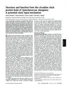
FIG. 6. A, model of CLOCK and BMAL evolution. The tree represents the known phylogenetic relationships among the species indicated at the far right. Next to each species name are symbolic representations of the CLOCK and BMAL homologs present in that animal. CLOCK and BMAL symbols at other points on the tree represent the homologs believed to be present in the ancestral animal at that place in the phylogenetic scheme. Black boxes ⫽ transactivators; circle labeled “⫹” ⫽ transactivation domain; white boxes ⫽ binding partner lacking a transactivation domain. B, model of circadian clock function in A. pernyi. There are three E-boxes (black bars) in the 5⬘-regulatory region of the apPer gene (horizontal black line). apCLK (white box labeled “CLK”) and apBMAL (black box labeled “BMAL” with transactivation domain shown as a circle labeled “⫹”) bind and activate transcription specifically from the most proximal E-box. The apPer mRNA (wavy line) translocates to the cytoplasm (above the curved line), where it is translated into apPER protein (gray boxes labeled “PER” with inhibitory domain shown as a circle labeled “⫺”). Some apPER enters the nucleus (below the curved line), where it can inhibit apCLK:apBMAL, thus shutting off its own transcription. Our inability to detect apPER in the nucleus of A. pernyi brain neurons may be due to high rates of nucleusspecific degradation and/or nuclear export (white arrows) and/or cytoplasmic sequestration of apPER.
inhibition of CLK:BMAL-driven transcription in S2 cells (3, 8) (Fig. 4). dTIM may augment the activity of dPER by facilitating dPER nuclear entry (25, 47), but this cannot be how apTIM enhances the inhibitory activity of apPER, because apPER is already nuclear in S2 cells (Fig. 5B). apTIM differs from dTIM in at least two ways. First, in S2 cells, apTIM shows little ability to inhibit and at high doses may even enhance apCLK:apBMAL-activated transcription (Fig. 4A), whereas dTIM clearly inhibits dCLK:CYC to a moderate level (3, 8, 48). Second, apTIM is nuclear in S2 cells and Sf9 cells (Table II), whereas dTIM is cytoplasmic in the same cell lines (25) (Table II). The “CLD” in apTIM is thus unable to function as a CLD in S2 or Sf9 cells. Although sequences similar to the CLD are present in most TIM proteins, such a
A Silkmoth Circadian Feedback Loop region is absent in Drosophila hydei TIM (45). Furthermore, the homology in this CLD region is very weak outside of the Drosophilidae, with only 14% identity to dTIM in A. gambiae TIM. Thus, the generality of the TIM CLD for cytoplasmic localization is unclear. PERIOD Evolution and Inhibitory Mechanism—Functional similarity of apPER to dPER was predicted based on the ability of an apPer transgene to rescue behavioral rhythms in dper null mutant flies (49). Although apPER is only two-thirds the size of dPER, its function is well conserved in our S2 cell assays. Both PERs possess a homologous C-terminal domain that inhibits CLK:BMAL-mediated transcription, and a conserved bipartite NLS that is functional in S2 cells (8) (Fig. 5). The PAS domain of apPER also resembles that of dPER (50); however, it remains unknown whether the apPER PAS domain can bind apTIM as the dPER PAS domain binds dTIM. Interestingly, the PAC of apPER, which is homologous to the CLD of dPER, does not override the NLS to produce predominantly cytoplasmic immunostaining in S2 cells (Fig. 5 and Table II) or Sf9 cells (Table II). In S2 cells, apPER robustly inhibits not only apCLK:apBMAL (Fig. 4A) but also dCLK:CYC (Supplementary Fig. S2), even though the mechanism of transcriptional activation (BCTR of apBMAL versus the glutamine-rich C terminus of dCLK) is different. The versatility of the inhibitory activity of apPER suggests that its CCID does not negate a specific transactivation domain. Instead, the C-terminal inhibitory domain of apPER may be a general transcriptional repressor domain, or it may act on the DNA binding and/or dimerization of CLK: BMAL, which is mediated by the bHLH and PAS domains and is therefore likely to be independent of the transactivation domain(s). In vitro gel-shift assays have shown that dPER binding to dCLK:CYC can inhibit DNA binding without disrupting dCLK:CYC heterodimer formation (48), suggesting that dPER can interfere with the bHLH domains of dCLK:CYC. However, the inhibitory activity of apPER is dependent on the C terminus of apCLK (Fig. 4B). Thus apPER cannot act only through the bHLH domains of apCLK:apBMAL. Toward a General Insect Circadian Clock Model—Our data in S2 cells indicate that a transcriptional feedback loop resembling that found in the circadian clock of Drosophila can be constructed from A. pernyi CLOCK, BMAL, PERIOD, and TIMELESS proteins, and the apPeriod gene (Fig. 6B). Such a feedback loop could explain the previously detected in vivo rhythms in apPer RNA and protein levels (15, 26). However, it remains unclear why apPER is always primarily cytoplasmic in A. pernyi brain neurons, when it possesses an evolutionarily ancient, classic bipartite NLS (Fig. 5B). We propose that apPER may indeed enter the nucleus, if only transiently or at low levels. The conservation of the NLS in insect PER proteins (Supplementary Fig. S1) suggests that, although PER has been detected only in the cytoplasm in most insects, PER nuclear entry may be widely true. The regulation of PER subcellular localization in Drosophila differs dramatically from that of A. pernyi and most other insects (15, 16). The tobacco hawkmoth, Manduca sexta, is also atypical in that PER nuclear immunostaining (but not PER rhythmicity) is readily detectable in brain neurons (51). However, regardless of how PER subcellular transport is controlled, a Drosophila-like feedback loop may form the core of the circadian clockwork in insects. Acknowledgments—We thank Sriram Sathyanarayanan for help generating some of the constructs. We thank Aditi V. Chavda, Kurtis N. Gray, and Kasia Macko for expert technical assistance. REFERENCES 1. Young, M. W., and Kay, S. A. (2001) Nat. Rev. Genet. 2, 702–715 2. Williams, J. A., and Sehgal, A. (2001) Annu. Rev. Physiol. 63, 729 –755
38157
3. Darlington, T. K., Wager-Smith, K., Ceriani, M. F., Staknis, D., Gekakis, N., Steeves, T. D. L., Weitz, C. J., Takahashi, J. S., and Kay, S. A. (1998) Science 280, 1599 –1603 4. Allada, R., White, N. E., So, W. V., Hall, J. C., and Rosbash, M. (1998) Cell 93, 791– 804 5. Rutila, J. E., Suri, V., Le, M., So, W. V., Rosbash, M., and Hall, J. C. (1998) Cell 93, 805–914 6. Huang, Z. J., Edery, I., and Rosbash, M. (1993) Nature 364, 259 –262 7. Ponting, C. P., and Aravind, L. (1997) Curr. Biol. 7, R674-R677 8. Chang, D. C., and Reppert, S. M. (2003) Curr. Biol. 13, 758 –762 9. Reppert, S. M., and Weaver, D. R. (2001) Annu. Rev. Physiol. 63, 647– 676 10. Lee, C., Etchegaray, J.-P., Cagampang, F. R., Loudon, A. S., and Reppert, S. M. (2001) Cell 107, 855– 867 11. Gotter, A. L., Manganaro, T., Weaver, D. R., Kolakowski, Jr., L. F., Possidente, B., Sriram, S., MacLaughlin, D. T., and Reppert, S. M. (2000) Nat. Neurosci. 3, 755–756 12. Benna, C., Scannapieco, P., Piccin, A., Sandrelli, F., Zordan, M., Rosato, E., Kyriacou, C. P., Valle, G., and Costa, R. (2000) Curr. Biol. 10, R512–R513 13. Curtin, K. D., Huang, Z. J., and Rosbash, M. (1995) Neuron 14, 365–372 14. Shafer, O., Rosbash, M., and Truman, J. W. (2002) J. Neurosci. 22, 5946 –5954 15. Sauman, I., and Reppert, S. M. (1996) Neuron 17, 889 –900 16. Za´ vodska´ , R., Sauman, I., and Sehnal, F. (2003) J. Biol. Rhythms 18, 106 –122 17. Shearman, L. P., Sriram, S., Weaver, D. R., Maywood, E. S., Chaves, I., Zheng, B., Kume, K., Lee, C. C., van der Horst, G. T., Hastings, M. H., and Reppert, S. M. (2000) Science 288, 1013–1019 18. Froy, O., Chang, D. C., and Reppert, S. M. (2002) Curr. Biol. 12, 147–152 19. King, D. P., Zhao, Y., Sangoram, A. M., Wilsbacher, L. D., Tanaka, M., Antoch, M. P., Steeves, T. D., Vitaterna, M. H., Kornhauser, J. M., Lowrey, P. L., Turek, F. W., and Takahashi, J. S. (1997) Cell 89, 641– 653 20. Hogenesch, J. B., Gu, Y.-Z., Jain, S., and Bradfield, C. A. (1998) Proc. Natl. Acad. Sci. U. S. A. 95, 5474 –5479 21. Gekakis, N., Staknis, D., Nguyen, H. B., Davis, F. C., Wilsbacher, L. D., King D. P., Takahashi, J. S., and Weitz, C. J. (1998) Science 280, 1564 –1569 22. Takahata, S., Ozaki, T., Mimura, J., Kikuchi, Y., Sogawa, K., and FujiiKuriyama, Y. (2000) Genes Cells 5, 739 –747 23. Jeon, M., Gardner, H. F., Miller, E. A., Deshler, J., and Rougvie, A. E. (1999) Science 286, 1141–1146 24. Vodovar, N., Clayton, J. D., Costa, R., Odell, M., and Kyriacou, C. P. (2002) Curr. Biol. 12, R610 –R611 25. Saez, L., and Young, M. W. (1996) Neuron 17, 979 –990 26. Gotter, A. L., Levine, J. D., and Reppert, S. M. (1999) Neuron 24, 953–965 27. Blau, J., and Young, M. W. (1999) Cell 99, 661– 671 28. Jin, X., Shearman, L. P., Weaver, D. R., Zylka, M. J., de Vries, G. J., and Reppert, S. M. (1999) Cell 96, 57– 68 29. Kume, K., Zylka, M. J., Sriram, S., Shearman, L. P., Weaver, D. R., Jin, X., Maywood, E. S., Hastings, M. H., and Reppert, S. M. (1999) Cell 98, 198 –205 30. Gu, Y.-Z., Hogenesch, J. B., and Bradfield, C. A. (2000) Annu. Rev. Pharmacol. Toxicol. 40, 519 –561 31. Go¨ rlich, D., and Kutay, U. (1999) Annu. Rev. Cell Dev. Biol. 15, 607– 660 32. Holt, R. A., Subramanian, G. M., Halpern, A., Sutton, G. G., Charlab, R., Nusskern, D. R., Wincker, P., Clark, A. G., Ribeiro, J. M., Wides, R., Salzberg, S. L., Loftus, B., Yandell, M., Majoros, W. H., Rusch, D. B., Lai, Z., Kraft, C. L., Abril, J. F., Anthouard, V., Arensburger, P., Atkinson, P. W., Baden, H., de, Berardinis, V., Baldwin, D., Benes, V., Biedler, J., Blass, C., Bolanos, R., Boscus, D., Barnstead, M., Cai, S., Center, A., Chaturverdi, K., Christophides, G. K., Chrystal, M. A., Clamp, M., Cravchik, A., Curwen, V., Dana, A., Delcher, A., Dew, I., Evans, C. A., Flanigan, M., GrundschoberFreimoser, A., Friedli, L., Gu, Z., Guan, P., Guigo, R., Hillenmeyer, M. E., Hladun, S. L., Hogan, J. R., Hong, Y. S., Hoover, J., Jaillon, O., Ke, Z., Kodira, C., Kokoza, E., Koutsos, A., Letunic, I., Levitsky, A., Liang, Y., Lin, J. J., Lobo, N. F., Lopez, J. R., Malek, J. A., McIntosh, T. C., Meister, S., Miller, J., Mobarry, C., Mongin, E., Murphy, S. D., O’Brochta, D. A., Pfannkoch, C., Qi, R., Regier, M. A., Remington, K., Shao, H., Sharakhova, M. V., Sitter, C. D., Shetty, J., Smith, T. J., Strong, R., Sun, J., Thomasova, D., Ton, L. Q., Topalis, P., Tu, Z., Unger, M. F., Walenz, B., Wang, A., Wang, J., Wang, M., Wang, X., Woodford, K. J., Wortman, J. R., Wu, M., Yao, A., Zdobnov, E. M., Zhang, H., Zhao, Q., Zhao, S., Zhu, S. C., Zhimulev, I., Coluzzi, M., della Torre, A., Roth, C. W., Louis, C., Kalush, F., Mural, R. J., Myers, E. W., Adams, M. D., Smith, H. O., Broder, S., Gardner, M. J., Fraser, C. M., Birney, E., Bork, P., Brey, P. T., Venter, J. C., Weissenbach, J., Kafatos, F. C., Collins, F. H., and Hoffman, S. L. (2002) Science 298, 129 –149 33. Markova, E. P., Ueda, H., Sakamoto, K., Oishi, K., Shimada, T., and Takeda, M. (2003) Comp. Biochem. Physiol. B 134, 535–542 34. Hogenesch, J. B., Gu, Y. Z., Moran, S. M., Shimomura, K., Radcliffe, L. A., Takahashi, J. S., and Bradfield, C. A. (2000) J. Neurosci. 20, RC83 35. Maemura, K., de la Monte, S. M., Chin, M. T., Layne, M. D., Hsieh, C. M., Yet, S. F., Perrella, M. A., and Lee, M. E. (2000) J. Biol. Chem. 275, 36847–36851 36. Cermakian, N., Whitmore, D., Foulkes, N. S., and Sassone-Corsi, P. (2000) Proc. Natl. Acad. Sci. U. S. A. 97, 4339 – 4344 37. Ishikawa, T., Hirayama, J., Kobayashi, Y., and Todo, T. (2002) Genes Cells 7, 1073–1086 38. Gaunt, M. W., and Miles, M. A. (2002) Mol. Biol. Evol. 19, 748 –761 39. The Caenorhabditis elegans Sequencing Consortium. (1998) Science 282, 2012–2018 40. International Human Genome Sequencing Consortium (2001) Nature 409, 860 –921 41. Venter, J. C., Adams, M. D., Myers, E. W., Li, P. W., Mural, R. J., Sutton, G. G., Smith, H. O., Yandell, M., Evans, C. A., Holt, R. A., Gocayne, J. D., Amanatides, P., Ballew, R. M., Huson, D. H., Wortman, J. R., Zhang, Q., Kodira, C. D., Zheng, X. H., Chen, L., Skupski, M., Subramanian, G.,
38158
A Silkmoth Circadian Feedback Loop
Thomas, P. D., Zhang, J., Gabor Miklos, G. L., Nelson, C., Broder, S., Clark, A. G., Nadeau, J., McKusick, V. A., Zinder, N., Levine, A. J., Roberts, R. J., Simon, M., Slayman, C., Hunkapiller, M., Bolanos, R., Delcher, A., Dew, I., Fasulo, D., Flanigan, M., Florea, L., Halpern, A., Hannenhalli, S., Kravitz, S., Levy, S., Mobarry, C., Reinert, K., Remington, K., Abu-Threideh, J., Beasley, E., Biddick, K., Bonazzi, V., Brandon, R., Cargill, M., Chandramouliswaran, I., Charlab, R., Chaturvedi, K., Deng, Z., Di, Francesco, V., Dunn, P., Eilbeck, K., Evangelista, C., Gabrielian, A. E., Gan, W., Ge, W., Gong, F., Gu, Z., Guan, P., Heiman, T. J., Higgins, M. E., Ji, R. R., Ke, Z., Ketchum, K. A., Lai, Z., Lei, Y., Li, Z., Li, J., Liang, Y., Lin, X., Lu, F., Merkulov, G. V., Milshina, N., Moore, H. M., Naik, A. K., Narayan, V. A., Neelam, B., Nusskern, D., Rusch, D. B., Salzberg, S., Shao, W., Shue, B., Sun, J., Wang, Z., Wang, A., Wang, X., Wang, J., Wei, M., Wides, R., Xiao, C., Yan, C., Yao, A., Ye, J., Zhan, M., Zhang, W., Zhang, H., Zhao, Q., Zheng, L., Zhong, F., Zhong, W., Zhu, S., Zhao, S., Gilbert, D., Baumhueter, S., Spier, G., Carter, C., Cravchik, A., Woodage, T., Ali, F., An, H., Awe, A., Baldwin, D., Baden, H., Barnstead, M., Barrow, I., Beeson, K., Busam, D., Carver, A., Center, A., Cheng, M. L., Curry, L., Danaher, S., Davenport, L., Desilets, R., Dietz, S., Dodson, K., Doup, L., Ferriera, S., Garg, N., Gluecksmann, A., Hart, B., Haynes, J., Haynes, C., Heiner, C., Hladun, S., Hostin, D., Houck, J., Howland, T., Ibegwam, C., Johnson, J., Kalush, F., Kline, L., Koduru, S., Love, A., Mann, F., May, D., McCawley, S., McIntosh, T., McMullen, I., Moy, M., Moy, L., Murphy, B., Nelson, K., Pfannkoch, C., Pratts, E., Puri, V., Qureshi, H., Reardon, M., Rodriguez, R., Rogers, Y. H., Romblad, D., Ruhfel, B., Scott, R., Sitter, C., Smallwood, M., Stewart, E., Strong, R., Suh, E., Thomas, R., Tint, N. N., Tse, S., Vech, C., Wang, G., Wetter, J., Williams, S., Williams, M., Windsor, S., Winn-Deen, E., Wolfe, K., Zaveri, J., Zaveri, K., Abril, J. F., Guigo, R., Campbell, M. J., Sjolander, K. V., Karlak, B., Kejariwal, A., Mi, H., Lazareva, B., Hatton, T., Narechania, A., Diemer, K., Muruganujan, A., Guo, N., Sato, S., Bafna, V., Istrail, S., Lippert, R., Schwartz, R., Walenz, B., Yooseph, S., Allen, D., Basu, A., Baxendale, J., Blick, L., Caminha, M., Carnes-Stine, J., Caulk, P., Chiang, Y. H., Coyne, M., Dahlke, C., Mays, A., Dombroski, M., Donnelly, M., Ely, D., Esparham, S., Fosler, C., Gire, H., Glanowski, S., Glasser, K., Glodek, A., Gorokhov, M., Graham, K., Gropman, B., Harris, M., Heil, J., Henderson, S., Hoover, J., Jennings, D., Jordan, C., Jordan, J., Kasha, J., Kagan, L., Kraft, C., Levitsky, A., Lewis, M., Liu, X., Lopez, J., Ma, D., Majoros, W., McDaniel, J., Murphy, S., Newman, M., Nguyen, T., Nguyen, N., Nodell, M., Pan, S., Peck, J., Peterson, M., Rowe, W., Sanders, R., Scott, J., Simpson, M., Smith, T., Sprague, A., Stockwell, T., Turner, R., Venter,
42.
43.
44. 45. 46. 47. 48. 49. 50. 51.
E., Wang, M., Wen, M., Wu, D., Wu, M., Xia, A., Zandieh, A., and Zhu, X. (2001) Science 291, 1304 –1351 Aparicio, S., Chapman, J., Stupka, E., Putnam, N., Chia, J. M., Dehal, P., Christoffels, A., Rash, S., Hoon, S., Smit, A., Gelpke, M. D., Roach, J., Oh, T., Ho, I. Y., Wong, M., Detter, C., Verhoef, F., Predki, P., Tay, A., Lucas, S., Richardson, P., Smith, S. F., Clark, M. S., Edwards, Y. J., Doggett, N., Zharkikh, A., Tavtigian, S. V., Pruss, D., Barnstead, M., Evans, C., Baden, H., Powell, J., Glusman, G., Rowen, L., Hood, L., Tan, Y. H., Elgar, G., Hawkins, T., Venkatesh, B., Rokhsar, D., and Brenner, S. (2002) Science 297, 1301–1310 Dehal, P., Satou, Y., Campbell, R. K., Chapman, J., Degnan, B., De Tomaso, A., Davidson, B., Di Gregorio, A., Gelpke, M., Goodstein, D. M., Harafuji, N., Hastings, K. E., Ho, I., Hotta, K., Huang, W., Kawashima, T., Lemaire, P., Martinez, D., Meinertzhagen, I. A., Necula, S., Nonaka, M., Putnam, N., Rash, S., Saiga, H., Satake, M., Terry, A., Yamada, L., Wang, H. G., Awazu, S., Azumi, K., Boore, J., Branno, M., Chin-Bow, S., DeSantis, R., Doyle, S., Francino, P., Keys, D. N., Haga, S., Hayashi, H., Hino, K., Imai, K. S., Inaba, K., Kano, S., Kobayashi, K., Kobayashi, M., Lee, B. I., Makabe, K. W., Manohar, C., Matassi, G., Medina, M., Mochizuki, Y., Mount, S., Morishita, T., Miura, S., Nakayama, A., Nishizaka, S., Nomoto, H., Ohta, F., Oishi, K., Rigoutsos, I., Sano, M., Sasaki, A., Sasakura, Y., Shoguchi, E., Shin-i, T., Spagnuolo, A., Stainier, D., Suzuki, M. M., Tassy, O., Takatori, N., Tokuoka, M., Yagi, K., Yoshizaki, F., Wada, S., Zhang, C., Hyatt, P. D., Larimer, F., Detter, C., Doggett, N., Glavina, T., Hawkins, T., Richardson, P., Lucas, S., Kohara, Y., Levine, M., Satoh, N., and Rokhsar, D. S. (2002) Science 298, 2157–2167 Myers, M. P., Wager-Smith, K., Wesley, C. S., Young, M. W., and Sehgal, A. (1995) Science 270, 805– 808 Ousley, A., Zafarullah, K., Chen, Y., Emerson, M., Hickman, L., and Sehgal, A. (1998) Genetics 148, 815– 825 Goto, S. G., and Denlinger, D. L. (2003) J. Insect Physiol. 48, 803– 816 Vosshall, L. B., Price, J. L., Sehgal, A., Saez, L., and Young, M. W. (1994) Science 263, 1606 –1609 Lee, C., Bae, K., and Edery, I. (1999) Mol. Cell. Biol. 19, 5316 –5325 Levine, J. D., Sauman, I., Imbalzano, M., Reppert, S. M., and Jackson, F. R. (1995) Neuron 15, 147–157 Reppert, S. M., Tsai, T., Roca, A. L., and Sauman, I. (1994) Neuron 13, 1167–1176 Wise, S., Davis, N. T., Tyndale, E., Noveral, J., Folwell, M. G., Bedian, V., Emery, I. F., and Siwicki, K. K. (2002) J. Comp. Neurol. 447, 366 –380