Jun 26, 1991 - ERIKA MARSILIO,1 SENG H. CHENG,2 BRIAN SCHAFFHAUSEN,3 EVA PAUCHA,1t. AND DAVID ..... Clayton, C. E., D. Murphy, M. Lovett, and P. W. J. Rigby. 1983. .... J. W. Ludlow, A. K. Arthur, E. Fanning, I. Bikel, and D. M..
JOURNAL OF VIROLOGY, Oct. 1991, p. 5647-5652 0022-538X/91/105647-06$02.00/0 Copyright © 1991, American Society for Microbiology
Vol. 65, No. 10
The T/t Common Region of Simian Virus 40 Large T Antigen Contains a Distinct Transformation-Governing Sequence ERIKA MARSILIO,1 SENG H. CHENG,2 BRIAN SCHAFFHAUSEN,3 EVA PAUCHA,1t AND DAVID M. LIVINGSTON'* Dana-Farber Cancer Institute and Departments of Medicine and Pathology, Harvard Medical School, Boston, Massachusetts 021151; Laboratory of Cellular Regulation, Genzyme Corporation, Framingham, Massachusetts 017012; and Department of Biochemistry, Health Science Campus, Tufts University, Boston, Massachusetts 02J 13 Received 12 December 1990/Accepted 26 June 1991
Simian virus 40 large T antigen (T)
can
transform cultured cells, but the mechanisms by which it functions
are not entirely understood. Several lines of evidence have suggested that the amino-terminal 130 residues of T may be sufficient to confer the transforming capability. Oligonucleotide-directed mutagenesis was used to generate a series of deletion and substitution mutants within the amino-terminal 82 residues of T, the segment
which is shared with simian virus 40 small t antigen (t). Results of stability and transformation assays of these mutants strongly suggest that the 1-to-82 region of T contains sequences which govern T transforming activity and affect in vivo stability. Instability and a defect in transforming activity could be separated from one another genetically. Thus, the 1-to-82 region appears to contain a specific region that contributes to the transforming function of the protein. This segment operates by means other than the simple binding of pRb and/or p107.
Simian virus 40 large T antigen (T) has been shown by many laboratories to elicit the neoplastic transformation of cultured rodent cells (45). However, although there is new information showing that its transforming function likely depends on its ability to modulate the activities of at least two tumor suppressor gene products, pRb and p53, and a third protein, p107, the detailed mechanism by which T transforms is still largely unknown (7, 8, 9, 25, 46, 47). What seems clear is that T must perform more than one biochemical act to elicit the appearance of neoplastic properties. In this regard, it appears that at least some T transforming functions are the products of blocks of sequences located in the amino-terminal half of the molecule (1, 3, 4, 5, 37, 38). Multiple reports indicate that various amino-terminal T fragments extending from residues 1 to 127 to 300 can establish early-passage rat cells in culture, although at frequencies lower than those of wild type (1-5, 37, 38). Moreover, these truncated species can induce certain primary strains and established cell lines to manifest anchorageindependent growth (3, 5, 37, 38). Therefore, the aminoterminal third of the protein can perform biochemical functions, which, taken together, are sufficient for the transformation of some cells. Other functions which have been shown genetically to depend on sequences within the amino-terminal region of T are binding to the viral replication origin (32) and nuclear localization (19, 20). However, the abilities to bind the origin and to localize in the nucleus are dispensable for transformation (3, 12, 18, 21, 26, 32, 34). Another activity associated with the amino-terminal half of T is the ability to stimulate DNA synthesis in quiescent cells (14, 31, 39). The role of this activity in the T-dependent transformation mechanism remains to be fully determined, although, in the case of two other DNA tumor virus species, adenoviruses and bovine papillomavirus, transformation function cannot be separated from the ability to initiate DNA synthesis in quiescent cells (24, 29, 30, 48).
Recent reports indicate that mutations within the C-terminal -60% of large T, which disrupt the p53 binding region, render the molecule incapable of transforming REF52 but not C3H1Ot1/2 cells (33, 40). The former is a continuous line of rat cells, and the latter is a continuous line of murine cells. Some of the aforementioned mutations also suppressed the immortalization of primary C57BL/6 mouse cells by T (43, 44). Therefore, although T-p53 complex formation seems not to be involved in the transformation of C3H1Ot1/2, it does appear to be required for the transformation of REF52 cells (40). The formation of T-p53 complexes may also play a significant role in supporting the immortalizing function of T in certain primary murine cells (33, 43). Given the additional fact that the amino-terminal -130 residues of T are capable of immortalizing other rodent cell species (1), the protein may be able to exert its immortalizing function by at least two independent biochemical routes, one of which involves p53 binding and the other which does not. Moreover, there may be significant host range limitations on the expression of these functions. A large number of amino-terminal T mutants which are either partly or fully defective for transforming activity have been identified (6, 12, 21, 34, 35, 37, 38, 42). For example, Kalderon and Smith (21) have shown that numerous mutants with lesions involving the sequence extending from residues 105 to 115 are transformation defective. Recently, it was shown that these mutants fail to transform cells to anchorage-independent growth (3). Taken together, the results suggest that the 105-to-115 segment constitutes a functional domain devoted to T transforming function. This region bears strong similarity in both primary sequence and predicted secondary structure to a transforming domain of the adenovirus ElA product (10, 41). In adenovirus ElA, this region has been implicated in the stimulation of host cell DNA synthesis and the maintenance of viral transforming activity (24, 30, 36, 48, 50, 51). This segment is now known to operate in both T and ElA by serving as a binding site for at least two cell-encoded proteins, pRb and p107 (7-9, 46, 47). The former is a known tumor suppressor gene product (22, 23). Although there is presently no direct evidence to
Corresponding author. t Deceased. *
5647
5648
J. VIROL.
NOTES
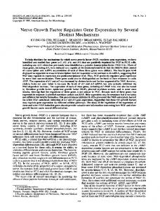
A
D2C2 a
PvB-Q 55-70 59-64 46-51 40-45 34-3 A B
A B
A B
AB A B
d l 135 A B
A B
.-
180
116 T Ag
_ 84
__^-_o
_.
N
58 48
36.5 Or i.-WIM
,:::::
'-willoo'111",wlO'lItqHW*$ftw--
-!.
7is!
,,illlllllf
;.
B EMdI59-64 0
2
4
8
11 23
EMsb59-64
pBR328
Pvu-O
0 2 4 8 11 23 0 2 4 8 11 23
0
2 4 8 11 23
_T 1debes
FIG. 1. Western blot and pulse-chase analysis of amino-terminal deletion mutants of T. (A) Western blot analysis of T deletion mutants. All cells were grown at 37°C in a humidified, 10% C02-containing atmosphere in Dulbecco's modified Eagle's medium (GIBCO) supplemented with 10% fetal calf serum (Flow Laboratories). Dishes (100 mm) of nearly confluent CV-1P cells were transfected in duplicate (lanes labelled A and B) with 20 p.g of plasmid DNA by the calcium phosphate precipitation method, and cell lysates were prepared 24 h later. Equal amounts of total protein extract (800 ,ug) were immunoprecipitated with pAB423 (7). The immune complexes were separated by sodium dodecyl sulfate gel electrophoresis, transferred to nitrocellulose, and assayed in a Western blot by using pAB423 and anti-mouse immunoglobulin G conjugated to alkaline phosphatase. The lane marked D2C2 contains a pAB423 immunoprecipitate of a clonal CV-1P cell line which constitutively synthesizes T. pBR328 DNA was used as a negative control, while pPVU-O, a plasmid which encodes wild-type T, was used as a positive control. The designations 65-70, 59-64, 46-51, 40-45, and 34-39 represent the newly generated internal deletion mutants described in the text, and the numbers represent the specific residues which have been removed in each case. d11135 is a T mutant (generously donated by J. Pipas) in which residues 17 to 27 have been deleted. The new deletion mutants described were generated by using the gapped heteroduplex method essentially as described by Kalderon et al. (19) and Harvey et al. (16). The synthetic oligonucleotides used to generate the variant T species were each 30 nucleotides long. Following annealing of the mutagenic oligonucleotides to the gapped heteroduplexes, Escherichia coli DH5a was transformed by each DNA heteroduplex. Colonies bearing the mutant DNAs were first identified by colony hybridization and then verified by DNA sequencing. (B) Pulse-chase analysis of deletion and substitution mutants of T. Dishes (60 mm)
VOL. 65,
1991
support the suspicion, the latter is suspected of having analogous properties. Moreover, genetic analysis strongly suggests that T and ElA binding to pRb and/or p107 contributes to their transforming properties (7, 8, 46, 47). T and the other simian virus 40 early region product, small t antigen (t), share an 82-residue amino-terminal segment. This region lies upstream of the pRb-p107 binding segment and has not been extensively studied for potential functions. In support of the possibility that this region might contribute to T transforming function, we recently found that its complete elimination from T was linked to the failure of T to induce anchorage-independent growth (28). Given that the 1-to-82 sequence represents more than 50% of the mass of the smallest truncated T species with overt 'rI.nsforming activity, we chose to evaluate it further as a potential contributor to the transforming function of the protein. The initial strategy was to generate a set of T mutants containing serial, colinear deletions of six amino acids located within the 1-to-82 region. Specifically, the deletions extended from residues 34 to 70. Mutagenesis was performed by a specific oligonucleotide-directed method, with an intact simian virus 40 early region-containing plasmid, pPVU-O, as template. pPVU-O contains the entire early region of simian virus 40 and the viral early promoter cloned in pBR328 (21). To check for expression of these various mutants and of another deletion mutant, d11135 from the laboratory of J. Pipas (34), which lacks residues 17 to 27, an equivalent amount of each mutant DNA and the wild-type plasmid was transfected into CV-1P cells. The transfections were carried out in duplicate. The resulting viral proteins produced in these transient assays were immunoprecipitated with pAB423, a monoclonal antibody which recognizes an epitope in the carboxy terminus of T (15). The various mutant T species were analyzed by Western immunoblotting (7). All immunoprecipitations were performed in antibody excess with a standard quantity of cell extract. The results are shown in Fig. 1A. In duplicate sets of transfections (labelled A and B), each mutant plasmid gave rise to the synthesis of an immunoreactive T species which comigrated with the wild-type protein. Similar results were obtained following transfection of BALB/c/3T3 cI A-31, Rat-1, or CREF cells (data not shown). Given that cells transfected with all but the d165-70 mutant yielded significantly less T than the wild-type control, pulse-chase labelling analysis of these various proteins was carried out over a period of 24 h in an effort to assess the stability of each of the mutant proteins. All the mutants, except for d165-70, were characterized by less than normal half-lives. Only the results of the pulse-chase analysis of one deletion mutant are shown here as an example (Fig. 1B). As shown in the left panel of Fig. 1B, the half-life of the deletion mutant d159-64 ranged from 2
h, compared with more than 12 h for the wild-type counterpart. Thus, the abnormally low steady-state levels of mutant T present in these extracts are due, at least in part, to rapid protein turnover. The instability notwithstanding, each of these unstable mutants, as shown earlier, retained the ability to bind pRb, p107, and p53 readily (9), suggesting no
to 4
NOTES
5649
TABLE 1. Focus-forming activity of various T deletion mutants' Mutant
Pvu-O d11135 EMdl34-39 EMdl40-45 EMdl46-51 EMdl59-64 EMdl65-70
% FF CREFb
100
0 0 2.4 0.3 0 19.3
% FF REFc
100
0 0.1 5.1 5.6 0 25.7
% Neoc
DNA
replication
100
+
ND 0 5.9 9.6 0 42.8
+
a Sparse 60-mm dishes of either REF (Whittaker, M.A. Bioproducts) or CREF cells (11) were transfected with 10 pLg of plasmid DNA. Cells were maintained in culture for a period of 3 to 4 weeks, after which time they were fixed and the foci were stained with anti-T pAB423 and goat anti-mouse immunoglobulin G conjugated to alkaline phosphatase (Promega). Values are shown as percentages of wild-type activity. Neo, percent Neo-resistant colonies obtained after transfection of REF cells with equal amounts of wild-type or mutant plasmid DNA (10 p.g) and plasmid DNA containing a Neo-resistant gene (10 ,ug). FF, focus formation on the indicated cell lines. Cells were subsequently maintained in culture and fed with G418 (400 p.g/ml) for an equal amount of time. The replication assay was performed with CV-1P cells (33). Typically, 1 to 2 ,ug of DNA was transfected onto a 60% confluent dish. Three to four days following transfection, the DNA was isolated by the Hirt method (17), digested with DpnI, separated by electrophoresis in agarose gels, and then blotted onto nitrocellulose (34). Random-primed, 32P-labelled wild-type plasmid DNA was used as a probe (Boehringer Mannheim randomprimed DNA labelling kit was used essentially as described by the manufacturer). ND, not determined. b Values represent averages for 12 experiments. c Values represent averages for six experiments.
major defect in these functions. Therefore, they do not appear to be wholly inert. Given that all mutant proteins could bind pRb, p107, and p53, we asked whether any of the plasmids encoding them could transform an established rat cell line, CREF (11), as measured in a focus formation assay. The results of these focus formation assays are summarized in Table 1 and are expressed as percentages of the wild-type effects. In these experiments, all mutants, with the exception of d165-70, failed to transform CREF cells to any degree. Similarly, they failed to transform primary rat embryo fibroblasts (REF). In this case, the identity of a given focus as an authentic T transformant was checked by in situ immunoperoxidase staining with pAB423. Only those foci which tested positive by staining in this assay were scored here. An activated ras allele has been shown to partially complement transformation-defective T mutants (27). In view of this, we also cotransfected an activated ras-containing plasmid, PM1 (generously donated by Michael Corbly), along with the different mutant DNAs into REF and CREF cells. The activated ras, however, failed to rescue our deletion mutants in this transformation assay (data not shown). Similarly, when each of the mutant plasmids was cotransfected with a plasmid containing a G418 resistance gene into REF in search of T-containing immortal colonies, none of the mutants functioned normally in comparison with the wild type (Table 1). However, as in the previously described
of CV-1P cells were transfected with 10 ,ug of DNA. Twenty-four hours later the cells were labelled for 3 h with 200 ,uCi of [35S]methionine per ml in serum-free medium. They were then refed with complete medium lacking radioactivity. Cell extracts were prepared and immunoprecipitated with the monoclonal antibody pAB423 (7) at the indicated times. The designations 0 to 23 indicate the time in hours after removal of radioactivity, when the relevant cell lysates were prepared. The origin of the T doublet in all of the relevant lanes is unclear, although, given the known susceptibility of T to proteolytic degradation after extraction, there is the suspicion that the lower band may be a degradation product of the upper band.
5650
J. VIROL.
NOTES
TABLE 2. Focus formation activity of substitution and deletion mutants'
180
Mutant Mutant
Pvu-O 0. Al'".'f
T-
ANkAWL
-AL .401,1 1.k.
-
116
-
84
-
58
-
48
-
36.5
FIG. 2. Immunoprecipitation of the mutants EMdl59-64 and EMsb59-64. Cells were maintained in culture as described in the legend to Fig. 1. CV-1P cells were transfected with 20 ,ug of plasmid DNA. Twenty-four hours posttransfection, the cells were labelled for 3 h with 200 ,uCi of [35S]methionine per ml in serum-free medium. Cell extracts were prepared and immunoprecipitated with the monoclonal antibody pAB423 (7). Lanes 1 and 2 contain two independent immunoprecipitates obtained from cells transfected with the substitution mutant EMsb59-64; lane 3 contains the immunoprecipitates obtained from cells transfected with EMdl59-64; lane 4 contains the immunoproducts from cells transfected with pPVU-O (encodes wild-type T); and lane 5 contains the immunoproducts from cells which have been mock transfected. Arrows (from top to bottom) indicate the respective migration positions of p107, pRb, and p53.
assays, d165-70 functioned better than the rest of the mutants tested. Finally, we also investigated whether the mutant T
species retained DNA replicating activity by assaying the mutant genomes for their ability to replicate in CV-1P cells. With the exception of d165-70, all the mutants were judged to be defective in the ability to replicate DNA as measured in a blotting assay after DNA transfection. This failure could be a result of insufficient functional T, the short half-life of the relevant mutant proteins, or a defect in their intrinsic DNA replication function. To determine whether the inability of these deletion mutants to transform cells was due to the short half-life of the protein within the cell and not to the disruption of a potential transforming domain, we sought to engineer a mutant within the 1-to-82 region which was as stable as wild-type T. The sequence NAAIRS has been found to assume different secondary structural motifs in different proteins (49). As a result, others suggested that this sequence is inherently flexible and that this characteristic allows it the freedom to assume different conformations in different environments; hence, it could be used in mutagenesis experiments in which the aim is to minimize conformational disruption following gross changes in primary protein structure (17a, 49). Since deleting sequences between residues 17 and 64 led to clear instability, we suspected that loss of sequence in this region
EMdl59-64 EMsb59-64
~~~Expt
1 (%(% FFFF CREF) CREF)
100 0
10.8
Expt Ep 2
% FF REF 100
0 10.4
% Neo 100
0 13
a Two separate experiments measuring the focus formation activity of the substitution mutant, EMsb59-64, and the deletion mutant, EMdl59-64, were performed. Each was performed under the same conditions described for the experiment whose results are shown in Table 1. In experiment 1, CREF cells were studied. In experiment 2, an analogous experiment was performed with primary REF. All cells were maintained under the same conditions described in the legend to Fig. 1. Values represent averages for three experiments. For abbreviations, see Table 1, footnote a.
was responsible for this defect. To test this hypothesis, an oligonucleotide encoding the NAAIRS sequence was inserted in frame at the site of the dl59-64 mutation. The resulting substitution mutant protein, sb59-64, comigrated with both wild-type and d159-64 T, but, unlike the latter, which was the least stable of all the mutants tested, this product exhibited nearly wild-type stability in pulse-chase analysis (Fig. 1B) and accumulated to wild-type levels in a transient expression assay (Fig. 2), unlike its progenitor, dl59-64. In this experiment, we suspect that the latter was present in an amount below that needed to detect coprecipitating proteins. Indeed, in the past it was readily shown to bind to pRb, p107, and p53 (9). Furthermore, the sb59-64 protein retained the ability to bind pRb and p107 and bound p53 like wild-type T (Fig. 2). This notwithstanding, like the d159-64 parental mutant, it was clearly defective in focusforming activity when assayed on CREF cells and REF cells, as shown in Table 2. Therefore, the introduction of NAAIRS at the site of the former deletion restored wild-type stability but failed to restore wild-type transforming activity. Indeed, the product is clearly still defective in both focus-forming and REF colony-forming activity. From these findings, one can argue that elements within the 1-to-82 sequence affect two aspects of T behavior, stability and transforming activity. Indeed, from the data presented here, these two functions appear to be separable. Given these and prior observations (9), it seems reasonable to suggest that the 1-to-82 region may constitute a transformation-controlling segment, at least in part, distinct from that which gives rise to pRb, p107, and p53 binding activity (9). Conceivably, it plays a role in the T-pRb and/or T-p107 interaction, not so much by affecting the affinity of either protein for T but rather by altering the functional effect(s) of T binding on one or more of these elements. Another possibility, among several, is suggested by the existence of some elements of sequence homology within this region and transforming domain 1 of ElA. Harlow and coworkers have shown that elements of this transformation-controlling region of ElA are responsible for the p300 binding activity of ElA (47, 48). From the genetics of the ElA-p300 binding reaction, one can argue that p300 binding contributes to ElA transforming activity. Although there is no direct supporting evidence at the moment, it remains possible that this region of T also binds p300 or an analogous protein which recognizes the segment in T homologous to that in ElA. Experiments aimed at testing this possibility are in progress.
VOL. 65,
NOTES
1991
We are grateful to Margaret Bradley, Beth Weiner, and John Ludlow for helpful suggestions; Janet Merrill for synthesis of the oligonucleotides; and Ann Desai for expert help in preparing the manuscript. The work was supported by grants from the National Cancer Institute to D.M.L., S.H.C., and E.P. E.M. was supported by a research fellowship from the National Cancer Institute.
REFERENCES 1. Asselin, C., and M. Bastin. 1985. Sequences from polyomavirus and simian virus 40 large T genes capable of immortalizing primary rat embryo fibroblasts. J. Virol. 56:958-968. 2. Chang, L. S., S. Pan, M. M. Pater, and G. Di Mayorca. 1985. Differential requirement for SV40 early genes in immortalization and transformation of primary rat and human embryonic cells. Virology 146:246-261. 3. Chen, S., and E. Paucha. 1990. Identification of a region of simian virus 40 large T antigen required for cell transformation. J. Virol. 64:3350-3357. 4. Clayton, C. E., D. Murphy, M. Lovett, and P. W. J. Rigby. 1983. A fragment of the SV40 large T antigen gene transforms. Nature (London) 299:59-61. 5. Colby, W. W., and T. Shenk. 1982. Fragments of the simian virus 40 transforming gene facilitate transformation of rat embryo cells. Proc. Natl. Acad. Sci. USA 79:5189-5193. 6. Cole, C. N., J. Tornow, R. Clark, and R. Tjian. 1986. Properties of the simian virus 40 (SV40) large T antigens encoded by SV40 mutants with deletions in gene A. J. Virol. 57:539-546. 7. DeCaprio, J. A., J. W. Ludlow, J. Figge, J.-Y. Shew, C.-M. Huang, W.-H. Lee, E. Marsilio, E. Paucha, and D. M. Livingston. 1988. SV40 large tumor antigen forms a complex with the product of the retinoblastoma susceptibility gene. Cell 54:275283. 8. Dyson, N., K. Buchkovich, P. Whyte, and E. Harlow. 1989. The cellular 107K protein that binds to adenovirus ElA also associates with the large T antigens of SV40 and JC virus. Cell 58:249-255. 9. Ewen, M. E., J. W. Ludlow, E. Marsilio, J. A. DeCaprio, R. C. Millikan, S.-H. Cheng, E. Paucha, and D. M. Livingston. 1989. An N-terminal transformation-governing sequence of SV40 large T antigen contributes to the binding of both pllO Rb and a second cellular protein, p120. Cell 58:257-267. 10. Figge, J., T. Webster, T. F. Smith, and E. Paucha. 1988. Prediction of similar transforming regions in simian virus 40 large T, adenovirus ElA, and myc oncoproteins. J. Virol.
62:1814-1818.
11. Fischer, P. B., L. E. Babiss, B. I. Weinstein, and H. S. Ginsberg. 1982. Analysis of type 5 adenovirus transformation with a cloned rat embryo cell line (CREF). Proc. Natl. Acad. Sci. USA
79:3527-3531. 12. Fischer-Fantuzzi, L., and C. Vesco. 1985. Deletion of 43 amino acids in the NH2-terminal half of the large tumor antigen of simian virus 40 results in a non-karyotic protein capable of transforming established cells. Proc. Natl. Acad. Sci. USA 82:1891-1895. 13. Gluzman, Y., and B. Ahrens. 1982. SV40 early mutants that are defective for viral DNA synthesis but competent for transformation of cultured rat and simian cells. Virology 123:78-92. 14. Graessmann, M., and A. Graessmann. 1976. Early SV40 specific RNA contains information for T-antigen formation and chromatin replication. Proc. Natl. Acad. Sci. USA 73:366-370. 15. Harlow, E., L. V. Crawford, D. C. Pim, and N. M. Williamson. 1981. Monoclonal antibodies specific for simian virus 40 tumor antigens. J. Virol. 39:861-869. 16. Harvey, R., K. M. Hehir, A. E. Smith, and S. H. Cheng. 1989. pp60c-src variants containing lesions that affect phosphorylation at tyrosines 416 and 527. Mol. Cell. Biol. 9:3647-3659. 17. Hirt, B. 1967. Selective extraction of polyoma DNA from infected mouse cell cultures. J. Mol. Biol. 26:365-369. 17a.Holman, P., and B. Schaffhausen. Unpublished data. 18. Jat, P. S., and P. A. Sharp. 1986. Large T antigens of simian virus 40 and polyomavirus efficiently establish primary fibroblasts. J. Virol. 59:746-750.
5651
19. Kalderon, D., W. D. Richardson, A. F. Markham, and A. E. Smith. 1984. Sequence requirements for nuclear location of simian virus 40 large T antigen. Nature (London) 311:33-38. 20. Kalderon, D., B. L. Roberts, W. D. Richardson, and A. E. Smith. 1984. A short amino acid sequence able to specify nuclear location. Cell 39:499-509. 21. Kalderon, D., and A. E. Smith. 1984. In vitro mutagenesis of a putative DNA binding domain of SV40 large T. Virology 139: 109-137. 22. Klein, G. 1987. The approaching era of the tumor suppressor gene. Science 238:1539-1545. 23. Knudson, A. G., Jr. 1971. Mutation and cancer: statistical study of retinoblastoma. Proc. Natl. Acad. Sci. USA 68:820-823. 24. Lillie, J. W., P. M. Loewenstein, M. R. Green, and M. Green. 1987. Functional domains of adenovirus type 5 Ela proteins. Cell 50:1091-1100. 25. Livingston, D. M., and M. K. Bradley. 1987. The simian virus 40 large T antigen: a lot packed into a little. Mol. Biol. Med.
4:63-80.
26. Manos, M. M., and Y. Gluzman. 1985. Genetic and biochemical analysis of transformation-competent, replication-defective simian virus 40 large T antigen mutants. J. Virol. 53:120-127. 27. Michalovitz, D., L. Fischer-Fantuzzi, C. Vesco, J. M. Pipas, and M. Oren. 1987. Activated Ha-ras can cooperate with defective simian virus 40 in the transformation of nonestablished rat embryo fibroblasts. J. Virol. 61:2648-2654. 28. Montano, X., R. C. Millikan, J. M. Milhaven, D. A. Newsome, J. W. Ludlow, A. K. Arthur, E. Fanning, I. Bikel, and D. M. Livingston. 1990. SV40 small t and an amino terminal domain of large T share a common transforming function. Proc. Natl. Acad. Sci. USA 87:7448-7452. 29. Moran, E., and M. B. Mathews. 1987. Multiple functional domains in the adenovirus Ela gene. Cell 48:177-178. 30. Moran, E., B. Zerler, T. M. Harrison, and M. B. Mathews. 1986. Identification of separate domains in the adenovirus ElA gene for immortalization activity and the activation of virus early genes. Mol. Cell. Biol. 6:3470-3480. 31. Mueller, C., A. Graessmann, and M. Graessmann. 1978. Mapping of early SV40-specific functions by microinjection of different early viral DNA fragments. Cell 15:579-585. 32. Paucha, E., D. Kalderon, R. W. Harvey, and A. E. Smith. 1986. Simian virus 40 origin DNA-binding domain on large T antigen. J. Virol. 57:50-64. 33. Peden, K. W. C., A. Srinivasan, J. M. Farber, and J. M. Pipas. 1989. Mutants with changes within or near a hydrophobic region of simian virus 40 large tumor antigen are defective for binding cellular protein p53. Virology 168:13-21. 34. Pipas, J. M., K. W. C. Peden, and D. Nathans. 1983. Mutational analysis of simian virus 40 T antigen: isolation and characterization of mutants with deletions in the T-antigen gene. Mol. Cell. Biol. 3:203-213. 35. Rutila, J. E., M. J. Imperiale, and W. W. Brockman. 1986. Replication and transformation functions of in vitro-generated simian virus 40 large T antigen mutants. J. Virol. 58:526-535. 36. Schneider, J. F., F. Fischer, C. R. Goding, and N. C. Jones. 1987. Mutational analysis of the adenovirus Ela gene: the role of transcriptional regulation in transformation. EMBO J. 6:2053-2060. 37. Sompayrac, L. M., and K. J. Danna. 1982. Isolation and characterization of simian virus 40 early region deletion mutants. J. Virol. 43:328-331. 38. Sompayrac, L. M., and K. J. Danna. 1985. The simian virus 40 sequences between 0.169 and 0.423 map units are not essential to immortalize early-passage rat embryo cells. Mol. Cell. Biol. 5:1191-1194. 39. Soprano, K. J., N. Galanti, G. J. Jonak, S. McKercher, J. M. Pipas, K. W. C. Peden, and R. Baserga. 1983. Mutational analysis of simian virus 40 T antigen: stimulation of cellular DNA synthesis and activation of rRNA genes by mutants with deletions in the T-antigen gene. Mol. Cell. Biol. 3:214-219. 40. Srinivasan, A., K. W. C. Peden, and J. M. Pipas. 1989. The large tumor antigen of simian virus 40 encodes at least two distinct transforming functions. J. Virol. 63:5459-5463.
5652
NOTES
41. Stabel, S., P. Argos, and L. Phillipson. 1985. The release of growth arrest by microinjection of adenovirus Ela DNA. EMBO J. 4:2329-2336. 42. Sugano, S., and N. Yamaguchi. 1984. Two classes of transformation-deficient, immortalization-positive simian virus 40 mutants constructed by making three-base insertions in the T antigen gene. J. Virol. 52:884-891. 43. Tevethia, M. J., J. M. Pipas, T. Kierstead, and C. Cole. 1988. Requirements for immortalization of primary mouse embryo fibroblasts probed with mutants bearing deletions in the 3' end of SV40 gene A. Virology 162:76-89. 44. Thompson, D. L., D. Kalderon, A. E. Smith, and M. J. Tevethia. 1990. Dissociation of Rb-binding and anchorage-independent growth from immortalization and tumorigenicity using SV40 mutants producing N-terminally truncated large T antigens. Virology 178:15-34. 45. Tooze, J. (ed.). 1980. DNA tumor viruses, 2nd ed., part II. Cold Spring Harbor Laboratory, Cold Spring Harbor, N.Y. 46. White, P., K. J. Buchkovich, J. M. Horowitz, S. H. Friend, M. Raybuck, R. A. Weinberg, and E. Harlow. 1988. Association between an oncogene and an anti-oncogene: the adenovirus Ela
J. VIROL.
47.
48. 49.
50.
51.
proteins bind to the retinoblastoma gene product. Nature (London) 334:124-129. White, P., N. M. Williamson, and E. Harlow. 1989. Cellular targets for transformation by the adenovirus Ela proteins. Cell 56:67-75. Whyte, P., H. E. Ruley, and E. Harlow. 1988. Two regions of the adenovirus early region 1A proteins are required for transformation. J. Virol. 62:257-265. Wilson, I. A., D. H. Haft, E. D. Getzoff, J. A. Tainer, R. A. Lerner, and S. Brenner. 1985. Identical short peptide sequences in unrelated proteins can have different conformations: a testing ground for theories of immune recognition. Proc. Natl. Acad. Sci. USA 82:5255-5259. Zerler, B., E. Moran, K. Maruyama, J. Moomaw, T. Grodzicker, and H. E. Ruley. 1986. Adenovirus ElA coding sequences that enable ras and pmt oncogenes to transform cultured primary cells. Mol. Cell. Biol. 6:887-899. Zerler, B., R. J. Roberts, M. B. Mathews, and E. Moran. 1987. Different functional domains of the adenovirus ElA gene are involved in regulation of host cell cycle products. Mol. Cell. Biol. 7:821-829.