Content-based image retrieval in medical applications for picture archiving and communication systems Thomas M. Lehmann1, Mark O. Güld, Christian Thies, Benedikt Fischer, Daniel Keysersa, Michael Kohnenb, Henning Schubertb, Berthold B. Weinb Department of Medical Informatics a Chair of Computer Science VI b Department of Diagnostic Radiology Aachen University of Technology (RWTH), Aachen, Germany
ABSTRACT Picture archiving and communication systems (PACS) aim to efficiently provide the radiologists with all images in a suitable quality for diagnosis. Modern standards for digital imaging and communication in medicine (DICOM) comprise alphanumerical descriptions of study, patient, and technical parameters. Currently, this is the only information used to select relevant images within PACS. Since textual descriptions insufficiently describe the great variety of details in medical images, content-based image retrieval (CBIR) is expected to have a strong impact when integrated into PACS. However, existing CBIR approaches usually are limited to a distinct modality, organ, or diagnostic study. In this state-of-the-art report, we present first results implementing a general approach to content-based image retrieval in medical applications (IRMA) and discuss its integration into PACS environments. Usually, a PACS consists of a DICOM image server and several DICOM-compliant workstations, which are used by radiologists for reading the images and reporting the findings. Basic IRMA components are the relational database, the scheduler, and the web server, which all may be installed on the DICOM image server, and the IRMA daemons running on distributed machines, e.g., the radiologists’ workstations. These workstations can also host the web-based front-ends of IRMA applications. Integrating CBIR and PACS, a special focus is put on (a) location and access transparency for data, methods, and experiments, (b) replication transparency for methods in development, (c) concurrency transparency for job processing and feature extraction, (d) system transparency at method implementation time, and (e) job distribution transparency when issuing a query. Transparent integration will have a certain impact on diagnostic quality supporting both evidence-based medicine and case-based reasoning. Keywords: Content-Based Image Retrieval (CBIR), Picture Archiving and Communication Systems (PACS), Digital Imaging and Communication in Medicine (DICOM), Image Classification Code, Distributed System, Workflow Integration
1. INTRODUCTION Referring to the pioneers in medical informatics, information logistics is one of the most important aims of hospital information systems (HIS) [1]. More detailed, HAUX has postulated the general aim for a HIS from the functional point of view, as (a) to present information suitably (information presentation), (b) to record and to distribute information suitably so that the information needed is available at the right place and at the right time (information logistics), and, if necessary, (c) to provide support for decisions – a support, more than a pure information presentation does (decision support) [2]. In other words, implementing a HIS aims to present its users the right information at the right place and the right time. With respect to diagnostic imaging, picture archiving and communication systems (PACS) intend to provide images in a suitable quality for diagnosis to the radiologists in a fast and efficient way. In the past decades, large efforts have been 1
[email protected]; phone +49 241 80-88793; fax +49 241 80-82426; http://www.irma-project.org; Department of Medical Informatics, Aachen University of Technology, Pauwelsstr. 30, D - 52057 Aachen, Germany.
made addressing the right-place and right-time paradigms. For instance, gigabit networking, sophisticated data storage or pre-fetching mechanisms, as well as efficient and fast computable compression techniques enable rapid image communication and increase storage efficiency [3]. However, the right-information paradigm still lacks sufficient fulfillment. Although recently emerging technologies of flat panel x-ray detectors in combination with the ongoing development of large, high-contrasted monitors for viewing digital radiographs contribute to a more reliable detection and diagnosis of diseases, the physicians still have limited abilities to address and select the images. Modern standards for digital imaging and communication in medicine (DICOM) comprise standardized textual descriptions of study, patient, body region examined, and technical parameters related to the imaging modality. Currently, this is the only information used to select the right information within PACS, i.e., the complete set of relevant images. Since alphanumerical descriptions insufficiently describe the great variety of local details in medical images, content-based image retrieval (CBIR) is expected to have a strong impact when integrated into PACS [4]. Global color, texture, or shape analyses are used in common or commercial CBIR systems, e.g., the query by image content (QBIC) approach [5]. Such systems model two semantic layers of visual information processing: the raw data layer representing the images and a feature layer which is used to process retrieval requests. Later generations added a third layer to the semantic model. For the content-based retrieval engine (CORE), objects and spatial relationships are described by "concepts" within the so called interpretation layer [6]. In Blobworld, this layer is referred to as scheme layer. It is build from ellipsoids ("blobs") representing image regions of uniform color or texture on an abstract level of interpretation [7]. However, two or three semantic layers are insufficient to model medical knowledge for image retrieval and consequently, results are rather poor when common CBIR systems are used to retrieve medical images [8,9]. In recent reports, some approaches for content-based retrieval specially designed to support medical tasks have been published. KORN et al. describe a system for fast and effective retrieval of tumor shapes in mammogram x-rays [10]. This approach has certain restrictions on both the images (mammography only) and the features (tumor shapes only) that are supported by the system. Likewise, the automatic search and selection engine with retrieval tools (ASSERT) operates only on high resolution computed tomographies of the lung [11]. A physician delineates the region bearing a pathology and marks a set of anatomical landmarks when the image is entered into the database. Hence, ASSERT has extremely high data entry costs, which prohibit its application for clinical routine. CHU et al. present a knowledge-based image retrieval system with spatial and temporal constructs [12]. Brain lesions are extracted automatically within threedimensional data sets from computed tomography and magnetic resonance imaging. Their representation model consists of an additional knowledge-based fourth layer within the semantic model. This layer provides a mechanism for accessing and processing spatial, evolutionary, and temporal queries. However, those concepts for medical image retrieval are task-specific, i.e., limited to a distinct modality, organ, or diagnostic study and, hence, usually not transferable to other medical applications. In a previous work, we have presented a more general approach to content-based image retrieval in medical applications (IRMA). In particular, the semantic layers that are needed for medical imagery and the processing steps that are required to extract information on different semantic layers were discussed [13]. In this paper, we present first results implementing the IRMA approach and discuss its interfaces and benefits to PACS environments.
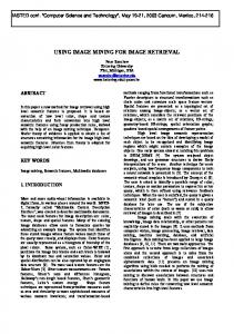
2. METHODS The basic principle of any CBIR system is to describe the extensive pixel information of images by only a few but representative numerical features, which are efficiently applicable for query processing. Note that the proper selection of such features again corresponds to the right-information paradigm. 2.1. IRMA processing steps and semantic layers Figure 1 summarizes the stepwise data processing within the IRMA system. In total, six semantic layers are modeled in order to incorporate medical a-priori knowledge. The first processing step categorizes images with respect to their modality, orientation, body region examined, and biological system imaged. For that, a detailed hierarchical coding scheme was developed [14], which exceeds the complexity of existing DICOM tags such as (0018/0015) "body part examined" or (0018/5100) "patient position" [15]. Automatic categorization by means of global features (i.e., a few
images raw data layer
categorization categories registration
registered data layer
RST-parameters feature extraction feature vectors feature selection
feature layer
indexing
scheme layer
blob-trees identification retrieval
object layer
query knowledge layer
query results
Figure 1: IRMA processing steps and semantic layers
feature values represent the entire image) is based on a reference database of 10,000 images selected arbitrarily from clinical routine and classified manually by experienced radiologists using a PACS-integrated web interface [16]. Several likely categories are supported. Furthermore, prototypes for each category allow the registration of images into a standardized position, and the parameters for rotation, scaling, and translation (RST) are determined. This transfers the images from the raw data layer to the registered data layer. Note, however, that the images are not re-transformed at this time of processing, but only the RST parameters are assigned. Within IRMA, extraction and selection of local features (i.e., each image pixel is represented by a feature vector) are separated. This enables adaptation to both, image category and query content. Adaptive query processing is supported by pre-defined feature sets computed at data entry time. For instance, texture-based features are used for queries concerning the tissue structure (e.g., bone tumor), while contour-based features extracted from the same image support a query on bone fractures. Computing local features, the feature layer of data abstraction results in an explosion of data volume, which must be reduced efficiently for effective query processing. Therefore, the indexing step is used to compute a causal and hierarchical multi-scale segmentation of images. Likewise the Blobworld concept, image segments are represented by a best fitting ellipsoid (blob) and a mean feature vector [7]. In contrast to Blobworld, the causal multi-resolution approach of IRMA results in hierarchically structured blob trees. With respect to the registration parameters computed earlier, the blob trees are transformed in the standard positions according to likely image categories. This results in the IRMA scheme layer. While some queries can already be answered on the scheme layer, further extraction of knowledge from the images is possible. Based on a-priori knowledge coded in the labeled prototypes of each category, correspondence of blobs and anatomical regions and/or biological objects can be established automatically. This identification leads to the object layer. Blob identification supports semantic queries, which concern defined organs or other well-known structures in the image. Further abstraction for query processing is provided within the knowledge layer. For instance, methods must be provided to extend query formulation with qualitative descriptors like "larger", "more intense", or "between these two examples", and combination of these [4]. The formulation of such queries requires intensive work on the design of the user interface. However, current state of IRMA development is not focused on this work. 2.2. IRMA system design In the previous section, the model of information processing resulting in several processing steps was described. Images, local and global features, segmentation and hierarchical blob trees must be computed, addressed, and handled. However, this variety of semantic layers and processing modules significantly reduces when the level of implementation is concerned. In other words, the same basic mechanisms of the system are used to handle information on different semantic layers. Doing so, the entire system is built from three central components, a database, a
.cpp
images, features & tree data
SQL
sources of programs
web server PHP
http
IRMA GUIs
IRMA scheduler
sockets
IRMA daemons
IRMA database SQL
jobs
methods, networks & experiments
administrative information (cluster)
server
clients
Figure 2: IRMA system design
scheduling service communicating with IRMA daemons on connected workstations, and a web server providing graphical user interfaces (GUIs) to client processes (Fig. 2). Central database. The core of the IRMA system is a relational database. The database is used to store administrative information about (a) physical entities, i.e., image data (images, global and local features), tree data (hierarchical image representation obtained by segmentation), and source code (user-implemented programs), and (b) logical entities, i.e., image processing algorithms, image or feature sets, methods, experiments, and the infrastructure of the workstation cluster. Large physical entities are stored outside the database and can be hosted by any computer within the distributed system. Using information about the cluster infrastructure, transparent access to and automatic replication of all physical entities are implemented. Global feature vectors are stored directly in the database. Integer, float, character string and region graphs are valid data types. Images and local features can be replicated for improved performance on the distributed file system and unambiguous references to all data locations are kept within the database. Since the IRMA cluster is composed of workstations with heterogeneous hardware and operating systems, the C and C++ sources of all procedures are provided by the database. New routines for image analysis can be added and used at any time, in particular, when the system is already in use. Image processing algorithms are decomposed into consecutive steps operating on features. Each step is performed by an executable program that consists of user-implemented subroutines linked to a generic main routine. This structured composition allows programs to be used in various contexts. All programs for feature extraction and evaluation are regarded uniquely as a transformation of features. Hence, only three types of programs exist: (a) transformation of a set of features into a single feature (T → 1, e.g., determining a PCA matrix), (b) transformation of one feature into another (1 → 1, e.g., computing a binary threshold of an image), and (c) transformation of a single feature into a set of features (1 → T, e.g., generating multiple representations of one image via small transformations). A program with one of its parameterizations results in a so-called method. Directed graphs with a set of sources and destinations build networks of methods. They model the dataflow within image processing algorithms. Each inner node of the network is either a method or a control element. So far, the IRMA system provides an IF element and a
corresponding MERGE element to allow conditional execution of network parts. A WHILE element can be used to execute loops. Furthermore, a control element for the manipulation of feature sets is offered. Source and destination nodes provide an interface to the feature data, either a single feature or a feature set for any such node. For a retrieval task, the system allows to define experiments, which assign features or sets of features to a subset of source nodes. The idea is to model a query as the combination of an experiment and a query element. The experiment defines the algorithm, its parameters, and the reference set (the knowledge) while the query element assigns features to the remaining source nodes. This extended query-by-example approach is suitable for images, sketches, regions of interest, and blob trees. Processing management. Each computer within the system runs the IRMA daemon, a background process that automatically installs new programs on its host and starts them on demand. Due to the heterogeneous cluster architecture, programs are transferred as source code and compiled with machine-specific make files. Once a program has been started, it awaits orders to execute its feature transformation subroutine on input features. These orders, which are issued by the central scheduling service, are called jobs. The IRMA daemon also transfers the required input data if it is not accessible within the workstation’s local area network and protocols the location of the output data in the IRMA database. The IRMA scheduler, a central service, manages the execution of all feature transformations for data entry and query processing. For each task, the scheduler creates a data structure to log the progress during the execution of the corresponding network of methods. Using socket-based communication with all IRMA daemons and all running programs, the scheduler identifies, dispatches, and monitors all jobs among the cluster. The scheduler also provides load balancing between all available computers. Web interface. Adequate GUIs are required for CBIR applications. With respect to platform independence, distributed system architecture, and easy-to-use design, web-based front-ends were implemented within IRMA for data entry and retrieval. According to the general approach of IRMA, there is no specific interface. Rather, self-standing GUIs are designed to support certain applications. However, all GUIs are combined from a small set of modular components. Each type of module covers a fundamental aspect of GUIs. Output modules visualize image data, features, parameters, or blob trees. For instance, whenever an image is displayed in an IRMA GUI, a frame hosting an icon that fits a squared bounding box of about 100 pixels in size, a heading with the name or identifier of the image, and a footing with relevant parameters (e.g., the IRMA code of the image) is generated by the module. Clicking the icon opens a window displaying the full-sized image. Output modules are also used to present relevance facts, i.e., information for the user why the images result from a certain query. Parameter modules provide GUI elements, which allow the user to enter parameters, options and selections regarding a query. Hence, parameter modules support relevance feedback, i.e., information for the system whether resulting images are intended or unwanted. Process modules allow the combination of results from prior queries. Boolean operators such as AND, OR, and NOT are provided. Although these elements have great impact to query refinement, they are missing in common CBIR concepts. In addition to these modules, transaction logging provides UNDO/REDO-functionality. The log keeps track of the user's refinement process and enables an easy data exploration of the stored images, because a selection process can be transferred directly to other sets of source data. Note that this modularization also separates the content of information from its visualization. The client side of the GUIs utilizes JavaScript, hypertext markup language (HTML), and the document object model (DOM) to implement interactive modules. Browser requests are sent via the hypertext transfer protocol (HTTP) to the web server. Using a hypertext preprocessor (PHP), the web server inserts the request into the database using the structured query language (SQL). After communicating a request to IRMA, the scheduler automatically determines the processing steps required, controls the job list, and signals completion via the database. 2.3. IRMA – PACS integration Coupling of CBIR and PACS can be done on different levels of hard- and software architectures. For the sake of compatibility, both CBIR and PACS must maintain their autonomy as self-standing applications. Information exchange between both components must rely on standardized protocols and should be independent from the actual systems and computer hardware in use. System integration. In general, a DICOM-PACS connects imaging modalities, viewing stations, printers and other devices to an archive, which stores both image data and alphanumerical descriptions of study, patient, imaging modality and setup, body region examined, and other information usually used to access the images. In this architecture, the
DICOM-PACS environment
IRMA system
imaging imaging imaging modalities imaging modalities modalities modalities
IRMA daemon
operating on any client
system interface (sockets)
server level PACS archive
DICOM network
communication interface (DICOM, HL7)
viewing application
application programming interface (API)
browser interface (HTML, PHP)
IRMA core (database, scheduler, web server) database interface (XML-RPC, SQL)
server level operating on the physician‘s workstation
GUIs for data retrieval
IRMA handle
browser interface (HTML, PHP)
GUI for data entry
Figure 3: IRMA – PACS integration
PACS archive is one among other system components, which are all connected within a DICOM network (left hand side in Figure 3). The core of the IRMA system is linked directly to the PACS archive by means of the DICOM and health level seven (HL7) protocols. In other words, the IRMA core is built as a DICOM compliant service class user ordering all information from the service class provider (PACS archive) autonomously, while storage location of image data and patient-related information remains unchanged. An IRMA handle is designed to run on the hardware that is in use for PACS viewing stations. This handle is addressed via the application programming interface (API) provided by the PACS core. By means of this API, the unique DICOM identifier (ID) of the image object currently displayed is passed to IRMA and stored in the IRMA database using SQL. By means of remote procedure calls within the extended markup language (XML-RPC), feature extraction and other IRMA methods, networks, or experiments can be initiated. Only this handle needs adaptation when IRMA is integrated into another PACS environment. Figure 3 exemplifies the system integration of PACS and IRMA. The Aachen University Hospital uses a PACS core implemented by Sectra Imtec AB, Sweden (shown on the left hand side). Hence, the PACS-API in use is the Clinical Applications Interface™ (CAI™). On the right hand side, the IRMA core is based on a central relational database, the scheduler, and the web interface. Since all IRMA GUIs rely on HTML which is dynamically generated by means of PHP, they can be displayed in any internet browser that is installed on the radiologists’ workstation. Workflow integration. Using the CAI interface, an IRMA data entry option is available within the menu of the PACS viewing software. When a radiologist reads an image that has been acquired by a DICOM modality and he wants to enter it into the IRMA reference database, the standard procedure for data entry is started by selecting this menu option and the API passes the image ID to the IRMA handle that communicates it to the IRMA core. If reference categorization and labeling was selected by the physician, the IRMA handle also starts the corresponding server programs and the IRMA GUI for data entry, which is opened in a window on the physician’s workstation. The image ID is used by the IRMA core to request the image itself as well as alphanumerical information (e.g., the diagnosis), which then is displayed in the IRMA GUI for data entry. Like this application for manual data entry, all applications for image retrieval are also decoupled from the PACS program for viewing and reporting. The standard procedure for queries by examples can be run stand alone but also initiated from the PACS application via the API. In the latter case, the same mechanisms as described before are started.
3. RESULTS The IRMA project is ongoing research and its implementation is still in progress. The latest information is available via the project’s web site http://irma-project.org. Nonetheless, some IRMA GUIs are already in use for data browsing and
Figure 4: IRMA GUI for data entry
reference categorization. Based on the continuously growing test-bed of labeled data, first experiments lead to encouraging results. In addition, they prove the IRMA system’s transparency. 3.1. Graphical user interfaces Based on the GUI modules for output and parameter selection, the IRMA code editor allows data entry and reference categorization according to the IRMA code [14]. The GUI displays selected radiographs and offers the radiologist standardized alphanumerical descriptors for classification. Based on the selection of primary code positions, the selection windows for the sub-codes are adapted automatically to valid code entities. All code alterations are recorded by the transaction logging mechanism and stored in the IRMA database. They can be viewed via the protocol icon on the right hand side of the browser window (Fig. 4). Other GUIs exist for the definition of the IRMA code and browsing the IRMA reference database. 3.2. Categorization and retrieval experiments Although manual labeling of the IRMA reference database is still in progress, the system was already used for processing primitive queries, i.e., queries regarding the category of images. Based on a subset of 1,617 images from six body regions (i.e., abdomen, limbs, chest, breast, skull, and spine) acquired with various modalities, in several orientations, and from different biological systems, a tangent distance classifier performed with as low as 8 % error rate [17]. In another study, 1,867 chest radiographs were separated automatically with error rate less than 1 % into frontal (posteroanterior/anteroposterior) and lateral orientation by a simple correlation measure computed from substantially size-reduced icons of 8 x 8 pixels [18]. These leaving-one-out experiments were performed automatically within the IRMA cluster and controlled by the IRMA scheduler via corresponding job lists. 3.3. System transparency The experiments have shown that the IRMA framework offers transparency regarding several aspects from the viewpoint of a distributed system and that of the user. In particular, (a) (b) (c) (d) (e)
location and access transparency for data, methods, and experiments are established by the IRMA daemons, replication transparency for methods in development is obtained from automatic source code transfer, concurrency transparency for job processing and feature extraction is guaranteed by the central scheduling service, system transparency at method implementation time is provided by the central database, and job distribution transparency when issuing a query results from the schedule service.
4. DISCUSSION In contrast to specific applications, a general CBIR approach for medical images is presented and implemented combining a central database with a distributed system architecture suitable for large image databases such as within a PACS. Resulting from its modular concept, the IRMA system supports rapid prototyping and quick integration of novel image analysis methods. So far, the IRMA system was already used to successfully answer primitive queries on the registered data layer. These experiments have proven the validity and applicability of the IRMA concept. Resulting from its system transparency, IRMA is suitable for sophisticated image processing without user interaction. In other words, CBIR principles are made available for a variety of applications including PACS. The current approach for integration of PACS and IRMA leaves both systems autonomous. However, further integration is easily possible. Usually, a PACS consists of a DICOM image server and several DICOM-compliant workstations for reading the images and reporting the findings. Basic IRMA components are the relational database, the scheduler, and the web server, which all may be installed on the host of the PACS archive. Further components of IRMA are the daemons running on distributed machines. Note that idle computational capacities of PACS workstations can be used directly for content-based query processing within the IRMA approach, independent of whether the communication is based on the proprietary IRMA protocol or is integrated into standards such as DICOM. Further integration is easy to realize. For instance, global image analysis may be used to fill or control DICOM header information regarding the imaging modality, orientation, body region examined, or biological system imaged. This information may be wrong in more than 15 % of the cases if it is preset automatically by the DICOM modality [19]. Furthermore, the detailed IRMA code can be consistently integrated to supplement the DICOM standard. The interconnection of the IRMA processing management and the web-based query interfaces needs further investigation. Note that the system’s back-end primarily aims at off-line processing. Especially during the interactive parameter optimization of an algorithm, it will be necessary to calculate certain branches for several parameterizations in advance to provide acceptable system responses. However, a full query refinement process results in relevance feedback which alters an algorithm's input only but not its parameterization, and hence, it can be pre-computed at data entry time.
5. CONCLUSION Usually, a PACS consists of a DICOM image server and several DICOM-compliant workstations for reading the images and reporting the findings. Central IRMA components are the relational database, the scheduler, and the web server while the daemons are running on distributed machines. Two interfaces are designed to connect CBIR and PACS. The IRMA core is implemented as a DICOM service class user, and the API of the PACS core is used to connect PACS applications for viewing and reading of radiographs to an IRMA handle interfacing the IRMA core. Note that only this handle must be adopted when IRMA is integrated to another PACS environment. As a result, PACS and IRMA run autonomously but transparently. Hence the combination of both enables improved image selection in clinical routine supporting both information logistics (right-information paradigm) and decision support (case-based reasoning and evidence-based medicine). Regarding the right-information-paradigm, local image analysis in subsequent IRMA processing steps will have further impact on HIS and PACS.
ACKNOWLEDGEMENT This work was performed within the image retrieval in medical applications (IRMA) project, which is supported by the German Research Community (Deutsche Forschungsgemeinschaft, DFG) grants Le 1108/4 and Le 1108/6.
REFERENCES 1.
Reichertz PL: Towards systematisation. Methods of Information in Medicine 1977; 16: 125-30.
2. 3. 4. 5. 6. 7.
8. 9. 10. 11.
12. 13. 14. 15.
16.
17. 18. 19.
Haux R: Knowledge-based decision support for diagnosis and therapy – On the multiple usability of patient data. Methods of Information in Medicine 1989; 28: 69-77. Kim Y, Horii SC (eds): Handbook of Medical Imaging – Volume 3: Display and PACS. SPIE Press, Bellingham, WA, 2000. Tagare HD, Jaffe CC, Duncan J: Medical image databases – A content-based retrieval approach. Journal of the American Medical Informatics Association JAMIA 1997; 4: 184-198. Niblack W, Barber R, Equitz W, Flickner M, Yanker P, Ashley J: The QBIC-project – Querying images by content using color, texture and shape. Proceedings SPIE 1993; 1908: 173-187. Wu KJ, Narasimhalu AD, Mehtre BM, Lam CP, Gao YJ: CORE – A content-based retrieval engine for multimedia information systems. Multimedia Systems 1995; 3: 25-41. Carson C, Belongie S, Greenspan H, Malik J: Blobworld – Image segmentation using expectation-maximization and its application to image querying. IEEE Transactions on Pattern Analysis and Machine Intelligence 2002; 24(8): 1026-1038. Nappi M, Polese G, Tortora G: FIRST –Fractal indexing and retrieval system for image databases. Image and Vision Computing 1998; 16: 1019-1031. Pentland A, Picard RW, Scarloff S: Photobook – Content-based manipulation of image databases. Proceedings SPIE 1994; 2185: 34-47. Korn P, Sidiropoulos N, Faloutsos C, Siegel E, Protopapas Z: Fast and effective retrieval of medical tumor shapes. IEEE Transactions on Knowledge and Data Engineering 1998; 10(6): 889-904. Shyu CR, Brodley CE, Kak AC, Kosaka A, Aisen AM, Broderick LS: ASSERT – A physician-in-the-loop contentbased retrieval system for HRCT image databases. Computer Vision and Image Understanding 1999; 75(1/2): 111132. Chu WW, Hsu CC, Cárdenas AF, Tiara RK: Knowledge-based image retrieval with spatial and temporal constructs. IEEE Transactions on Knowledge and Data Engineering 1998; 10(6): 872-888. Lehmann TM, Wein B, Dahmen J, Bredno J, Vogelsang F, Kohnen M: Content-based image retrieval in medical applications – A novel multi-step approach. Proceedings SPIE 2000; 3972: 312-320. Lehmann TM, Schubert H, Keysers D, Kohnen M, Wein BB: The IRMA code for unique classification of medical images. Proceedings SPIE 2003; 5033: in press in this issue. National Electrical Manufacturers Association (NEMA) ed: Digital Imaging and Communications in Medicine (DICOM) – Part 3, PS 3.3-2001. NEMA Publishing, Rosslyn, VA, 2001. (http://medical.nema.org/dicom/2001/01_03PU.PDF) Wein B, Lehmann TM, Keysers D, Schubert H, Kohnen M: Detailed image classification code for image retrieval of medical images (IRMA). In: Lemke HU, Vannier MW, Inamura K, Farman AG, Doi K, Reiber JHC (eds): CARS 2002 – Computer Assisted Radiology and Surgery. Proceedings of the 16th International Congress and Exhibition Paris, Springer-Verlag, Berlin, 2002; 513–517. Keysers D, Dahmen J, Ney H, Wein BB, Lehmann TM: A statistical framework for model-based image retrieval in medical applications. Journal of Electronic Imaging, 2003; 12(1): in press. Lehmann TM, Güld MO, Keysers D, Schubert H, Wenning A, Wein BB: Automatic detection of the view position of chest radiographs. Proceedings SPIE 2003, 5032: in press. Güld MO, Kohnen M, Keysers D, Schubert H, Wein BB, Bredno J, Lehmann TM: Quality of DICOM header information for image categorization. Proceedings SPIE 2002; 4685: 280-287.