Contour-Based Image Registration Using Mutual Information* Nancy A. Álvarez1, José M. Sanchiz2, Jorge Badenas2, Filiberto Pla2, and Gustavo Casañ2 1 Universidad
de Oriente, Santiago, Cuba
[email protected] 2 Universidad Jaume I, Castellón, Spain {sanchiz,badenas,pla,ncasan}@uji.es
Abstract. Image registration is a problem that arises in many image processing applications whenever information from two or more scenes have to be aligned. In image registration the use of an adequate measure of alignment is a crucial issue. Current techniques are classified in two broad categories: area based and feature based. All methods include some similarity measure. In this paper a new measure that combines mutual information ideas, spatial information and feature characteristics, is proposed. Edge points are used as features, obtained from a Canny edge detector. Feature characteristics like location, edge strength and orientation are taken into account to compute a joint probability distribution of corresponding edge points in two images. Mutual information based on this function is minimized to find the best alignment parameters. The approach has been tested with a collection of portal images taken in real cancer treatment sessions, obtaining encouraging results.
1 Introduction Image registration techniques find applications in several medical fields, like tissue or injury evolution monitoring. In some medical applications there is a need of integrating complementary information from different imaging sensors, that is, different radiological imaging modalities, and also in matching images from the same modality taken at different times. Portal imaging consists of sensing therapeutic radiation applied from electron accelerators in cancer treatment [1]. They are formed by the projections of anatomical structures over the sensing area after it goes through the body. Due to the high energy of the radiation, there is a poor contrast in portal images compared to x-ray, axial tomography or magnetic resonance images. Introduction of electronic portal imaging devices has increased the quality of portal images. Detection of patient pose errors during or after treatment is the main use of portal images. For patient pose monitoring, portal images are compared to higher quality simulated portal images used as reference, or to a reference portal image taken at the first therapy session. Any misalignment has to be detected and corrected. Misalign*
Work partially supported by the Spanish Ministry of Science and Technology under Project TIC2003-06953, and by Fundació Caixa Castelló under project P1-1B2002-41.
J.S. Marques et al. (Eds.): IbPRIA 2005, LNCS 3522, pp. 227–234, 2005. © Springer-Verlag Berlin Heidelberg 2005
228
Nancy A. Álvarez et al.
ments are traditionally detected manually after the session. Automatic misalignment detection before the session, using an innocuous dose, is desirable. Several registration methods with some degrees of automation, designed to compare portal images among them and with their corresponding simulation images, have been reported in the literature [2, 3, 4, 5]. This work is being developed as part of a project between the Radiotherapeutical Oncology Department at Provincial Hospital of Castellón, Spain, and University Jaume I, Castellón. It is aimed at automating and improving quality control in radiotherapy, mainly focused at patient positioning. We describe a registration method based on ideas of mutual information. Instead of a joint probability distribution derived from grey levels, used in conventional mutual information registration, we propose a joint probability function derived from the spatial localization of features, and features similarity. The minimization of the mutual information based on this function provides the alignment parameters between two images. The method has been tested with portal and magnetic resonance images.
2 Related Work Registration algorithms have applications in many fields. They are valuable tools in medical imaging, remote sensing, computer vision, etc. Currently, research is directed to multimodal registration and to cope with region deformations [6]. Many different registration algorithms have been proposed, and almost all share a common framework: optimizing a cost function that measures the alignment between images [7]. In feature-based approaches the cost function is computed from characteristics of features (edges, ridges) extracted before registration. In the case of portal images, features from the irradiation field geometry have been used [8], where the distance measure is based on the Hausdorff distance modified by using a voting scheme that is expressed as a parameter introduced in the expression of this distance. This modification makes the method tolerant to small position errors like those that occur with automatic edge detectors. Techniques that use manually selected landmarks to be matched have been also used, [9]. In this work contours of the irradiation field are manually selected and their points used for registration using chamfer matching [10]. Pixel-based approaches use all the pixels of an image. A Fourier transform-based cross correlation operator was used in [4] to find the optimal registration, accounting for translations and rotations. A new image alignment measure was introduced in [11, 12] based on entropy concepts developed as part of Shannon´s information theory: mutual information. It was used to measure the statistical dependence between image intensities of corresponding pixels in two images. Hybrid techniques that combine both approaches have been proposed. In [13] mutual information is computed using feature points locations instead of image intensity. In [14] the registration function includes spatial information by combining mutual information with image gradient. Our method uses edges detected from portal images from conventional edge extractors. The registration function is derived from the mutual information concept, and combines three attributes of edges: edge point location, edge strength and edge orien-
Contour-Based Image Registration Using Mutual Information
229
tation. These attributes provide spatial information, and are used to build a probability estimate of the possible correspondence of two edge points in two images. A joint probability table is computed for all possible correspondences, and minimization of the mutual information is applied to obtain the best match and the alignment parameters.
3 Registration Based on Entropy Minimization Mutual Information Mutual Information is a concept from information theory, and is the basis of one of the most robust registration methods [15]. The underlying concept of mutual information is entropy, which can be considered a measure of dispersion of a probability distribution. In thermology, entropy is a measure of the disorder of a system. A homogeneous image has a low entropy while a high contrast image has a high entropy. If we consider as a system the pairs of aligned pixels in two images, disorder, and joint entropy, increases with misregistration, and correct alignment gives a minimum of the mutual information of the two images. Given two images A and B, the definition of the mutual information I(A,B) is: I(A,B) = H(A) + H(B) – H(A,B) , (1) H(A) and H(B) being the entropies of images A and B, and H(A,B) being the joint entropy. Correct registration corresponds with maximization of the mutual information. Following Shannon´s information theory, the entropy of a probability distribution P is computed as: H = − ∑ p log p .
(2)
p∈P
Typically, the joint probability distribution of two images is estimated as a normalized joint histogram of the intensity values [12]. The marginal distributions are obtained by summing over the rows or over the columns of the joint histogram. Including Feature Information We propose a new measure of mutual information computed only from features. We use edge points as features, and point location, edge strength and edge orientation as feature characteristics. Edge points are a significant source of information for image alignment, they are present in portal images and in simulated radiographies obtained from a treatment planner, so they are useful for intra and inter modality registration. In optimal alignment position edge points from one image should match their corresponding points in location and also in edge strength and orientation. In [13] a new mutual information-based measure was introduced. Instead of using image intensity for estimation of mutual information it uses feature points location information. Let {a1,a2,...,aN} and {b1,b2,...,bM} be two sets of feature points in two images A and B. The mutual information is a function of the joint probability:
I ( p ) = ∑∑ p ijT log i
j
p ijT
∑ p kjT ∑ pilT k
l
,
(3)
230
Nancy A. Álvarez et al.
where pij represents the joint probability between feature point i in A and feature point j in B: pijT =
(− λD ) T ij
e (−λD ) , ∑e ij
(4)
T ij
T stands for the spatial mapping (rigid, similarity, affine) applied for aligning one point set with the other. DijT is a distance measure between two points ai and bj (e.g. T
Euclidean distance). p ij is a measure of the correspondence likelihood between those two feature points, while
∑p
T ij
i
and
∑p
T ij
are the marginal probabilities.
j
In [14] the mutual information measure is extended to include spatial information. Locations with valuable spatial information (e.g. transition of tissues) are denoted by strong gradients. The extension is accomplished by multiplying the mutual information extracted from grey level probability distributions with a gradient term. This term includes the gradient magnitude and orientation. The mutual information measure proposed in [14] is: Inew(A,B) = G(A,B) I(A,B) , (5) with G(A,B) being the gradient term obtained as: G(A,B)= ∑ α (∇ai , ∇b j )min ∇ai , ∇b j , (6) ( ai ,b j )∈A∩ B
(
)
ai and bj denote two points in images A and B, and α is the angle between two gradient vectors. When the two images are registered, point ai will be located close to its matching point bj. If a joint probability table is built considering the distances from each ai to all the bj with j=1, 2, …, M, in one of the M cells of the i-th column, there will the a maximum of that column, point bj, so having the biggest likelihood of being the match of ai. Re-computing the table for different spatial mappings T, one of the joint probability tables obtained will be the best, having the smallest distances of matched points. Similarly, with the images registered, an edge point ai will match some bj having similar edge strength since they represent the same edge point. The edge orientation after the mapping has to be also similar. Denoting as Dij the distance between ai and bj, Φij the difference in edge strength, and Οij the difference in edge orientation after the mapping, we can base the mutual information measure on these feature points characteristics: I(A,B) = f(Dij, Φij, Οij) . (7) Our main contribution is the use of several feature attributes to estimate the joint probabilities. We use the gradient magnitude at a feature point as an estimation of the edge strength and the gradient direction as an estimation of the edge orientation: ∇ai ∇b Tj 2 T −1 DijT = ai − b Tj , Φij = ∇ai − ∇b j , . (8) Oij = cos ∇ai ∇b Tj Gradient magnitude at edge points can be different in corresponding edges detected in different images due the possibly different sensing devices used to take the images.
Contour-Based Image Registration Using Mutual Information
231
This can be overcome by scaling the gradient magnitude at edges in both images, giving, for example, a relative measure between zero and one. To estimate the joint probability of match between two edge points in two images we introduce an exponential function based on the feature attributes. If DijT , Φij, OTij are small, there is a higher probability of correspondence between those edge points. The proposed joint probability is expressed as follows: Φ + O D exp − + T
T
ij
ij
ij
γ γ2 γ 3 1 pijT = T T Dij Φij Oij exp − + + ∑∑ γ2 γ 3 γ1 i j
(9)
with γk being constants. Using the probability distribution function given in (9), mutual information is computed as described in (3). The main advantage of our approach compared to the classical mutual information is that this latter method does not use the neighbouring relations among pixels at all, all spatial information is lost, while our approach is precisely based on spatial information. Compared to the method reported in [13], we propose a combination of feature attributes, compared to the method in [14], our approach is only based on feature points. Edge Detection Extraction of edges can be done by several methods, first derivative-based methods (Sobel masks), or second derivative-based, like Laplacian of a Gaussian or Canny [16]. In this work we have used the Canny edge detector, that selects edge points at locations where zero-crossings of the second derivative occur. Optimization Optimization of the registration function is done by exhaustive search over the search space. We assume a rigid transformation to align one image with the other, a rotation followed by a translation, both in 2D, so the search space is three-dimensional. A revision of optimization strategies can be found in [17]: Powell´s method, and simplex method, conjugate-gradient and Levenberg-Marquardt methods. Since the principal purpose of our work is to prove the feasibility of a new form of obtaining the joint probability used for the computation of the mutual information, no analysis on the convenience of using a certain optimization has been made. Exhaustive search is a sufficiently simple method for a bounded three-dimensional search space, and it finds a global optimum, avoiding the main drawback of other optimization algorithms of converging to a local optimum.
4 Results We have tested our approach with about fifteen pairs of medical images of different sources, portal images provided from sessions of radiotherapy treatments at Provincial Hospital of Castellón, and Magnetic Resonance (MR) images obtained from the internet [18].
232
Nancy A. Álvarez et al.
For portal image registration, image alignment parameters were determined by human operators and compared to the results of our approach. For MR images the alignment parameters were available along with the images. The influence of each feature in (9) was tested by making several experiments T
where each term: Dij ,
T ij
T
, Oij , was included or not. The overall best performance
was observed when the three characteristics are used. In the computation of pijT the values of γ1, γ2 and γ3 were fixed heuristically (20, DT
ΦT
OT
10, and 1). They were selected by computing e ij , e ij and e ij using the edge sets without applying any transformation, and observing the graphical representation of T
these functions. As our intention was that small values of Dij ,
T ij
T
, Oij represent a
high correspondence probability, we fixed γ1, γ2 and γ3 as values close to the time constants of the exponential functions.
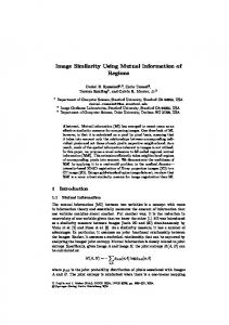
a)
c)
e)
b)
d)
f)
Fig. 1. Portal image of a hip with patient in a) initial position and in c) wrong position. Edges detected in each image, b) and d). Both sets of overimposed edges e) before and f) after the registration
Figure 1 shows the registration of two portal images. 2a) shows a portal image of a hip taken in an initial radiotherapy session. c) shows an image from another session that must be aligned with a). b) and d) show the same images with its edges superimposed. These edges correspond mainly to hip bones. e) shows the same image as a) with edges overimposed from both b) and d) before registration, and f) reflects the
Contour-Based Image Registration Using Mutual Information
233
arrangement of edges after registration with our approach. Note the improvement in alignment. Figure 2 compares the joint probability functions obtained after applying the classical mutual information approach using a joint histogram of grey levels, and our feature-based method. Both functions are computed for the best alignment parameters obtained from the registration, that is, lowest entropy. High intensity values correspond to high likelihood of correspondence.
Fig. 2. Joint probability functions computed with classical grey level-based mutual information approach (left) and our feature-based method (right)
We have tested our approach with several portal images and compared the results with the registration given manually by several human observers, and with the classical mutual information method based on grey level values. The manual registration was made by identifying common features in both images and registering them. The errors of our method with the human results were within acceptable levels, often less than a pixel in translation and less than a degree in rotation, and always better than the classical method. Although we assumed a rigid transformation in our tests, there is no a priori restriction to a particular type of transformation.
5 Conclusions and Further Work The inclusion of spatial data in the computation of the mutual information is a subject under current investigation. In this paper we propose a new measure of registration that combines mutual information with spatial data obtained from feature attributes, like edge points. Instead of a joint histogram of grey levels, the classical approach, we estimate a joint probability distribution based on feature points. We introduce a probability estimate that two feature points match based on points similarity. An optimization algorithm is then applied to find the best registration parameters where a minimum of the mutual information based on joint entropy occurs. The proposed approach can be used to register images from different sources, multimodal registration, since it can combine different features as needed. One has to provide a way to compute the probability that two features in two images correspond. The new approach improves the classical mutual information method, based only on intensity values, which obtains poor performance in low contrast images like portal
234
Nancy A. Álvarez et al.
images. Furthermore, the number of points used to build the probability function is significantly smaller, only feature points, than the number used to build the joint histogram in the classical approach, all points in the images. Further work is addressed at investigating the use of other features in the approach, as boundaries of regions in segmented images, or the overlapping area of segmented regions. The key question is which attributes to include in the computation of the joint probability table, and how to combine them.
References 1. Langmack, A.,: Portal Imaging. Br J Radiol 74 (2001) 789-804 2. Gottesfeld, L., A survey of image registration techniques. ACM Computing Surveys 24 (1992) 325-376. 3. Plattard, D., Soret, M., Troccaz, J., Vassal, P., Giraud, JY., Champleboux, G., Artignan, X., Bolla, M.: Patient Set-Up using Portal Images: 2D/2D Image Registration Using Mutual Information. Computer Aided Surgery (2000) 246-262 4. Hristov, DH., Fallone, BG.: A gray-level image alignment algorithm for registration of portal images and digitally reconstructed radiographs. Med Phys 23 (1996) 75-84 5. Van Elsen, PA., Pol, E., Viergever, M.A.: Medical image matching – a review with classification. ACM Computing Surveys 24 (1992) 325-376 6. Lester, H., Arrige, S.R.: A survey of hierarchical non-linear medical image registration. Pattern Recognition 32 (1999) 129-149 7. Zitová, B., Flusser, J.: Image registration methods: a survey. Image and Vision Computing 21 (2003) 977-1000 8. Chmielewski, L., Kukolowicz, P.F., Gut, P., Dabrowski, A.: Assesment of the quality of radiotherapy with the use of portal and simulation images – the method and the software. Journal of Medical Informatics & Technologies 3 (2002) 171-179 9. Leszczynski, K., Loose, S., Dunscombe, P.: Segmented chamfer matching for the registration of field borders in radiotherapy images. Phys Med. Biol 40 (1995) 83-94 10. Borgefors, G.: Hierarchical chamfer matching: a parametric edge matching algorithm. IEEE Trans PAMI 10 (1988) 849-865 11. Maes, F., Collignon, A., Vandermeulen, D., Marchal, G., Suetens, P.: Multimodality image registration by maximization of mutual information. IEEE Transactions on Medical Imaging 16 (1997) 187-198 12. Viola, P., Wells, W.M.: Alignment by maximization of mutual information. International Journal of Computer Vision 24 (1997) 137-154 13. Rangarajan, A., Chui, H., Duncan, J.: Rigid point feature registration using mutual information. Medical Image Analysis 4 (1999) 1-17 14. Pluim J., Maintz, J.B., Viergever, M.A.: Image registration by maximization of combined mutual information and gradient information. IEEE Transactions on Medical Imaging 19 (2000) 809-814 15. West, J. et al.: Comparison and evaluation of retrospective intermodality brain image registration techniques. J. Comput. Assited Tomography 21(1997) 554-566. 16. Canny, J.F: A Computational Approach to Edge Detection. IEEE TPAMI 8 (1986) 679-698 17. Maes, F., Vandermeulen, D., Suetens, P.: Comparative evaluation of multiresolution optimization strategies for multimodality image registration by maximization of mutual information. Medical Image Analysis 3 (1999) 373-386 18. http://www.itk.org