focus on cell polarity
REVIEWS Coordinated protein sorting, targeting and distribution in polarized cells Ira Mellman* and W. James Nelson‡
Abstract | The polarized distribution of functions in polarized cells requires the coordinated interaction of three machineries that modify the basic mechanisms of intracellular protein trafficking and distribution. First, intrinsic protein-sorting signals and cellular decoding machineries regulate protein trafficking to plasma membrane domains; second, intracellular signalling complexes define the plasma membrane domains to which proteins are delivered; and third, proteins that are involved in cell–cell and cell–substrate adhesion orientate the three-dimensional distribution of intracellular signalling complexes and, accordingly, the direction of membrane traffic. The integration of these mechanisms into a complex and dynamic network is crucial for normal tissue function and is often defective in disease states. Epithelial–mesenchymal transition Phenotypic and functional changes in epithelial cells, usually associated with the loss of cell–cell adhesion and increased cell migration, as cells are induced to become fibroblasts.
*Genentech, Inc., 1 DNA Way, South San Francisco, California 94080, USA. ‡ Department of Biology, the Department of Molecular and Cellular Physiology and the Bio‑X Program, Stanford University, Stanford, California 94305, USA. e‑mails: mellman.ira@gene. com;
[email protected] doi:10.1038/nrm2525
Cell polarity is a structural and functional specialization that is ubiquitous in biology. The commonality of polarity across the phyla reflects a fundamental requirement of individuals to localize different activities to distinct regions of cells, especially when individual cells come together to form complex multicellular tissues. The specialized domains of the plasma membrane that result from polarization determine cell orientation, function and fate. For example, polarization enables long-range communication by neurons and short-range communication in the immune system, vectorial transport of ions across epithelial cells and niche-specific orientation of stem-cell division, which specifies the developmental fate of daughter cells. At first glance, the fact that different cell types exhibit diverse polarized phenotypes implies that a diverse array of specialized machineries has evolved. However, it seems that simple variations of common mechanistic themes result in the unique shapes, asymmetries and functions that characterize polarized cells and tissues. First, intrinsic protein-sorting codes are recognized and segregated by cytoplasmic adaptor complexes that regulate protein trafficking to plasma membrane domains. Second, signalling complexes and scaffolds become differentially associated with the cytosolic face of the membrane, where they define and stabilize the biochemical features of resulting domains. Third, adhesion receptors that detect neighbouring cells and the extracellular matrix (ECM) provide cues that orientate cells in three-dimensional (3D) space. Two considerations support the idea that cell polarity is achieved through the integration of these three conserved molecular mechanisms. First, all eukaryotic cells
nATurE rEvIEWS | molecular cell biology
share common cellular machineries for post-translational protein trafficking and compartmentalization1. Second, cells can adopt different shapes and functions in response to specific physiological contexts. For example, during embryogenesis, a single cell can change its shape and function as it migrates, according to morphogenetic gradients, and can then repolarize on detecting transcriptionally specified cell–cell interactions2. In disease states, such as cancer, epithelial cells lose polarity (through epithelial– mesenchymal transition (EMT)), disengage from multicellular interactions, migrate and then reintegrate into a second tissue, in which they undergo structural and functional reorganization to reside at the new site3,4. Thus, the dynamics and plasticity of the loss and re-establishment of polarity suggest that common machineries of membrane traffic are used at all times, but are deployed differently depending on the physiological context. Here, we summarize the basic cellular machineries and biochemical rules that control the delivery of protein components to different plasma membrane domains in a generic polarized cell. We then describe the spatial cues and signalling pathways that organize these basic machineries to produce various cell shapes and functions. Finally, we consider how component asymmetry at the single-cell level is orientated in 3D space to define polarity in complex tissues. We emphasize how these different pathways generate plasticity in the forms of cell polarity, and how defects underlie important pathological states. Although much has been learnt about each of these three mechanisms in isolation, understanding how they are integrated and coordinated into a network remains a central challenge — one that is fundamental voluME 9 | novEMbEr 2008 | 833
reVieWs Plasma membrane COPI COPI
Tether, SNARE
Adaptors, clathrin
Cytoskeleton Adaptors, clathrin
COPII
Adaptors, clathrin
Tether, SNARE
ER
Golgi
E TGN
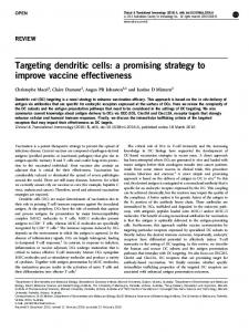
Figure 1 | a generic post-translational pathway for protein trafficking to the plasma Nature Reviews | Molecular Cell Biology membrane. After synthesis in the endoplasmic reticulum (ER), membrane proteins are sorted into vesicles by the coatomer protein complex-II (COPII) machinery and delivered to the Golgi complex by vesicle-tethering and SNARE machineries. Intra-Golgi transport and retrograde transport from the Golgi to the ER are regulated by the COPI machinery. At the trans-Golgi network (TGN), proteins are sorted into vesicles by intrinsic sorting motifs and cytoplasmic adaptor complexes, and are transported along cytoskeletal elements to the plasma membrane. Protein delivery to the plasma membrane is mediated by vesicletethering and SNARE machineries. Some proteins (for example, ligand–receptor complexes) are internalized through another set of adaptors and delivered to an endosome (E), from which they might be recycled back to the plasma membrane.
to understanding organogenesis, tissue function and various pathological states.
COPII A specific coat protein complex that initiates the vesicle budding process from the endoplasmic reticulum.
COPI A specific coat protein complex that initiates the vesicle budding process from membranes of the Golgi complex, and is involved in intra-Golgi and Golgi-toendoplasmic reticulum vesicle trafficking.
AP–clathrin complex A complex of proteins that comprises adaptor proteins and structural clathrin (which forms a coat that initiates vesicle budding from membranes).
Rab GTPases A large family of Ras-like GTPases that have key roles in the secretory and endocytic pathways.
Controlling protein sorting and trafficking The generic post-translational pathway of membraneprotein trafficking to the plasma membrane involves the sequential transport of proteins between different membrane compartments (for example, the endoplasmic reticulum (Er), the Golgi complex, endosomes and the plasma membrane1). Membrane proteins are modified in these membrane compartments, usually through the assembly and processing of complex oligosaccharide chains (FIG. 1). Transport is mediated by vesicular intermediates that bud from one compartment and fuse with the next. Genetics and in vitro reconstitution experiments have identified three classes of protein complexes that form these vesicular intermediates5–7: coatomer protein complex-II (COPII) complexes are crucial for Er–Golgi trafficking; COPI complexes are crucial for Golgi–Er trafficking and intra-Golgi transport; and the adaptor protein (AP)–clathrin complex (the AP–clathrin complex) is crucial for transport between the Golgi, the plasma membrane and endosomes. Certain proteins of each complex recognize and selectively recruit different cargo proteins into transport vesicles (non-recognized proteins are excluded from vesicles), whereas other complex proteins deform and sculpt the membrane to produce the membrane vesicle or a tubule. The AP–clathrin complex also has accessory proteins that modulate the basic functions of the complex, increasing the complexity of cargo-selection strategies and affecting
834 | novEMbEr 2008 | voluME 9
membrane curvature8. regardless of how or where transport vesicles are formed, vesicle trafficking between compartments is mediated by the actin and microtubule cytoskeleton9, and vesicle fusion with the target plasma membrane is regulated by organelle-specific Rab GTPases, vesicle-tethering complexes10 and SNAREs5. These generic mechanisms are the core machineries of trafficking in all cells, and are modified in polarized cells to sort proteins into separate plasma membrane domains. below, we focus on how machineries that are involved in the Golgi–plasma membrane–endosome pathways control membrane-protein sorting to different plasma membrane domains in polarized cells. Protein sorting in the exocytic and endocytic pathways. There are three major sites of protein sorting in the exocytic and endocytic pathways: the Golgi complex, the plasma membrane and endosomes (FIG. 1). At each site, proteins can be sorted into separate vesicle carriers on the basis of intrinsic sorting signals and the cellular machineries that recognize those signals. A major component of protein sorting at all three sites is provided by the AP-complex family, which not only recognizes proteinsorting signals but also regulates the assembly of clathrin scaffolds, which sculpt the membrane to form vesicles. Four AP complexes (AP-1–4) have been identified, and these localize to different membrane compartments between the Golgi complex, the plasma membrane and endosomes11. Each AP complex is composed of two large subunits (α, γ, ε, δ or β1–β4; ~100 kDa each), one small subunit (σ1–σ4; ~20 kDa) and one medium subunit (µ1–µ4; ~50 kDa). Together with other proteins (such as GGA, AP180 (assembly protein of 180 kDa), epsin-1, epsin-2, EPS15, β-arrestin and ArH) that interact with clathrin, the AP subunits recognize and bind specific amino-acid motifs in the cytoplasmic domain of membrane proteins and cluster these proteins into patches on the membrane by assembling a clathrin cage8,11. one of the best-studied interactions is between the AP-2– clathrin complex and the transferrin receptor (Tfnr), which occurs on the plasma membrane. The µ subunit of AP-2 (µ2) recognizes a degenerate tetrapeptide cytoplasmic domain sorting motif (YXXØ; in which Ø represents any hydrophobic amino-acid residue), resulting in the clustering of Tfnr into clathrin-coated pits on the plasma membrane12. These coated pits then bud into the cytoplasm and are delivered to early endosomes. This sorting motif can be recognized in endosomes, probably by the AP-1–clathrin complex, and the receptor is recycled back to the plasma membrane by an endosome-derived vesicle population11 (FIG. 2). In general, protein recycling from the endosome to the plasma membrane can be signal dependent or signal independent. It is difficult to distinguish these two mechanisms in non-polarized cells, which exhibit a single, nondifferentiated plasma membrane. However, the situation is different in polarized cells. For example, in epithelial cells and neurons, Tfnr and other well-characterized recycling receptors (such as low-density lipoprotein receptor (lDlr)) are localized to a specialized domain of the plasma membrane: the basolateral membrane in epithelia www.nature.com/reviews/molcellbio
f o c u s o n c e l l proelVaire iW ty s Plasma membrane domain I (apical or axonal) COPI
Galectin, SNARE
Lipid rafts, lectin
COPI
Cytoskeleton
E AP or CL
COPII
AP-1B or CL AP-1, AP-4 or CL Exocyst, SNARE
Tether, SNARE
ER
Golgi
TGN Cytoskeleton Plasma membrane domain II (basolateral or somatodendritic)
Figure 2 | Superimposing protein sorting demands on the generic trafficking Nature Reviews Cell Biology pathway. In polarized cells, such as epithelia and neurons, protein| Molecular processing occurs along the generic pathway between the endoplasmic reticulum (ER) and the trans-Golgi network (TGN; see also FIG. 1). However, proteins in the TGN might be sorted into various different vesicles through the recognition of different intrinsic sorting motifs (TABLE 1) and cytoplasmic adaptor complexes. These vesicles are then targeted either directly or indirectly, through an endosome (E), to different plasma membrane domains (designated as domains I and II) along cytoskeletal elements. These cytoskeletal elements might have different orientations (or polarity) relative to the different membrane domains. Vesicle delivery to each plasma membrane domain is mediated by different vesicle-tethering and SNARE complexes. Some proteins are internalized through another set of adaptors and delivered to an endosome, from which they might be recycled back to the original plasma membrane domain, or to the other domain by trancytosis, depending on the presence (or activation or inactivation) of specific protein-sorting motifs. AP, adaptor protein; CL, clathrin; COP, coatomer protein complex.
Vesicle-tethering complex A large protein complex that localizes to various sites of vesicle delivery in the secretory pathway and facilitates the capture, docking and fusion of specific vesicles with different membranes (for example, the exocyst complex at the plasma membrane).
SNARE (Soluble N-ethyl-maleimidesensitive fusion protein attachment-protein receptor). These proteins comprise a large protein superfamily and mediate the fusion of transport vesicles with membranes.
Transferrin receptor A membrane protein that binds soluble transferrin, which is required for the cellular import of iron.
and the somatodendritic membrane in neurons13,14. In such cases, the recognition of specific targeting information in the cytoplasmic domain of Tfnr is essential to maintain the polarized localization of the receptor to the appropriate domain. Polarity sorting motifs and adaptors for basolateral proteins. In polarized cells, cytoplasmic domain-sorting signals are not only required for endocytosis, they are also required for the polarized delivery of newly synthesized receptors and the maintenance of recycling receptors in their appropriate membrane domains15,16 (FIG. 2). Canonical internalization signals have a role in polarized endocytosis and recycling, and they are assisted by additional sorting signals and decoding adaptor complexes. lDlr is a well-studied example. It has two Tyr-based sorting motifs — a membrane-proximal motif and a C-terminal motif — in its cytoplasmic domain, and these are required for post-Golgi delivery and the maintenance of lDlr to the basolateral or somatodendritic domains13,14. The membrane-proximal motif is important for lDlr endocytosis, and although the C-terminal motif is not required for endocytosis, it encodes a signal for sorting to the basolateral or
nATurE rEvIEWS | molecular cell biology
somatodendritic domains13,14. Many other basolateral membrane proteins (TABLE 1) have Tyr-based targeting motifs16, although dileu-based signals have also been identified (dileu motifs can also be responsible for clathrin-mediated endocytosis)17. other motifs, such as the sorting motif in Tfnr18, seem to be devoid of crucial Tyr or leu residues18,19. The observation that many basolateral proteins have a Tyr- or dileu-based motif for basolateral polarity has long suggested that AP–clathrin complexes are important in one or more steps in specifying protein trafficking in polarized cells. The only AP complex to be identified to date that is definitively associated with basolateral protein trafficking is an epithelial cell-specific variant of the clathrin-associated AP-1 complex (designated AP-1b), in which the ubiquitously expressed µ1A subunit is replaced by a closely related µ1b subunit 20,21. AP-1b clearly functions to determine the polarity of Tyr-based signals (and some non-Tyr signals, such as that in Tfnr), as the absence of µ1b leads to a non-polarized or apically localized protein, a similar effect to that of eliminating the sorting motif. However, it is unlikely that all such signals are decoded by AP-1b. Dileu-based signals, for example, programme basolateral polarity, but in an AP-1b-independent manner 20,21 (TABLE 1). A second AP complex, AP-4, also has a role in basolateral targeting, although its cargo and signal specificity remain poorly defined22. AP-1b is not expressed in all polarized cells, indicating that other adaptors or protein-sorting pathways exist. Many neurons, for example, do not express µ1b, but protein transport to the somatodendritic plasma membrane domain involves the same targeting signals that are used for transport to the basolateral membrane in most epithelial cells14. Hepatocytes also do not express µ1b, but they use the same Tyr-based signals on basolateral proteins (such as lDlr) as µ1b-positive epithelial cells23. The identities of these alternative basolateral adaptors remain elusive. Additionally, do AP-1b or any alternative adaptor proteins work in conjuction with clathrin? This is possible, but the removal of clathrin by small interfering (si)rnA has a surprising range of effects on basolateral membrane-protein sorting 24. Proteins such as vSvG (the major surface-coat protein of the stomatitis virus, which is a Tyr-dependent protein), nCAM (neural cell adhesion molecule, which is Tyr-independent) and CD147 (which is leu dependent) are delivered in a 1:2 ratio to the basolateral and apical membranes, whereas others are delivered in a 2:1 ratio (Tfnr, which is Tyr independent), a 3:1 ratio (epithelial (E)-cadherin, which is dileu dependent) or are unaffected (na+/K+-ATPase). by comparison, deletion of both basolateral sorting motifs in lDlr results in the almost complete mistargeting of mutant lDlr to the apical membrane13. The observation that some proteins are less affected than others by clathrin knockdown further indicates that there is a degree of redundancy and overlap in sorting signals and decoding elements, only some of which are likely to be clathrin dependent. For example, ankyrins are adaptor proteins that bind to a subclass of membrane proteins (such as E-cadherin and na+/K+-ATPase) and to the cytoskeletal protein spectrin25. Importantly, decreased expression of voluME 9 | novEMbEr 2008 | 835
reVieWs Table 1 | Intrinsic protein sorting codes and the cellular decoding machinery* Protein
Protein code
Decoding machinery
Protein sorting pathway A (apical plasma membrane and axonal membrane) Occludin and ERGIC53
N-linked glycosylation
Unknown (lectin?)
p75, sucrase-isomaltase and podocalyxin (or GP135)
O-linked glycosylation
Unknown (lectin?)
Basolateral membrane
PLAP, THY1 and DAF
GPI-anchor (oligomerization)
Lipid raft
A domain of the plasma membrane that comprises the basal and lateral membranes. It is orientated towards cell–extracellular matrix (basal membrane) and cell–cell contacts (lateral membrane).
Haemagglutinin and neuraminidase
Transmembrane domain
Lipid raft
Rhodopsin and Na+-dependent bile acid transporter
Cytoplasmic domain
Unknown
Podocalyxin (or GP135)
Cytoplasmic PDZ domain
Unknown
LDLR, TfnR, VSVG and AGPR-HI
Cytoplasmic domain (Tyr based)
AP-1B
Somatodendritic membrane
ErB2
Unknown
AP-1B
H /K -ATPase
Cytoplasmic domain (Tyr based)
Unknown
FcII B2R
Cytoplasmic domain (diLeu based)
Unknown
E-cadherin
Juxtamembrane 40 amino acids
Ankyrin
CD147
Cytoplasmic domain (Leu based)
Unknown
Na+/K+-ATPase, poly-IgAR and NCAM
Cytoplasmic domain
Unknown
A part of neuronal cells that comprises the dendritic membranes and soma and excludes the axon.
Apical membrane A domain of the plasma membrane in polarized epithelial cells that is usually orientated on the luminal side of epithelial tubes (for example, the intestine).
Ankyrin A large adaptor protein that was originally found in erythrocytes but is ubiquitously expressed in nucleated cells. Ankyrin binds to various membrane proteins, spectrin and actin-binding proteins.
Spectrin A large protein that comprises subunits (α and β) that form a heterotetramer, (αβ)2. Spectrin binds to ankyrin and to the actin cytoskeleton.
Sucrase-isomaltase A glycosidase that comprises activities of a sucrase and an isomaltase. Sucrase-isomaltase is found in the apical membrane of intestinal epithelial cells.
Glycosyl phosphoinositol A modification at the C terminus of membrane proteins that occurs in the endoplasmic reticulum. This modifcation allows the protein to insert into the outer leaflet of the lipid bilayer.
Lipid raft A membrane subdomain that is enriched in glycosphingolipids, sphingomyelin and cholesterol.
Transcytosis The delivery of transport vesicles between the apical and basolateral membrane domains of polarized cells.
Protein sorting pathway B (basolateral plasma membrane and somatodendritic membrane)
+
+
*Two generic sorting pathways are described that could represent the apical or axonal (pathway A) and basolateral or somatodendritic (pathway B) plasma membrane domains of polarized cells. See the main text for experimental evidence that these proteins belong to different sorting pathways, and for selected references. AP, adaptor protein; E-cadherin, epithelial cadherin; GPI, glycosyl phosphoinositol; LDLR, low-density lipoprotein receptor; NCAM, neural cell adhesion molecule.
ankyrin b or ankyrin G reduces E-cadherin trafficking from the Golgi to the basolateral membrane26. Further studies are required to uncover additional mechanisms and pathways of protein sorting and trafficking. Polarized sorting of apical proteins. The sorting motifs and the cellular machineries that sort proteins to the apical membranes of epithelial cells are different from those that sort proteins to basolateral membranes. In contrast to the cytoplasm-orientated basolateral sorting motif, apical sorting motifs are localized in the extracellular or transmembrane domains of proteins27 (TABLE 1). Extracellular domain motifs contain N- and O-linked oligosaccharide chains (such as those found in p75 (REF. 28) and sucrase-isomaltase29), although it is unclear whether these oligosaccharide chains are directly recognized by sorting machinery or whether they control protein conformation and hence the display of a sorting motif in the amino-acid backbone of the protein. The membrane-associated signal can be the transmembrane domain itself (as occurs in some viral glycoproteins, such as haemagglutinin and neuraminidase), but the best described signal is the glycosyl phosphoinositol (GPI) lipid anchor 30. GPI-anchored proteins are sorted into the apical pathway in the Golgi complex. This occurs by GPI-anchored protein oligomerization in lipid rafts31 that are enriched in glycosphingolipids, sphingomyelin and cholesterol30. Interestingly, large clusters of GPI-anchored proteins fail to form in a lectin-resistant Madin–Darby Canine Kidney (MDCK) cell line that missorts GPI-anchored proteins. This suggests that carbohydrates are involved in this clustering event and are necessary for apical sorting 32. Several other proteins in lipid rafts might have roles in lipid-raft synthesis (such
836 | novEMbEr 2008 | voluME 9
as FAPP2 (REF. 33)) and stabilization (such as MAl30), and in clustering of apical proteins (such as the lipidraft protein galectin-4 (REF. 34)). It is unknown, however, how apical protein clustering, by lipid rafts or any other mechanism, leads to deformation of the membrane and to the formation of a transport vesicle that can deliver its cargo to the apical plasma membrane. In general, the cytoplasmic domain of apical proteins is not thought to be important in protein trafficking. The protein rhodopsin, however, is targeted to the apical membrane of photoreceptors by a sorting determinant that is localized to its cytoplasmic C terminus35. This sorting determinant interacts with dynein36. Importantly, missorting of rhodopsin leads to retinitis pigmentosa (BOX 1). basolateral (and somatodendritic) sorting signals are often ‘dominant’ over apical sorting signals. In other words, if both a functional basolateral and an apical signal are present, basolateral targeting will ensue. Transcytosis of membrane proteins from the basolateral to apical domains can occur if the basolateral signal is inactivated after newly synthesized proteins are included into basolaterally directed transport vesicles (FIG. 2); transcytosis involves endocytosis from one membrane domain, delivery to endosomes, resorting and trafficking to the other membrane domain. For example, the adhesion-signalling protein neuronal-glial (ng)-CAM has a Tyr-containing, AP-1b-dependent basolateral targeting signal that is inactivated by phosphorylation on reaching the basolateral surface. In the absence of a functional basolateral signal, ng-CAM is not captured by AP-1b in endosomes after internalization and is instead transcytosed to the apical surface37. variations on this simple theme probably explain other examples of trancytosis (such as the extensively studied polymeric Ig receptor 38). www.nature.com/reviews/molcellbio
f o c u s o n c e l l proelVaire iW ty s Box 1 | When cell polarity goes awry Various disease states (see table) show that cells can dissolve or reorganize the cellular machinery that generates polarized distributions of proteins4. This process can occur at the three layers of regulation of cell polarity that are discussed in the main text. Mutations in the intrinsic protein-sorting motifs that sort proteins to specific membrane domains are found in genetic disorders of major organs, such as the kidney, lungs, intestine and nervous system125,131. These mutations can affect protein transport through the secretory pathway, and can cause missorting of proteins to the wrong membrane domain or cause loss of protein localization at the plasma membrane. In these cases, overall polarity might not be affected, but the key role of the mutated protein in a biological process can result in the loss of cell function. Mutations in key components that detect extrinsic orientational cues have strong effects on cell organization and function. Many of these proteins are tumour suppressors, including phosphatase and tensin homologue (PTEN)127, and many oncogenic mutations activate phosphoinositide 3-kinase (PI3K) or AKT/protein kinase B (PKB) directly. Changes in the distributions of phosphatidylinositol-3,4,5-trisph osphate (PtdIns(3,4,5)P3) and phosphatidylinositol-3,4-bisphosphate (PtdIns(3,4)P2) might have additional effects on the structure and function of cells. Scribble, lethal giant larvae (LGL) and discs large (DLG) were isolated as neoplastic tumour-suppressor mutations in Drosophila melanogaster, based on the distinctive ‘giant larvae’ phenotype of zygotic mutant animals132. Little is known about their roles in human disease, but the loss or decreased expression of human homologues of Scribble and LGL have been found in colorectal cancers and malignant melanoma128. Changes in the orientational cues that specify cell polarity occur in a form of reversed cell polarity, termed epithelial– mesenchymal transition (EMT). EMT has an essential role in embryogenesis and homeostasis (during morphogenetic movements, epidermal wound healing and migration of neutrophils in chemotactic gradients) and in cancer-cell metastasis3. EMT is characterized by decreased expression of epithelial (E)-cadherin (which is mediated by the expression of transcriptional repressors such as SNAIL, ZEB and some basic helix–loop–helix factors133), the loss of cell–cell adhesion and epithelial characteristics, and altered cell–extracellular matrix (ECM) interactions, including ECM remodelling by secreted metalloproteinases and increased cell migration. It is conceivable that the ‘forward reaction’ can be re-initiated when a metastatic cell reaches the appropriate distal tissue site or niche, resulting in a partly differentiated tumour3.
Disease Bartter syndrome* (REF. 123)
Protein ROMK ClCK
Defect Missorted to apical membrane Missorted to basolateral membrane Recycling on apical membrane
Liddle syndrome* (REF. 124)
βENaC
Cystic fibrosis125
CFTR (∆F508) CFTR (C-terminal PDZ) Sucrase-isomaltase
ER exit
LDLR (FH-Turku)
Missorted to the apical membrane
Polycystin-1 and -2
Growth or mitotic defects; protein mislocalization (for example, Na+/K+-ATPase, EGFR) Cilium motility Unknown (protein transport to cilium?) Unknown Rhodopsin transport into cilium T-tubule organization of Na+/K+-ATPase, Na+/Ca2+ exchanger and IP3R complex; stress-induced sudden cardiac death Loss of plasma membrane localization, ventrical arrhythmia and sudden cardiac death Loss of axon and dendrite localization, corpus callosum hypoplasia, retardation and aphasia
Congenital sucraseisomaltase deficiency126 Familial hypercholesterolaemia23 Polycystic kidney disease‡ Situs invertus‡ Bardet–Biedl syndrome‡ Nephronophthisis‡ Retinitis pigmentosa‡ Cardiac arrhythmia25
Left–right dynein BBS4, BBS5 and BBS8 NPHP1–4 Kinesin-2 (IFT) Ankyrin B
Brugada syndrome25
Voltage-gated Na+ channel (E1053K mutation in the ankyrin-G binding site) L1 cell adhesion molcule (S1224L and Y1229H mutations in the ankyrin-B binding site) PTEN and PI3K127
CRASH syndrome25 Oncogenesis
Scribble and LGL128 Retinitis pigmentosa129 Rhodopsin 130 Peutz–Jeghers syndrome LKB1 (PAR4)
Golgi exit Missorted to basolateral membrane
Activates the AKT/PKB signalling pathway (promotes cell survival, proliferation and growth) Decreased expression; function is unknown but the defect is found in colorectal cancer and malignant melanoma Missorting to photoreceptor apical membrane Mutations; overgrowth of cells in the gastrointestinal tract (hamartomas)
*Bartter syndrome and Liddle syndrome contain defects in Na+ transport. ‡Polycystic kidney disease, situs invertus, Bardet–Biedl syndrome, nephronophthisis and retinitis pigmentosa are ciliopathies92. ER, endoplasmic reticulum; LDLR, low-density lipoprotein receptor; PAR4, partitioning-defective-4.
nATurE rEvIEWS | molecular cell biology
voluME 9 | novEMbEr 2008 | 837
reVieWs Sorting in the trans-Golgi network and recycling endosomes. The sorting of plasma membrane proteins to their appropriate destinations seems to be dependent on the general core mechanisms found in the endocytic and secretory pathways, but protein sorting is subject to slight modifications in polarized cells. In all cells, the endocytic and secretory pathways possess multiple sorting sites. In most cells, however, the sorting sites that are most relevant for generating and maintaining polarity — the trans-Golgi network (TGn) and recycling endosomes — are found in the perinuclear cytoplasm (FIG. 2). It is highly likely that signals for apical or basolateral sorting are recognized at either or both of these sites, with the TGn mediating the sorting of newly synthesized proteins in the secretory pathway and recycling endosomes responsible for polarized recycling or transcytosis after endocytosis37,38. It also seems likely that newly synthesized membrane proteins could pass through recycling endosomes after leaving the Golgi and before reaching the surface39. Thus, recycling endosomes might be a common site for protein sorting; perhaps adaptor complexes in these endosomes select cargo for inclusion in transport vesicles that are destined for specific plasma membrane domains.
Getting to, and staying in, membrane domains Sorting proteins during exit from the Golgi or recycling endosomes is one level of adaptive machinery for generating different protein distributions in polarized cells. However, molecular sorting must be coupled to mechanisms that specify vesicle delivery and fusion to different plasma membrane domains. These mechanisms involve the cytoskeleton, for long-range vesicle delivery, and (at short range) protein complexes at target plasma membrane domains that specify the docking and fusion of vesicles.
Trans-Golgi network The terminal region of the Golgi complex, in which proteins are sorted and packaged into transport vesicles for delivery to the plasma membrane.
Recycling endosome A membrane compartment in which proteins delivered by transport vesicles are resorted and packaged into vesicles for delivery to different membranes.
Exocyst complex An example of a vesicletethering complex that associates with the plasma membrane and regulates the delivery of transport vesicles to the basolateral membrane domain of polarized epithelial cells.
The cytoskeleton in vesicle trafficking. Most polarized cells have specialized organizations of actin and microtubule cytoskeletons. Meshworks of short actin filaments are localized underneath the entire plasma membrane and might provide linkages through the ankyrin–spectrin complex to membrane proteins25. The actin cytoskeleton might have several roles in cell polarity and protein trafficking 40 as it is regulated by CDC42. CDC42 is involved at several crucial control points that regulate cell polarity (see below), including myosin motor proteins, which could regulate vesicle trafficking, and the ankyrin–spectrin complex. Indeed, ankyrin knockdown results in the loss of lateral membrane surface area41, perhaps by decreasing protein trafficking from the Golgi (see above) or by structurally destabilizing the basolateral membrane in a manner that is analogous to spectrin deletion in erythrocytes. Microtubule arrays are normally organized in fibroblasts with the dynamic plus ends at the cortex and the minus ends near the centrosomes, close to the nucleus. In polarized cells, microtubules are generally bundled along the long axis of the cell (apico– basal in epithelia, soma–axon in neurons) with all
838 | novEMbEr 2008 | voluME 9
the plus ends at the base of the cell or end of the axon, respectively 42,43. Microtubules seem to regulate the efficiency rather than the fidelity of vesicle delivery to the apical and basolateral membrane in epithelial cells44. Plus-end microtubule kinesin motors are involved in apical protein delivery (for example, the delivery of the p75 protein, but not of GPI-anchored proteins)45, and are generally involved in the targeting of basolateral46 and axonal membrane proteins47 (see the review by li and Gunderson in this issue). Controlling vesicle fusion at plasma membrane domains. The fusion of vesicles with the correct membrane domains is a crucial step in the generation of polarity. Two interconnected mechanisms regulate the initial tethering of vesicles to the membrane and then the fusion of vesicles with the target-membrane domain. Members of the rab family of small GTPases seem to control many stages of vesicle docking and fusion, especially by having a role in tethering vesicles to their target membranes10. vesicle-tethering complexes are found at many stages of the exocytic pathway and are thought to increase the efficiency and specificity of vesicle delivery (FIG. 2). At the plasma membrane, the exocyst complex seems to regulate the docking of a subset of vesicles, including basolateral vesicles in epithelial cells48, neurite outgrowths and axonal synapseassembly domains49. The exocyst is a large complex of at least six proteins, some of which are localized on the target plasma membrane and others of which are localized to the transport vesicle, along with a rab family GTPase that helps to control tethering-complex assembly (rAb8 on basolateral vesicles of epithelia; rAb3 on synaptic vesicles)50,51. This suggests that vesicle docking at the plasma membrane might occur through the assembly of an exocyst holocomplex. Another type of vesicle-tethering complex comprises annexins and is present on other plasma membranes, such as the apical membrane of epithelial cells. Annexins bind to membranes in a phosphatidylinositol-3,4-bisphosphate (PtdIns(3,4)P2)-dependent and Ca2+-dependent manner and self-aggregate. As with components of the exocyst complex, annexins might be present on both vesicles and target membranes52. The delivery of some apical proteins in epithelial cells (for example, the delivery of sucrase-isomaltase53) requires annexin II, but the full repertoire of proteins that require annexins for plasma membrane delivery is not known. Fusion of vesicles with a target plasma membrane is mediated by the SnArE complex, which comprises vesicle (v)-SnArEs (such as vesicle-associated membrane protein (vAMP)) and target (t)-SnArEs (the syntaxins)5. In polarized epithelial cells, apical and basolateral vesicles contain different v-SnArEs (such as tetanus-insensitive (Ti)-vAMP and vAMP8, respectively54), and different t-SnArEs are localized to the apical (syntaxin-3) and basolateral (syntaxin-4) plasma membranes 55,56 (FIG. 2). loss-of-function or mislocalization of SnArEs leads to a concomitant disruption in the delivery of apical or basolateral vesicles to the plasma membrane54,57–59. www.nature.com/reviews/molcellbio
f o c u s o n c e l l proelVaire iW ty s a Apical membrane CRB/PALS1 PATJ STD
P
b PAR5
PAR3
+ PAR6 PAR3 aPKC –
PAR3 Cytoplasm PAR1 PAR5
LGL SCRB DLG
PAR1
aPKC, PAR4
P
Basolateral membrane
Figure 3 | Signalling complexes and scaffolds on the cytosolic of the membrane Nature Reviewsface | Molecular Cell Biology define and stabilize membrane domains. a | Three signalling complexes (Crumbs, partitioning defective (PAR) and Scribble) associate with the cytoplasmic surface of the plasma membrane around sites of cell adhesion, which demarcates different plasma membrane domains — in this example, the apical membrane (top) and the basolateral membrane (bottom). Crumbs protein (CRB) is a transmembrane protein, but mechanisms of binding of the PAR and Scribble complexes are poorly understood (see the main text). Proteins within each of these three complexes physically interact, as do PATJ (PALS1 (protein associated with LIN-7)-1-associated tight-junction protein) and PAR6 in the Crumbs and PAR complexes. Atypical protein kinase C (aPKC; in the PAR complex) genetically interacts with, and stabilizes, CRB (broken line, +), and phosphorylates and negatively regulates (broken line, –) lethal giant larvae (LGL) in the Scribble complex. Overall, the PAR complexes reinforce the localization and activity of the Crumbs complex (thick arrow), and the PAR and Scribble complexes mutually antagonize each other (inhibition lines). b | PAR3 phosphorylation by PAR1 results in the binding of phosphorylated PAR3 and PAR5, and dislocation of PAR3 from the membrane into the cytoplasm. Similarly, PAR1 phosphorylation by aPKC or PAR4 results in the binding of phosphorylated PAR1 and PAR5, and dislocation of PAR1 into the cytoplasm (see the main text for details). SCRB, Scribble protein; STD, stardust.
Immunological synapse The site of interaction between a lymphocyte and an antigen-presenting cell.
Protein retention and molecular fences. Polarized membrane-protein localization can also be achieved by asymmetric stabilization at a plasma membrane domain. Selective stabilization can occur in the absence of direct sorting in the exocytic or endocytic pathways60, or as a mechanism for further refining proteins that are sorted in these pathways61. Several classes of membrane proteins bind to the cytoplasmic scaffold complex of ankyrin– spectrin, including ion transporters and channels (such as the anion exchanger, na+/K+-ATPase, voltage-gated na+ channel and na+/Ca2+-exchanger), receptors (such as Ins(1,4,5)P3 receptor, ryanodine receptor and N-methyld-aspartate receptor (nMDAr)), and cell-adhesion proteins (such as E-cadherin, l1 (ng-CAM) and CD44)25. In epithelial cells and neurons, ankyrin–spectrin complexes are differentially localized to the basolateral membrane62, and to axons and dendrites, respectively 63. binding to these scaffolds can recruit membrane proteins that function in the same biological process (such as T-tubule clustering of na+/K+-ATPase, IP3r and na+/Ca2+-exchanger)25 and can sequester membrane proteins from the endocytic pathway, thereby increasing their residence time and accumulation60. Another essential feature for the development and maintenance of polarity is a diffusion barrier that separates membrane domains that are otherwise part
nATurE rEvIEWS | molecular cell biology
of a continuous lipid bilayer. In epithelial cells, this barrier is localized to the boundary between the apical and basolateral membrane domains and comprises the tight junction, which acts as a molecular fence to prevent the free diffusion of proteins from one domain to the other 64; regulated vesicular transport (transcytosis) is therefore required to bypass the barrier. Diffusion barriers are not always dependent on cell–cell contact, and can be constructed in single cells by the deposition of cytoskeletal scaffolds that selectively recruit high concentrations of membrane proteins, thereby impeding the movement of freely diffusing membrane proteins. Such a barrier occurs at the axon initial segment in neurons and restricts the movement of axonal membrane proteins into the soma, and vice versa65. This molecular fence involves localized arrays of actin and ankyrin, and might form the basis of diffusion barriers in other cell types25. other cytoskeletal arrays might restrict membrane-protein diffusion in other systems (such as in the immunological synapse, in the border between a nascent bud and a mother yeast cell, and in the membrane-associated cytosolic ‘necklace’ that is found at the base of cilia).
Intrinsic membrane domain orientational cues Although asymmetry in the localization of plasma membrane proteins is achieved by intrinsic sorting mechanisms, it remains unclear how these pathways are coupled to the overall orientation of cells in a multicellular context. below, we discuss evidence that intracellular complexes (partitioning defective (PAr), Crumbs and Scribble complexes) provide orientation cues that specify overall cell polarity, shape and other functional specialization (FIGS 3,4). The role of the PAR complex. Polarization in single cells and in multicellular organisms can be induced by a pathway that involves the Ser/Thr kinase and tumour suppressor lKb1 (the mammalian orthologue of PAr-4, one of six PAr proteins that are required to establish polarity in early Caenorhabditis elegans development 66; see below). Activation of lKb1 in epithelial cells results in apico–basal polarization of single cells, even in the absence of cellular adhesion67; inactivation of lKb1 in neurons inhibits axon formation in vitro and in vivo68. Here, we consider the roles of PAr-4 (lKb1) and other PAr proteins in cell polarity in multicellular contexts. Among the substrates of lKb1 are members of the AMP-activated protein kinase (AMPK) family, which have multiple roles in cells69. In addition to being a glucose or energy sensor, AMPKα and AMPKβ phosphorylate non-muscle myosin light-chain kinase, an activity that is required for the formation of junctional complexes in Drosophila melanogaster 70. AMPK members have several functional homologues: the kinase synapses of amphids defective (SAD), the Ser/Thr kinase PAr1 and the family of ElKl-motif kinases (EMKs; also known as microtubule affinity-regulating kinases (MArKs)), which destabilize microtubules 71. These functional homologues have important roles in the orientation of polarity in neurons and epithelial cells69. voluME 9 | novEMbEr 2008 | 839
reVieWs a
Apical membrane
PtdIns(3,4)P2
b RAC1
CDC42–GTP
PTEN
Actin LGL
PAR6 PAR3 RAC1–GTP αPKC P13K
Basolateral membrane
WASP
βPIX GIT CDC42
LGL SCRB DLG
ARP2/3
Exocyst
PtdIns(3,4,5)P3
Figure 4 | roles of signalling complexes on the cytosolic face of| the membrane Nature Reviews Molecular Cell Biology control phosphoinositide distribution and vesicle trafficking. a | The partitioning defective (PAR) complex structurally and functionally interacts with Rho family GTPases, either by direct binding of PAR6 to active CDC42 (CDC42–GTP) or regulation of the guanine nucleotide-exchange factor T-cell-lymphoma invasion and metastasis-1 (TIAM1), which locally activates RAC1 (forming RAC1–GTP). These GTPases locally regulate actin organization. The PAR complex and associated CDC42 and RAC1 also locally regulate phospholipid synthesis through the direct binding of PAR3 to phosphatase and tensin homologue (PTEN), which generates phosphatidylinositol-(3,4)bisphosphate (PtdIns(3,4)P2), and through activation of phosphoinositide 3-kinase (PI3K), which generates phosphatidylinositol-3,4,5-trisphosphate (PtdIns(3,4,5)P3). PtdIns(3,4)P2 and PtdIns(3,4,5)P3 might be localized to different domains of the plasma membrane — in this example, the apical membrane (top) and the basolateral membrane (bottom). b | The Scribble complex locally regulates actin organization by Scribble protein (SCRB) binding to the PAK-interacting exchange-factor-β–G protein-coupled receptor kinase-interactor (βPIX–GIT) complex, which locally regulates CDC4 and RAC1 in activating the actin-polymerization machinery (Wiskott–Aldrich syndrome protein (WASP) or WASP family verprolin-homologous protein (WAVE), and actin-related protein-2/3 (ARP2/3)). Local activity of the βPIX–GIT complex and actin polymerization affect the organization of the exocyst vesicle-tethering complex and vesicle delivery to the plasma membrane.
PDZ domain A common structural domain of ~80 amino acids that is found in many signalling proteins. It was first found in PSD95, DLG and ZO1.
14-3-3 The characteristic migration pattern of a family of proteins on electrophoretic gels. 14-3-3 proteins bind to kinases, phosphatases and transmembrane receptors.
RING-finger protein A specialized type of zinc finger of 40–60 residues that binds to 2 atoms of zinc, and is involved in mediating protein–protein binding.
Apical junctional complex A collection of cell–cell junctions (tight junctions, adherens junctions and junction-associated proteins) that localize to the apex of the lateral membrane of polarized epithelial cells.
overexpression of PAr1b (also known as EMK1 and MArK2) in MDCK cells results in the partial reorientation of the apical plasma membrane to intercellular lumens on the lateral membrane domain, similar to the orientation of the apical (bile canalicular) membrane in hepatocytes72. MDCK apical plasma membrane reorientation is accompanied by a change in microtubule organization, such that the minus ends of microtubules localize to the intercellular lumens instead of to the top of the cells. In addition, trafficking of post-Golgi transport vesicles to the apical surface is redirected into an indirect pathway (vesicles first appear at the basolateral domain and then transcytose to the apical domain), as also occurs in hepatocytes73. because induction of the transcytotic pathway of apical vesicle transport occurs independently of microtubule reorganization, PAr1 probably affects apical protein sorting into vesicles or the delivery of vesicles to the plasma membrane. Interestingly, the PAr1 homologues in budding yeast, Kin1 and Kin2, interact with the machinery for vesicle tethering (the rab proteins and the exocyst complex) and membrane fusion (the SnArEs)74. Thus, both PAr1 and PAr4 might regulate at least two key control points in post-Golgi vesicle delivery: microtubule remodelling and orientation and, hence, the directionality of postGolgi vesicle transport; and the docking and fusion of
840 | novEMbEr 2008 | voluME 9
vesicles at the plasma membrane. The actual mechanisms by which these kinases exert their effects are unclear, as D. melanogaster mutants that are defective in AMPK or lKb1 exhibit generalized defects in polarity that affect both the apical and basolateral domains70. In addition to PAr1 and PAr4, the PAr complex comprises three scaffolding proteins with either multiple PDZ domains (PAr3 and PAr6) or 14-3-3 homology (PAr5), and a RING-finger protein75,76 (PAr2) (FIG. 3). biochemical studies show that PAr3, PAr6 (which also binds active CDC42 (REF. 77), see below) and atypical protein kinase C (aPKC) form a subcomplex (FIG. 4) that localizes to the apical junctional complex in polarized epithelial cells78,79 and to the tips of neuron axons80. Genetic studies reveal that the PAr3–PAr6–aPKC subcomplex contributes to the establishment and maintenance of apico–basal polarity in embryonic epithelial cells79,81 and to axon formation in neurons80,82 (see the review by Iden and Collard in this issue). The role of Scribble and Crumbs complexes. The PAr3–PAr6–aPKC subcomplex interacts functionally with two other complexes that control cell polarity: the Scribble and Crumbs complexes (FIG. 3) . The Scribble complex also comprises lethal giant larvae (lGl) and discs large (DlG), regulates the identity of the basolateral membrane, and is localized below the apical junctional complex and along the membrane at cell–cell contacts79,81. The Crumbs complex comprises the PDZ-domain-containing proteins PAlS1 (protein associated with lIn-7)-1 (also known as stardust (STD)) and PATJ (PAlS1-associated tight-junction protein)83. Crumbs regulates the identity of the apical membrane81 and localizes to the apical side of the junctional complex in polarized epithelial cells81. loss of function of either the Crumbs or Scribble complexes results in defects in epithelial polarity that are due to the reduction of the surface area of the apical and basolateral plasma membrane domains, respectively 79,81. How the Crumbs and Scribble complexes regulate the identity of the apical and basolateral membranes is unknown. The PAr, Crumbs and Scribble complexes mutually regulate the localization and activity of each other (FIG. 3). Genetic analysis showed that aPKC is required in early D. melanogaster development to maintain the presence of the Crumbs complex at the apical membrane, perhaps by direct phosphorylation84 or by Crumbs binding, through its PDZ-interaction domain, to PAr6 (REF. 85). later in development, Crumbs is required to stabilize the PAr3–PAr6–aPKC complex at the apical junctional complex 79,81. aPKC can also phosphorylate PAr3, decreasing the affinity of PAr3 for aPKC and suggesting that the association of PAr3 with PAr6–aPKC is dynamic86. This is further supported by the finding that the AMPK family member PAr1 phosphorylates PAr3, causing PAr3 to bind PAr5 and thereby inhibiting the association of PAr3 at the basolateral membrane87 (FIG. 3). Interestingly, PAr1 itself is phosphorylated by aPKC, which causes PAr1 to bind to PAr5 and results in the inhibition of the association of PAr1 with the cortex at the apical membrane88. Finally, a member of the Scribble www.nature.com/reviews/molcellbio
f o c u s o n c e l l proelVaire iW ty s complex, lGl, is also a target for aPKC phosphorylation, and lGl phosphorylation inhibits its localization to the apical cortex 89. The activities of aPKC, PAr1 and PAr5 have the overall effect of ‘protecting’ the identities of the apical and basolateral membrane domains. This is achieved by maintaining the apical activities of the Crumbs (and PAr) complex by blocking the intrusion of components of the Scribble complex (lGl), and vice versa. The positioning of the PAr3–PAr6–aPKC complex at the crucial apical–basolateral junction might allow it to arbitrate the phosphorylation of components, such as lGl (preventing its apical association) and PAr3, and prevents the PAr complex from encroaching too far into the basolateral domain. The PAr complex and Crumbs-3, an isoform of Crumbs90,91, also colocalize in the primary cilium in vertebrate polarized epithelial cells. The primary cilium is involved in many vertebrate developmental pathways and in tissue homeostasis92. Crumbs-3 and the PAr complex are directly bound through PAr6, and PAr5 is also present in the complex 90. localization of the complex to the cilium requires association with the microtubule motor protein KIF3A and intact microtubules90. Deletion of Crumbs-3 inhibits ciliogenesis90, whereas deletion of PAr3 or inhibition of aPKC results in a decrease in cilium length91. Although it is not known whether localization of the Crumbs-3–PAr complexes to the primary cilium affects cell polarity, defects in genes for the assembly or function of the primary cilium have been associated with many genetic disorders that are characterized by defects in growth and polarity of epithelial cells, including polycystic kidney disease, left–right asymmetry defects and Bardet–Biedl syndrome92 (BOX 1).
Bardet–Biedl syndrome A complex human genetic disease that is characterized by obesity, retinitis pigmentosa, polydactyly, mental retardation, hypogonadism and renal failure.
Extrinsic membrane domain orientational cues Although the intrinsic cell mechanisms described above regulate sorting, targeting and separation of proteins to different membrane domains, it is important to note that these domains are not organized arbitrarily on the cell surface, but rather are topologically ordered in 3D space. Here, we consider how extracellular cues provide the template that orientates intracellular mechanisms of domain specification and protein sorting. Cell adhesion, both to the substratum (ECM) and to other cells, is important in establishing the polarized orientation of cells. In general, individual epithelial cells that are grown in suspension — that is, in the absence of cell–cell and cell–ECM adhesion — do not develop polarity, but instead undergo programmed cell death (anoikis). When grown on a surface, however, even single epithelial cells develop an apico–basal axis of polarity. When this surface has biological relevance, such as a surface of laminin-containing ECM, neurons specifically form an axon93, single mammary epithelial cells selectively secrete β-casein from the apical surface94 and 3D epithelial cysts polarize correctly 95. In this way, cell–substrate adhesion is coupled not only to post-translational control of membrane traffic and cytoskeletal reorganization, but also to the control of transcriptional activity that specifies the function of the resulting polarized cell types96.
nATurE rEvIEWS | molecular cell biology
Cell–cell contact, mediated by a range of celladhesion proteins, is intimately involved in cell polarization97. For example, epithelial cell–cell adhesion in suspension culture induces the segregation of basolateral membrane proteins to the cell–cell contacts and induces the trafficking of apical proteins to the free surface98. Perhaps the most important protein for cell–cell adhesion and cell polarization is E-cadherin, which is required for epithelial organization in early embryonic development 99. Homotypic E-cadherin interactions are necessary to form the junctional complexes that maintain the diffusion barrier between the apical and basolateral domains of epithelial cells. They are also required for localization of the exocyst complex 48 and to ensure that basolateral vesicles are delivered100 to sites of cell–cell adhesion and to the lateral membrane domain. Although cell–cell contacts can help control epithelial cell differentiation101, cell–cell adhesion is insufficient to trigger full apico–basal polarity in epithelia and, as discussed above, a set of signals must be supplied following adhesion to ECM.
Integrating extrinsic and intrinsic polarity cues We have discussed three main layers of regulation that control cell polarity. Intrinsic mechanisms sort membrane proteins into different vesicles (sorting motifs and decoding machinery), and deliver these vesicles to different membrane domains (cytoskeleton-mediated targeting, vesicle tethering through exocyst complexes and annexins) for membrane fusion (SnArE proteins). Protein complexes at the plasma membrane (the PAr, Crumbs and Scribble complexes) control the identity and distribution of functionally and structurally unique plasma membrane domains from the cytosolic face. Extrinsic cues provided by cell adhesion to the ECM and by other cells control the orientation of cell polarity. but how are these different levels of regulation of cell polarity integrated into a single network (FIG. 5)? Localization of PAR, Scribble and Crumbs complexes. As mentioned above, PAr3–PAr6–aPKC complexes are localized at sites that are important for cell-polarity decisions: the boundary between the apical and basolateral membranes of epithelial cells (demarcated by the apical junctional complex), the cilium and the axon tip in neurons (FIG. 3). The localization of PAr3–PAr6–aPKC complexes to these sites could involve direct binding to cell-adhesion complexes or indirect recruitment by other complexes that are locally activated by adhesion at those sites (FIG. 5). Genetic studies in D. melanogaster suggest that PAr3 functions upstream of the cell–cell adhesion signal that is mediated by D. melanogaster E-cadherin102, but membrane proteins that recruit PAr3 to the apical junctional complex in D. melanogaster have not been identified. by contrast, in mammalian cells, PAr3 binds directly to the cell–cell adhesion proteins JAM-A103 and nectin104, both of which colocalize with E-cadherin at the apical junctional complex. Protein–protein interactions between the PDZ-domain-containing proteins of the Crumbs complex (PAlS1 and PATJ) and proteins of the PAr complex (PAr3 and PAr6) provide links that voluME 9 | novEMbEr 2008 | 841
reVieWs
Polarized cell surface
Golgi
TGN
Endosome
PM domain II
Crumbs
Fence
Endoplasmic reticulum
PM domain I
PAR3
Extrinsic spatial cue, (for example, cell adhesion)
Scribble
Intracellular scaffolds
Figure 5 | Polarized cells form from the hierarchical integration of three fundamental mechanisms. Cell adhesion to Nature Reviews | Molecular Cell Biology the extracellular matrix (ECM) or to adjacent cells provides an extrinsic spatial cue (dark blue) that signals the assembly of intracellular scaffolds (light blue): Crumbs and PAR3 specify the apical domain and Scribble specifies the basolateral domain. Intracellular scaffolds define distinct plasma membrane (PM) domains and separate them by inserting a molecular fence that acts as a diffusion barrier. Finally, protein-sorting codes and cellular decoding machineries distinguish membrane components destined for distinct plasma membrane domains, sorting them into distinct vesicular transporters at the level of the trans-Golgi network (TGN) and endosomes. The transport vesicles are equipped to recognize either domain I or II, thus constructing the biochemical heterogeneity that is characteristic of the polarized cell surface (grey). The integration of these mechanisms is important for normal tissue function and is often defective in disease states.
GEF A protein that is involved in locally activating small GTPases, such as the Rab proteins and members of the Rho GTPase family, by catalysing the exchange of GDP for GTP.
in principle could allow co-recruitment of the PAr and Crumbs complexes to the pre-specified plasma membrane domain83 (FIG. 4a). PAlS1 is also required for the trafficking of E-cadherin to the plasma membrane and thereby the localization of the exocyst complex to the plasma membrane105, indicating that complex feedback mechanisms are involved in the organization of these protein complexes at sites of cell–cell adhesion. Interestingly, disruption of one of the exocyst complex subunits, EXo84, results in the loss of Crumbs from the epithelial cell surface of early D. melanogaster embryos106. This suggests that the exocyst complex has a role in regulating the polarized organization of proteins at the apical junctional complex. Mechanisms involved in the localization of the Scribble complex to plasma membrane domains are poorly understood. local recruitment of the PAr and Scribble complexes to the plasma membrane occurs following cell-adhesionmediated activation of CDC42 and rAC1 due to a complex regulatory network107 (FIG. 4a). CDC42 and rAC1 are members of the rho family of small GTPases. Although the precise mechanisms are not clear, loss of activity of rAC1 and CDC42 inhibits the normal polarization of neurons and epithelial cells by causing a lack of axonal differentiation82 and the misorientation of the apico– basal axis95,108, respectively. This feature undoubtedly reflects the fact that components of the PAr complex are linked to the local regulation of CDC42 activity: CDC42 binds and recruits PAr6 to the cortex77,109; PAr3 binds the rAC1 guanine nucleotide-exchange factor (GEF) T-celllymphoma invasion and metastasis-1 (TIAM1)110; and loss of PAr3 inhibits CDC42-induced rAC1 activation in neurons82. Thus, the localization and activities of the PAr complex, CDC42 and rAC1, and perhaps the junctional complex itself, seem to be regulated by a positive-feedback loop (FIG. 4a).
842 | novEMbEr 2008 | voluME 9
The role of phospholipids in defining membrane domains. local asymmetries in the phospholipid content of plasma membrane domains also effect localization of CDC42 and cell polarity (FIG. 4a). local changes in phosphoinositide synthesis are determined by the relative activities of phosphoinositide 3-kinase (PI3K) and phosphatase and tensin homologue (PTEn). PI3K generates phosphatidylinositol-3,4,5-trisphosphate (PtdIns(3,4,5)P3) and PTEn removes the 5′ phosphate from PtdIns(3,4,5)P3 to generate PtdIns(3,4)P2. Inhibition of PI3K activity affects the normal polarization of neurons, by causing a lack of axonal differentiation80, and of epithelial cells, by causing the misorientation of the apico–basal axis111. PtdIns(3,4,5)P3 is localized to the axon tip of neurons80. In polarized epithelial cells, PtdIns(3,4,5)P3 is enriched in the basolateral plasma membrane111 and is absent from the apical membrane in which PtdIns(3,4)P2 is enriched112 (FIG. 4a). How might different phospholipids regulate the generation of cell polarity? Interestingly, annexin II, a putative scaffolding protein that is required for the delivery of sucrase-isomaltase to the apical membrane53, also binds PtdIns(3,4)P2 (REF. 113). Artificially induced accumulation of PtdIns(3,4)P2 on the basolateral membrane causes apical membrane proteins and annexin II to mislocalize to that membrane domain; concomitantly, the former apical membrane is disrupted112. Annexin II might in turn recruit other polarity-inducing proteins, such as CDC42 and aPKC (possibly through CDC42 as part of the PAr3–PAr6 complex). However, this cannot be the only mechanism involved in positioning CDC42 and aPKC. Active CDC42 might also be excluded from cell–cell contacts by a homologue of PAC-1, a rho GTPase-activating protein (rhoGAP) that inactivates CDC-42 at cell–cell contacts in early C. elegans embryos114. It is interesting, however, that PAr3 also www.nature.com/reviews/molcellbio
f o c u s o n c e l l proelVaire iW ty s GTPase-activating protein A protein that stimulates the GTPase activity of small GTPases (for example, Rab proteins and Rho family GTPases), leading to their inactivation.
1. 2. 3. 4. 5. 6.
7. 8. 9.
binds PTEn115,116 (FIG. 4a). This suggests a more complex local regulation of phospholipid synthesis on either side of the apical junctional complex. How phosphoinositide asymmetry connects to the polarity of membrane proteins is not clear. For example, it is unknown whether differences in the distributions of PtdIns(3,4)P2 and PtdIns(3,4,5)P3 have a role in the fidelity of docking and fusion of Golgi and endosome-derived transport vesicles (FIG. 4a). CDC42 and rAC1 activate the ubiquitously distributed Wiskott–Aldrich syndrome protein neural isoform n-WASP and WASP family verprolin-homologous protein-1 (WAvE1), respectively, and together these proteins regulate actin polymerization by the actin-related protein-2/3 (ArP2/3) complex 117. The mammalian exocyst subunit EXo70 associates with the plasma membrane by binding to PtdIns(3,4)P2 and PtdIns(3,4,5)P3. This membrane association is required for the recruitment of other exocyst components into the complex and the docking and fusion of post-Golgi basolateral transport vesicles with the plasma membrane118. Scribble also interacts with proteins that locally regulate CDC42 and rAC1 and regulate vesicle exocytosis. Scribble binds to the PAK-interacting exchange-factor-β–G protein-coupled receptor kinase-interactor (βPIX–GIT) complex 119 (FIG. 4b), of which βPIX is a GEF for CDC42 and rAC1 (REF. 120), and GIT is a GAP121. The βPIX–GIT complex regulates Ca2+-dependent docking and fusion of vesicles with the plasma membrane, and βPIX–GIT function requires its GEF activity and membrane localization by Scribble119 (FIG. 4b). Importantly, Scribble mutants in the central nervous system result in the accumulation of vesicles at synaptic boutons and in decreased vesicle release, suggesting that Scribble is important for vesicle docking and fusion with the synaptic membrane122. Whether the Scribble–lGl–DlG complex directs local tethering and fusion of vesicles through local regulation of CDC42 and rAC1 activity, and through recruitment of cognate t-SnArEs, requires further study. Thus, control of vesicle trafficking by the exocyst, actin polymerization induced by CDC42 and complexes such as Scribble and βPIX–GIT could reinforce the overall polarized phenotype that is induced by the PAr complex.
Palade, G. Intracellular aspects of the process of protein synthesis. Science 189, 867 (1975). Shook, D. & Keller, R. Mechanisms, mechanics and function of epithelial–mesenchymal transitions in early development. Mech. Dev. 120, 1351–1383 (2003). Thiery, J. P. & Sleeman, J. P. Complex networks orchestrate epithelial–mesenchymal transitions. Nature Rev. Mol. Cell Biol. 7, 131–142 (2006). Wodarz, A. & Nathke, I. Cell polarity in development and cancer. Nature Cell Biol. 9, 1016–1024 (2007). Rothman, J. E. Mechanisms of intracellular protein transport. Nature 372, 55–63 (1994). Lee, M. C., Miller, E. A., Goldberg, J., Orci, L. & Schekman, R. Bi-directional protein transport between the ER and Golgi. Annu. Rev. Cell Dev. Biol. 20, 87–123 (2004). Pearse, B. M. & Robinson, M. S. Clathrin, adaptors, and sorting. Annu. Rev. Cell Biol. 6, 151–171 (1990). Edeling, M. A., Smith, C. & Owen, D. Life of a clathrin coat: insights from clathrin and AP structures. Nature Rev. Mol. Cell Biol. 7, 32–44 (2006). Hehnly, H. & Stamnes, M. Regulating cytoskeletonbased vesicle motility. FEBS Lett. 581, 2112–2118 (2007).
nATurE rEvIEWS | molecular cell biology
Such a mechanism shows how intrinsic cellular machinery for specifying cell polarity is intimately coupled to extrinsic spatial cues; these cues provide a template that coordinates the development of complex tissue organization in 3D space. Indeed, it seems likely that even in single polarized cells, the same intrinsic machinery can be used for different purposes, depending on whether a cell is generating polarity de novo or is maintaining a previously formed polarized phenotype. This, in turn, might simply reflect the fact that the spatial cues detected during and following polarity induction are, themselves, different.
Conclusions Three major layers of regulation that control cell polarity have been identified on the basis of genetic, biochemical and cell biological criteria (FIG. 5). Intrinsic mechanisms sort membrane proteins into different post-Golgi or endosomal vesicles and deliver those vesicles to different membrane domains for membrane fusion. Protein complexes at the plasma membrane (the PAr, Crumbs and Scribble complexes) control the identity of plasma membrane domains. Extrinsic cues that are provided by cell adhesion to the ECM and to other cells control the orientation of cell polarity. Although the basic biochemical rules that govern protein sorting in the exocytic or endocytic pathway have been described, and important signalling complexes that control cell polarity have been identified, many of the details remain poorly understood. How are apical vesicles formed? What is the full repertoire of adaptor proteins that is required for basolateral protein sorting? How are intrinsic protein sorting and trafficking pathways integrated with functions of the Crumbs, PAr and Scribble complexes in specifying membrane-domain identity? How are extrinsic cell-adhesion orientational cues linked to the intracellular machineries that define membrane-domain organization? Answers to these questions will shed new light on many fundamental aspects of normal development and on the mechanisms that underlie the induction and progression of many diseases, such as cancer.
10. Grosshans, B. L., Ortiz, D. & Novick, P. Rabs and their effectors: achieving specificity in membrane traffic. Proc. Natl Acad. Sci. USA 103, 11821–11827 (2006). 11. Robinson, M. S. Adaptable adaptors for coated vesicles. Trends Cell Biol. 14, 167–174 (2004). 12. Ohno, H. et al. Interaction of tyrosine-based sorting signals with clathrin-associated proteins. Science 269, 1872–1875 (1995). 13. Matter, K., Hunziker, W. & Mellman, I. Basolateral sorting of LDL receptor in MDCK cells: the cytoplasmic domain contains two tyrosinedependent targeting determinants. Cell 71, 741–753 (1992). Provides a detailed analysis of sorting of LDLR in polarized MDCK epithelial cells, and defines two Tyr-based basolateral sorting signals. 14. Jareb, M. & Banker, G. The polarized sorting of membrane proteins expressed in cultured hippocampal neurons using viral vectors. Neuron 20, 855–867 (1998). Provides evidence that sorting signals that are important for protein delivery to the apical and basolateral membranes of polarized epithelial cells also function in sorting to the axonal and
15. 16. 17.
18.
19.
somatodendritic domains, respectively, of polarized hippocampal neurons. Rodriguez-Boulan, E. & Musch, A. Protein sorting in the Golgi complex: shifting paradigms. Biochim. Biophys. Acta 1744, 455–464 (2005). Folsch, H. Regulation of membrane trafficking in polarized epithelial cells. Curr. Opin. Cell Biol. 20, 208–213 (2008). Hunziker, W. & Fumey, C. A di-leucine motif mediates endocytosis and basolateral sorting of macrophage IgG Fc receptors in MDCK cells. EMBO J. 13, 2963–2969 (1994). Odorizzi, G. & Trowbridge, I. S. Structural requirements for basolateral sorting of the human transferrin receptor in the biosynthetic and endocytic pathways of Madin–Darby canine kidney cells. J. Cell Biol. 137, 1255–1264 (1997). Mostov, K. E., de Bruyn Kops, A. & Deitcher, D. L. Deletion of the cytoplasmic domain of the polymeric immunoglobulin receptor prevents basolateral localization and endocytosis. Cell 47, 359–364 (1986). Early study of cytoplasmic domain sorting motifs in the poly-IgA-receptor, which is involved in basolateral targeting and transcytosis.
voluME 9 | novEMbEr 2008 | 843
reVieWs 20. Folsch, H., Ohno, H., Bonifacino, J. S. & Mellman, I. A novel clathrin adaptor complex mediates basolateral targeting in polarized epithelial cells. Cell 99, 189–198 (1999). Identification of AP-1B as an epithelial specific adaptor protein that is required for basolateral protein sorting. 21. Gan, Y., McGraw, T. E. & Rodriguez-Boulan, E. The epithelial-specific adaptor AP1B mediates postendocytic recycling to the basolateral membrane. Nature Cell Biol. 4, 605–609 (2002). Identification of the recycling endosome as a site for AP-1B control of basolateral sorting of LDLR. 22. Simmen, T., Honing, S., Icking, A., Tikkanen, R. & Hunziker, W. AP-4 binds basolateral signals and participates in basolateral sorting in epithelial MDCK cells. Nature Cell Biol. 4, 154–9 (2002). 23. Koivisto, U. M., Hubbard, A. L. & Mellman, I. A novel cellular phenotype for familial hypercholesterolemia due to a defect in polarized targeting of LDL receptor. Cell 105, 575–585 (2001). 24. Deborde, S. et al. Clathrin is a key regulator of basolateral polarity. Nature 452, 719–723 (2008). Analysis of the effects of siRNA knockdown of clathrin on apical and basolateral membrane protein sorting in polarized epithelial cells. 25. Bennett, V. & Healy, J. Organizing the fluid membrane bilayer: diseases linked to spectrin and ankyrin. Trends Mol. Med. 14, 28–36 (2008). 26. Kizhatil, K. et al. Ankyrin-G is a molecular partner of E-cadherin in epithelial cells and early embryos. J. Biol. Chem. 282, 26552–26561 (2007). 27. Schuck, S. & Simons, K. Polarized sorting in epithelial cells: raft clustering and the biogenesis of the apical membrane. J. Cell Sci. 117, 5955–5964 (2004). 28. Yeaman, C. et al. The O-glycosylated stalk domain is required for apical sorting of neurotrophin receptors in polarized MDCK cells. J. Cell Biol. 139, 929–940 (1997). 29. Spodsberg, N., Jacob, R., Alfalah, M., Zimmer, K. P. & Naim, H. Y. Molecular basis of aberrant apical protein transport in an intestinal enzyme disorder. J. Biol. Chem. 276, 23506–23510 (2001). 30. Simons, K. & Ikonen, E. Functional rafts in cell membranes. Nature 387, 569–572 (1997). Overview of the organization and functions of lipid rafts. 31. Paladino, S., Sarnataro, D., Tivodar, S. & Zurzolo, C. Oligomerization is a specific requirement for apical sorting of glycosyl-phosphatidylinositol-anchored proteins but not for non-raft-associated apical proteins. Traffic 8, 251–258 (2007). Analysis of the mechanisms that are involved in GPI-protein sorting in the secretory pathway in polarized MDCK epithelial cells, and evidence for oligomerization in the late Golgi. 32. Hannan, L. A., Lisanti, M. P., Rodriguez-Boulan, E. & Edidin, M. Correctly sorted molecules of a GPIanchored protein are clustered and immobile when they arrive at the apical surface of MDCK cells. J. Cell Biol. 120, 353–358 (1993). 33. Vieira, O. V., Verkade, P., Manninen, A. & Simons, K. FAPP2 is involved in the transport of apical cargo in polarized MDCK cells. J. Cell Biol. 170, 521–526 (2005). 34. Delacour, D. et al. Galectin-4 and sulfatides in apical membrane trafficking in enterocyte-like cells. J. Cell Biol. 169, 491–501 (2005). 35. Chuang, J. Z. & Sung, C. H. The cytoplasmic tail of rhodopsin acts as a novel apical sorting signal in polarized MDCK cells. J. Cell Biol. 142, 1245–1256 (1998). Explains a role for sorting motifs in the cytoplasmic domain of rhodopsin during apical trafficking in polarized cells. 36. Tai, A. W., Chuang, J. Z. & Sung, C. H. Cytoplasmic dynein regulation by subunit heterogeneity and its role in apical transport. J. Cell Biol. 153, 1499–1509 (2001). 37. Wisco, D. et al. Uncovering multiple axonal targeting pathways in hippocampal neurons. J. Cell Biol. 162, 1317–1328 (2003). 38. Casanova, J. E., Breitfeld, P. P., Ross, S. A. & Mostov, K. E. Phosphorylation of the polymeric immunoglobulin receptor required for its efficient transcytosis. Science 248, 742–745 (1990). Explains the role of cytoplasmic sorting motif phosphorylation in the poly-IgA-receptor during transcytosis in polarized MDCK epithelial cells.
844 | novEMbEr 2008 | voluME 9
39. Gravotta, D. et al. AP1B sorts basolateral proteins in recycling and biosynthetic routes of MDCK cells. Proc. Natl Acad. Sci. USA 104, 1564–1569 (2007). 40. Rodriguez-Boulan, E., Kreitzer, G. & Musch, A. Organization of vesicular trafficking in epithelia. Nature Rev. Mol. Cell Biol. 6, 233–247 (2005). Detailed review of the mechanisms involved in protein sorting in polarized cells. 41. Kizhatil, K. et al. Ankyrin-G and β2-spectrin collaborate in biogenesis of lateral membrane of human bronchial epithelial cells. J. Biol. Chem. 282, 2029–2037 (2007). Evidence of a role of the ankyrin–spectrin complex in protein sorting and trafficking in polarized epithelial cells. 42. Baas, P. W., Deitch, J. S., Black, M. M. & Banker, G. A. Polarity orientation of microtubules in hippocampal neurons: uniformity in the axon and nonuniformity in the dendrite. Proc. Natl Acad. Sci. USA 85, 8335–8339 (1988). 43. Bacallao, R. et al. The subcellular organization of Madin–Darby canine kidney cells during the formation of a polarized epithelium. J. Cell Biol. 109, 2817–2832 (1989). 44. Grindstaff, K. K., Bacallao, R. L. & Nelson, W. J. Apiconuclear organization of microtubules does not specify protein delivery from the trans-Golgi network to different membrane domains in polarized epithelial cells. Mol. Biol. Cell 9, 685–699 (1998). 45. Jaulin, F., Xue, X., Rodriguez-Boulan, E. & Kreitzer, G. Polarization-dependent selective transport to the apical membrane by KIF5B in MDCK cells. Dev. Cell 13, 511–522 (2007). 46. Lafont, F., Burkhardt, J. K. & Simons, K. Involvement of microtubule motors in basolateral and apical transport in kidney cells. Nature 372, 801–803 (1994). 47. Hirokawa, N. & Takemura, R. Molecular motors and mechanisms of directional transport in neurons. Nature Rev. Neurosci. 6, 201–214 (2005). 48. Grindstaff, K. K. et al. Sec6/8 complex is recruited to cell–cell contacts and specifies transport vesicle delivery to the basal-lateral membrane in epithelial cells. Cell 93, 731–740 (1998). Describes the localization of the SEC6–SEC8 (exocyst) complex in polarized epithelial cells and evidence for a role in the delivery of transport vesicles to the basolateral membrane. 49. Hazuka, C. D. et al. The Sec6/8 complex is located at neurite outgrowth and axonal synapse-assembly domains. J. Neurosci. 19, 1324–1334 (1999). 50. Folsch, H., Pypaert, M., Schu, P. & Mellman, I. Distribution and function of AP-1 clathrin adaptor complexes in polarized epithelial cells. J. Cell Biol. 152, 595–606 (2001). 51. Ang, A. L. et al. Recycling endosomes can serve as intermediates during transport from the Golgi to the plasma membrane of MDCK cells. J. Cell Biol. 167, 531–543 (2004). Provides evidence that protein delivery between the Golgi complex and plasma membrane might involve an intermediate step through the recycling endosome. 52. Gerke, V., Creutz, C. E. & Moss, S. E. Annexins: linking Ca2+ signalling to membrane dynamics. Nature Rev. Mol. Cell Biol. 6, 449–461 (2005). 53. Jacob, R. et al. Annexin II is required for apical transport in polarized epithelial cells. J. Biol. Chem. 279, 3680–3684 (2004). 54. Pocard, T., Le Bivic, A., Galli, T. & Zurzolo, C. Distinct v-SNAREs regulate direct and indirect apical delivery in polarized epithelial cells. J. Cell Sci. 120, 3309–3320 (2007). 55. Low, S. H. et al. Differential localization of syntaxin isoforms in polarized Madin–Darby canine kidney cells. Mol. Biol. Cell 7, 2007–2018 (1996). 56. Fujita, H., Tuma, P. L., Finnegan, C. M., Locco, L. & Hubbard, A. L. Endogenous syntaxins 2, 3 and 4 exhibit distinct but overlapping patterns of expression at the hepatocyte plasma membrane. Biochem. J. 329, 527–538 (1998). 57. Sharma, N., Low, S. H., Misra, S., Pallavi, B. & Weimbs, T. Apical targeting of syntaxin 3 is essential for epithelial cell polarity. J. Cell Biol. 173, 937–948 (2006). Functional analysis of apical vesicle trafficking and the role of the t-SNARE syntaxin-3 in specifying vesicle fusion at the (apical) plasma membrane.
58. ter Beest, M. B., Chapin, S. J., Avrahami, D. & Mostov, K. E. The role of syntaxins in the specificity of vesicle targeting in polarized epithelial cells. Mol. Biol. Cell 16, 5784–5792 (2005). 59. Fields, I. C. et al. v-SNARE cellubrevin is required for basolateral sorting of AP-1B-dependent cargo in polarized epithelial cells. J. Cell Biol. 177, 477–488 (2007). 60. Hammerton, R. W. et al. Mechanism for regulating cell surface distribution of Na+, K+-ATPase in polarized epithelial cells. Science 254, 847–850 (1991). 61. Mays, R. W., Beck, K. A. & Nelson, W. J. Organization and function of the cytoskeleton in polarized epithelial cells: a component of the protein sorting machinery. Curr. Opin. Cell Biol. 6, 16–24 (1994). 62. Nelson, W. J. & Veshnock, P. J. Dynamics of membrane-skeleton (fodrin) organization during development of polarity in Madin–Darby canine kidney epithelial cells. J. Cell Biol. 103, 1751–1765 (1986). 63. Nelson, W. J. & Lazarides, E. The patterns of expression of two ankyrin isoforms demonstrate distinct steps in the assembly of the membrane skeleton in neuronal morphogenesis. Cell 39, 309–320 (1984). 64. Shin, K., Fogg, V. C. & Margolis, B. Tight junctions and cell polarity. Annu. Rev. Cell Dev. Biol. 22, 207–235 (2006). A review of the molecular organization and function of tight junctions in polarized epithelial cells. 65. Winckler, B., Forscher, P. & Mellman, I. A diffusion barrier maintains distribution of membrane proteins in polarized neurons. Nature 397, 698–701 (1999). Evidence for a diffusion barrier at the axonal hillock that controls the diffusion of proteins between the axonal and somatodendritic membrane domains. 66. Kemphues, K. J., Priess, J. R., Morton, D. G. & Cheng, N. S. Identification of genes required for cytoplasmic localization in early C. elegans embryos. Cell 52, 311–320 (1988). The genetic screen that identified the identity and function of the PAR complex in early C. elegans development. 67. Baas, A. F. et al. Complete polarization of single intestinal epithelial cells upon activation of LKB1 by STRAD. Cell 116, 457–466 (2004). Evidence that activated LKB1 (PAR-4) can lead to polarization of epithelial cells in the absence of cell–cell and cell–extracellular matrix adhesion. 68. Shelly, M., Cancedda, L., Heilshorn, S., Sumbre, G. & Poo, M. M. LKB1/STRAD promotes axon initiation during neuronal polarization. Cell 129, 565–577 (2007). 69. Williams, T. & Brenman, J. E. LKB1 and AMPK in cell polarity and division. Trends Cell Biol. 18, 193–198 (2008). 70. Lee, J. H. et al. Energy-dependent regulation of cell structure by AMP-activated protein kinase. Nature 447, 1017–1020 (2007). 71. Illenberger, S. et al. Phosphorylation of microtubuleassociated proteins MAP2 and MAP4 by the protein kinase p110mark. Phosphorylation sites and regulation of microtubule dynamics. J. Biol. Chem. 271, 10834–10843 (1996). 72. Cohen, D., Brennwald, P. J., Rodriguez-Boulan, E. & Musch, A. Mammalian PAR-1 determines epithelial lumen polarity by organizing the microtubule cytoskeleton. J. Cell Biol. 164, 717–727 (2004). 73. Cohen, D., Rodriguez-Boulan, E. & Musch, A. Par-1 promotes a hepatic mode of apical protein trafficking in MDCK cells. Proc. Natl Acad. Sci. USA 101, 13792–13797 (2004). 74. Elbert, M., Rossi, G. & Brennwald, P. The yeast Par-1 homologs Kin1 and Kin2 show genetic and physical interactions with components of the exocytic machinery. Mol. Biol. Cell 16, 532–549 (2005). 75. Suzuki, A. & Ohno, S. The PAR–aPKC system: lessons in polarity. J. Cell Sci. 119, 979–987 (2006). 76. Goldstein, B. & Macara, I. G. The PAR proteins: fundamental players in animal cell polarization. Dev. Cell 13, 609–622 (2007). Recent review of the PAR complex, covering their protein interactions, regulation and functions. 77. Atwood, S. X., Chabu, C., Penkert, R. R., Doe, C. Q. & Prehoda, K. E. Cdc42 acts downstream of Bazooka to regulate neuroblast polarity through Par-6 aPKC. J. Cell Sci. 120, 3200–3206 (2007). 78. Izumi, Y. et al. An atypical PKC directly associates and colocalizes at the epithelial tight junction with ASIP, a mammalian homologue of Caenorhabditis elegans polarity protein PAR-3. J. Cell Biol. 143, 95–106 (1998).
www.nature.com/reviews/molcellbio
f o c u s o n c e l l proelVaire iW ty s 79. Bilder, D., Schober, M. & Perrimon, N. Integrated activity of PDZ protein complexes regulates epithelial polarity. Nature Cell Biol. 5, 53–58 (2003). 80. Shi, S. H., Jan, L. Y. & Jan, Y. N. Hippocampal neuronal polarity specified by spatially localized mPar3/mPar6 and PI 3-kinase activity. Cell 112, 63–75 (2003). 81. Tanentzapf, G. & Tepass, U. Interactions between the crumbs, lethal giant larvae and bazooka pathways in epithelial polarization. Nature Cell Biol. 5, 46–52 (2003). Along with reference 79, provides genetic evidence of the roles of the PAR, Crumbs and Scribble polarity complexes in defining apical and basolateral membrane identity in polarized epithelial cells during D. melanogaster embryogenesis. 82. Nishimura, T. et al. PAR-6–PAR-3 mediates Cdc42-induced Rac activation through the Rac GEFs STEF/Tiam1. Nature Cell Biol. 7, 270–277 (2005). 83. Roh, M. H. & Margolis, B. Composition and function of PDZ protein complexes during cell polarization. Am. J. Physiol. Renal Physiol. 285, F377–F387 (2003). 84. Sotillos, S., Diaz-Meco, M. T., Caminero, E., Moscat, J. & Campuzano, S. DaPKC-dependent phosphorylation of Crumbs is required for epithelial cell polarity in Drosophila. J. Cell Biol. 166, 549–557 (2004). 85. Lemmers, C. et al. CRB3 binds directly to Par6 and regulates the morphogenesis of the tight junctions in mammalian epithelial cells. Mol. Biol. Cell 15, 1324–1333 (2004). 86. Nagai-Tamai, Y., Mizuno, K., Hirose, T., Suzuki, A. & Ohno, S. Regulated protein–protein interaction between aPKC and PAR-3 plays an essential role in the polarization of epithelial cells. Genes Cells 7, 1161–1171 (2002). 87. Benton, R. & St. Johnston, D. Drosophila PAR-1 and 14-3-3 inhibit Bazooka/PAR-3 to establish complementary cortical domains in polarized cells. Cell 115, 691–704 (2003). 88. Hurov, J. B., Watkins, J. L. & Piwnica-Worms, H. Atypical PKC phosphorylates PAR-1 kinases to regulate localization and activity. Curr. Biol. 14, 736–741 (2004). 89. Betschinger, J., Mechtler, K. & Knoblich, J. A. The Par complex directs asymmetric cell division by phosphorylating the cytoskeletal protein Lgl. Nature 422, 326–330 (2003). 90. Fan, S. et al. Polarity proteins control ciliogenesis via kinesin motor interactions. Curr. Biol. 14, 1451–1461 (2004). 91. Sfakianos, J. et al. Par3 functions in the biogenesis of the primary cilium in polarized epithelial cells. J. Cell Biol. 179, 1133–1140 (2007). 92. Singla, V. & Reiter, J. F. The primary cilium as the cell’s antenna: signaling at a sensory organelle. Science 313, 629–633 (2006). 93. Esch, T., Lemmon, V. & Banker, G. Local presentation of substrate molecules directs axon specification by cultured hippocampal neurons. J. Neurosci. 19, 6417–6426 (1999). 94. Streuli, C. H. et al. Laminin mediates tissue-specific gene expression in mammary epithelia. J. Cell Biol. 129, 591–603 (1995). 95. O’Brien, L. E. et al. Rac1 orientates epithelial apical polarity through effects on basolateral laminin assembly. Nature Cell Biol. 3, 831–838 (2001). Evidence that the extracellular matrix component laminin and RAC1 control epithelial cell polarity in 3D space. 96. Schmidhauser, C. et al. A novel transcriptional enhancer is involved in the prolactin- and extracellular matrix-dependent regulation of β-casein gene expression. Mol. Biol. Cell 3, 699–709 (1992). 97. Halbleib, J. M. & Nelson, W. J. Cadherins in development: cell adhesion, sorting, and tissue morphogenesis. Genes Dev. 20, 3199–3214 (2006).
nATurE rEvIEWS | molecular cell biology
98. Wang, A. Z., Ojakian, G. K. & Nelson, W. J. Steps in the morphogenesis of a polarized epithelium. I. Uncoupling the roles of cell–cell and cell–substratum contact in establishing plasma membrane polarity in multicellular epithelial (MDCK) cysts. J. Cell Sci. 95, 137–151 (1990). 99. Larue, L., Ohsugi, M., Hirchenhain, J. & Kemler, R. E-cadherin null mutant embryos fail to form a trophectoderm epithelium. Proc. Natl Acad. Sci. USA 91, 8263–8267 (1994). 100. Nejsum, L. N. & Nelson, W. J. A molecular mechanism directly linking E-cadherin adhesion to initiation of epithelial cell surface polarity. J. Cell Biol. 178, 323–335 (2007). Direct analysis of vesicle trafficking between the Golgi complex and cell–cell contacts and the role of microtubules, the exocyst and SNARE complexes. 101. Halbleib, J. M., Saaf, A. M., Brown, P. O. & Nelson, W. J. Transcriptional modulation of genes encoding structural characteristics of differentiating enterocytes during development of a polarized epithelium in vitro. Mol. Biol. Cell 18, 4261–4278 (2007). 102. Harris, T. J. & Peifer, M. Adherens junction-dependent and -independent steps in the establishment of epithelial cell polarity in Drosophila. J. Cell Biol. 167, 135–147 (2004). Genetic dissection of the roles of cadherin-mediated cell–cell adhesion and the PAR complex in epithelial cell polarity in developing D. melanogaster. 103. Ebnet, K. et al. The junctional adhesion molecule (JAM) family members JAM-2 and JAM-3 associate with the cell polarity protein PAR-3: a possible role for JAMs in endothelial cell polarity. J. Cell Sci. 116, 3879–3891 (2003). 104. Takekuni, K. et al. Direct binding of cell polarity protein PAR-3 to cell–cell adhesion molecule nectin at neuroepithelial cells of developing mouse. J. Biol. Chem. 278, 5497–5500 (2003). 105. Wang, Q., Chen, X. W. & Margolis, B. PALS1 regulates E-cadherin trafficking in mammalian epithelial cells. Mol. Biol. Cell 18, 874–885 (2007). 106. Blankenship, J. T., Fuller, M. T. & Zallen, J. A. The Drosophila homolog of the Exo84 exocyst subunit promotes apical epithelial identity. J. Cell Sci. 120, 3099–3110 (2007). 107. Arimura, N. & Kaibuchi, K. Key regulators in neuronal polarity. Neuron 48, 881–884 (2005). 108. Martin-Belmonte, F. et al. Cell-polarity dynamics controls the mechanism of lumen formation in epithelial morphogenesis. Curr. Biol. 18, 507–513 (2008). 109. Joberty, G., Petersen, C., Gao, L. & Macara, I. G. The cell-polarity protein Par6 links Par3 and atypical protein kinase C to Cdc42. Nature Cell Biol. 2, 531–539 (2000). 110. Mertens, A. E., Rygiel, T. P., Olivo, C., van der Kammen, R. & Collard, J. G. The Rac activator Tiam1 controls tight junction biogenesis in keratinocytes through binding to and activation of the Par polarity complex. J. Cell Biol. 170, 1029–1037 (2005). 111. Gassama-Diagne, A. et al. Phosphatidylinositol-3,4, 5-trisphosphate regulates the formation of the basolateral plasma membrane in epithelial cells. Nature Cell Biol. 8, 963–970 (2006). 112. Martin-Belmonte, F. et al. PTEN-mediated apical segregation of phosphoinositides controls epithelial morphogenesis through Cdc42. Cell 128, 383–397 (2007). Analysis of PtdIns(3,4)P2 and PtdIns(3,4,5)P3 distributions in polarized epithelial cells in 3D cultures, and the effects of mislocalization of these phosphoinositides on apical and basolateral membrane-domain organization. 113. Rescher, U., Ruhe, D., Ludwig, C., Zobiack, N. & Gerke, V. Annexin 2 is a phosphatidylinositol (4,5)-bisphosphate binding protein recruited to actin assembly sites at cellular membranes. J. Cell Sci. 117, 3473–3480 (2004). 114. Anderson, D. C., Gill, J. S., Cinalli, R. M. & Nance, J. Polarization of the C. elegans embryo by RhoGAPmediated exclusion of PAR-6 from cell contacts. Science 320, 1771–1774 (2008).
115. von Stein, W., Ramrath, A., Grimm, A., Muller-Borg, M. & Wodarz, A. Direct association of Bazooka/PAR-3 with the lipid phosphatase PTEN reveals a link between the PAR/aPKC complex and phosphoinositide signaling. Development 132, 1675–1686 (2005). 116. Wu, H. et al. PDZ domains of Par-3 as potential phosphoinositide signaling integrators. Mol. Cell 28, 886–898 (2007). 117. Ridley, A. J. Rho GTPases and actin dynamics in membrane protrusions and vesicle trafficking. Trends Cell Biol. 16, 522–529 (2006). 118. Liu, J., Zuo, X., Yue, P. & Guo, W. Phosphatidylinositol 4,5-bisphosphate mediates the targeting of the exocyst to the plasma membrane for exocytosis in mammalian cells. Mol. Biol. Cell 18, 4483–4492 (2007). 119. Audebert, S. et al. Mammalian Scribble forms a tight complex with the βPIX exchange factor. Curr. Biol. 14, 987–995 (2004). 120. Manser, E. et al. PAK kinases are directly coupled to the PIX family of nucleotide exchange factors. Mol. Cell 1, 183–192 (1998). 121. Zhao, Z. S., Manser, E., Loo, T. H. & Lim, L. Coupling of PAK-interacting exchange factor PIX to GIT1 promotes focal complex disassembly. Mol. Cell Biol. 20, 6354–6363 (2000). 122. Roche, J. P., Packard, M. C., Moeckel-Cole, S. & Budnik, V. Regulation of synaptic plasticity and synaptic vesicle dynamics by the PDZ protein Scribble. J. Neurosci. 22, 6471–6479 (2002). 123. Naesens, M., Steels, P., Verberckmoes, R., Vanrenterghem, Y. & Kuypers, D. Bartter’s and Gitelman’s syndromes: from gene to clinic. Nephron Physiol. 96, 65–78 (2004). 124. Staub, O. et al. Regulation of stability and function of the epithelial Na+ channel (ENaC) by ubiquitination. EMBO J. 16, 6325–6336 (1997). 125. Bertrand, C. A. & Frizzell, R. A. The role of regulated CFTR trafficking in epithelial secretion. Am. J. Physiol. Cell Physiol. 285, C1–18 (2003). 126. Keiser, M., Alfalah, M., Propsting, M. J., Castelletti, D. & Naim, H. Y. Altered folding, turnover, and polarized sorting act in concert to define a novel pathomechanism of congenital sucrase-isomaltase deficiency. J. Biol. Chem. 281, 14393–14399 (2006). 127. Salmena, L., Carracedo, A. & Pandolfi, P. P. Tenets of PTEN tumor suppression. Cell 133, 403–414 (2008). 128. Gardiol, D., Zacchi, A., Petrera, F., Stanta, G. & Banks, L. Human discs large and scrib are localized at the same regions in colon mucosa and changes in their expression patterns are correlated with loss of tissue architecture during malignant progression. Int. J. Cancer 119, 1285–1290 (2006). 129. Sung, C. H. & Tai, A. W. Rhodopsin trafficking and its role in retinal dystrophies. Int. Rev. Cytol. 195, 215–267 (2000). 130. Jenne, D. E. et al. Peutz–Jeghers syndrome is caused by mutations in a novel serine threonine kinase. Nature Genet. 18, 38–43 (1998). 131. Kleta, R. & Bockenhauer, D. Bartter syndromes and other salt-losing tubulopathies. Nephron Physiol. 104, p73–p80 (2006). 132. Bilder, D. Epithelial polarity and proliferation control: links from the Drosophila neoplastic tumor suppressors. Genes Dev. 18, 1909–1925 (2004). 133. Peinado, H., Olmeda, D. & Cano, A. Snail, Zeb and bHLH factors in tumour progression: an alliance against the epithelial phenotype? Nature Rev. Cancer 7, 415–428 (2007).
Acknowledgements
W.J.N. was supported by a grant from the National Institutes of Health (GM35527).
DATABASES uniprotKB: http://www.uniprot.org AP180 | CDC42 | EPS15 | GGA | LDLR | LKB1 | NMDAR | PAR1b | VAMP all linkS are active in the online PDf
voluME 9 | novEMbEr 2008 | 845