brief communications Coordination of the random asynchronous replication of autosomal loci
© 2003 Nature Publishing Group http://www.nature.com/naturegenetics
Published online 10 February 2003; doi:10.1038/ng1102 Random monoallelic expression and asynchronous replication define an unusual class of autosomal mammalian genes. We show that every cell has randomly chosen either the maternal or paternal copy of each given autosome pair, such that alleles of these genes scattered across the chosen chromosome replicate earlier than the alleles on the homologous chromosome. Thus, chromosome-pair non-equivalence, rather than being limited to X-chromosome inactivation, is a fundamental property of mouse chromosomes.
Monoallelically expressed genes fall into three distinct classes. X inactivation in female cells is a random process resulting in half of the cells choosing the maternal X chromosome and half choosing the paternal X chromosome1. By contrast, autosomal imprinted genes such as Igf2 and H19 are monoallelically expressed according to the parent of origin2. The third class, randomly monoallelically transcribed autoso-
mal genes, includes the large family of odorant-receptor genes3 as well as genes encoding the immunoglobulins4, T-cell receptors5, interleukins6,7, natural killer– cell receptors8 and pheromone receptors9. All monoallelically expressed genes share the property of asynchronous replication3,7,10, defined as one allele replicating earlier in S phase than the other allele. For most other genes, both alleles replicate syn-
chronously at a defined portion of S phase. Asynchronous replication is an epigenetic mark that appears before transcription and may underlie the differential behavior of two alleles of identical sequence10. For those genes whose transcription is randomly monoallelic, the asynchronous replication is also random. The asynchronous replication seems to be established early in development before tissue-specific transcription is established10,11 and is therefore found even in tissues in which the genes are not expressed3. For example, the asynchronous replication of odorant-receptor genes has been observed in all cell types analyzed, including fibroblasts and lymphocytes. The presence of asynchronous replication in a variety of cell types allowed us to compare the replication timing of diverse monoallelically expressed genes that are expressed in different cells. Given that these genes are widely dispersed across autosomes12,13, we sought to establish the extent to which their replication asynchrony is coordinated. We focused on four autosome pairs, each containing distinct loci of randomly monoallelically expressed genes (Fig. 1a).
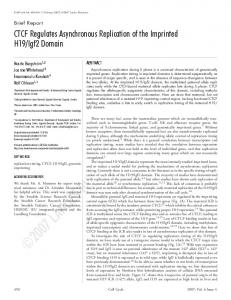
Fig. 1 Coordination of odorant-receptor asynchronous replication for individual mouse chromosome pairs. a, Diagram showing the relative positions of odorant-receptor genes (red) and Olfr3 other monoallelically expressed genes analyzed in Olfr5 Tcrb this study (blue) along with the location of control Olfr47 genes (black). Centromeric ends are at the top. Hoxa Dlx1 b–d, Two-color FISH analysis was done on a popuSnrpn Olfr10 Igk lation of mouse embryonic fibroblasts. Blue repreOlfr48 Il4 sents DAPI staining of chromatin. b, Analysis of D6Mit248 Tyr Myh4 Chromosome 11. The Cy3-labeled probe (red) V1rb1 identifies the Olfr1 odorant-receptor gene and Olfr1 Olfr41 Gt(ROSA)26Sor ( the FITC-labeled probe (green) identifies Olfr10. Cd4 The double-dot signals for the two probes in these Tg(Olfr19)Y11 images are on the same chromosome, indicating coordination of these two distant loci (30 of 30 cells counted). A control probe, Myh4, located 2 6 7 11 between the Olfr1and Olfr10 loci, is synchronously replicating (9% single dot–double dot pattern). c, Similar analysis of Chromosome 7 for two odorant-receptor genes from distinct clusters, Olfr5 (red) and Olfr41 (green), showing coordination (32 of 37 cells counted). Control probes between the Olfr5 and Olfr41 loci included the gene encoding tyrosinase, which is synchronously replicated in wild-type cells (13% single dot–double dot pattern), and the asynchronous but imprinted gene Snrpn. As expected, Snrpn did not show coordination with an odorant receptor (data not shown). Chr 11 30/30 Chr 7 32/37 Chr 2 33/36 d, Similar analysis showing that two odorantreceptor genes from distinct clusters of Chromosome 2 were coordinated (33 of 36 cells counted): Olfr48 (red), Olfr3 (green). A control probe, Dlx1, is synchronously replicating (10% single dot–double dot pattern). e,f, FISH analyses of line A.5 (see Supplementary Table 1 online) detected with a β-geo probe (green) that identifies the maternal Chromosome 11. Examples of cells probed with Olfr1 (red, e) and Olfr10 (red, f) are shown. Both of these odorant-receptor genes are maternal early-replicating in line A.5 (for data on all similar Olfr1 Olfr10 Chr 11 (pat) Chr 7 (mat) cell lines, see Supplementary Table 2 online). g,h, Lack of coordination between Chromosomes 7 and 11. In each case, the maternal chromosome is marked by the green probe. The maternal Chromosome 11 has a β-geo insertion and the paternal Chromosome 7 has a deletion at the tyrosinase locus. Line A.1 (see Supplementary Table 3 online) shows lack of coordination between paternally early-replicating Olfr1 on Chromosome 11 (g) and maternally early-replicating Olfr41 on Chromosome 7 (h).
a
b
e
nature genetics • volume 33 • march 2003
c
f
d
g
h
1
© 2003 Nature Publishing Group http://www.nature.com/naturegenetics
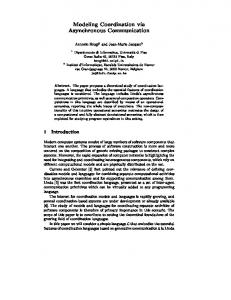
brief communications Fig. 2 A variety of monoallelically expressed genes show coordination. a–d, Analyses of a clonal cell line that has a marked maternal copy of Chromosome 6 (β-geo transgene, green). Probes for the Igk cluster (a), a large V1R pheromone receptor cluster (VNO-61; b), the Tcrb (c) and an odorant-receptor cluster containing Olfr47 (d) are shown (red). Line F.1 has all four genes maternally early replicating. Line C.1 (see Supplementary Fig. 2 online) has all Igk V1rb1 Tcrb Olfr47 four paternally early replicating. Two control probes, Hoxa and Cd4, each showed synchronous replication (12% and 18% single dot–double dot pattern, respectively). e, Analyses of uncloned fibroblasts (similar to the analyses shown in Fig. 1b–d) also showed coordination of an odorant-receptor gene cluster on Chromosome 6 containing Olfr47 (red) and the VNO V1R cluster (green; 30 of 32 cells). f, Same analysis as in e, but comparing coordination of Olfr47 with Tcrb (33 of 34 cells). g, A similar population analysis showed coordination of Il4 (red) and Olfr10 (green) on Chromosome 11 (25 of 26 cells). h, Example of coordination of endogenous Olfr48 (green) and transgenic Tg(Olfr19)Y11 (red) on Chromosome 2 (30 of 31 cells; P < 0.0000001).
Asynchronous replication can be assayed by fluorescence in situ hybridization (FISH) analysis of interphase nuclei14. Replicated loci are visualized as a doubledot hybridization signal, whereas unreplicated loci are visible as a single dot. Asynchronously replicating genes present a single dot–double dot pattern in 30–40% of S-phase cells, whereas synchronously replicating genes present this pattern in roughly 10–15% of S-phase cells14. Although the FISH assay is only an indirect assessment of replication timing, asynchronous replication observed with this assay has been corroborated by direct measurements of replication timing (refs. 10,11; see Supplementary Note 1 and Supplementary Fig. 1 online). To assess coordination of distant loci on a given chromosome, we used two-color FISH analysis to examine two genes simultaneously and scored cells that presented a single dot–double dot signal for both genes. If the two genes are coordinated, and are replicated during a overlapping portion of S phase, the double dots for both genes should be on the same chromosome (maternal or paternal) and therefore close to each other in the nucleus. If the two genes are not coordinated, the double dots for both genes should be on the same chromosome only 50% of the time. Using this approach we assessed the potential for coordination of asynchronous replication in wild-type primary mouse embryonic fibroblasts, analyzing two distinct odorant-receptor loci on Chromosome 11 that are 14 cM apart. Notably, we observed coordination in all of the 30 cells in which both probes presented the single dot–double dot pattern (Fig. 1b). Similarly, we observed coordination for two distant loci on Chromosome 7 (in 32 of 37 cells) and for two distant loci on Chromosome 2 (in 33 of 2
a
b
c
d
e
f
g
h
36 cells; Fig. 1c,d). As expected, in each case, genes between the distinct odorantreceptor loci replicated synchronously. These data indicate that odorant-receptor genes have long-range coordination of their replication asynchrony for the three autosomes examined. We examined whether coordinated asynchronous replication of odorantreceptor genes, once established, is heritable in the progeny of a given cell. We derived clonal cell lines from embryonic and adult mice with distinguishable maternal and paternal chromosomes for Chromosomes 7 and 11. We analyzed seven cell lines for Chromosome 7 and eight cell lines for Chromosome 11. In some cell lines we consistently observed early replication of the maternal allele (Fig. 1e), and in the other cell lines we consistently observed early replication of the paternal allele (see Supplementary Tables 1 and 2 online). These analyses indicate that for each odorant-receptor gene, the random choice of one of the two alleles to replicate early, once established, is heritable. Analyses of these clonal cell lines also confirmed coordination of asynchronous replication along a given chromosome. For all the cell lines analyzed, both loci replicated the same parental allele early (Fig. 1e,f and see Supplementary Tables 1 and 2 online). To test for genome-wide coordination, we analyzed clonal cell lines derived from mice carrying marks allowing us to distinguish the parental origins of two chromosome pairs at a time. We used FISH analysis to compare the replication timing of odorant-receptor genes on Chromosomes 6 and 11 with odorant-receptor genes on Chromosome 7 in pairwise comparisons. Odorant-receptor genes on different chromosomes were not coordinated in their replicative asynchrony; we
observed all possible outcomes (Fig. 1g,h and see Supplementary Table 3 online). Thus, rather than genome-wide coordination, asynchronous replication of odorantreceptor genes seems to be coordinated only at the level of each chromosome pair. To explore whether other randomly monoallelically transcribed genes are also coordinated in their replicative asynchrony, we examined clonal cell lines in which we can distinguish the maternal and paternal copies of Chromosome 6. We observed asynchronous replication of the Igk constant region, a V1R pheromonereceptor gene and Tcrb (Fig. 2a–c). Notably, all three of these loci were coordinated with each other as well as with an odorant-receptor gene cluster on Chromosome 6 (Fig. 2a–d). In some clonal cell lines the maternal alleles of all four genes replicated early (Fig. 2a–d), and in others the paternal alleles of all four genes replicated early (see Supplementary Fig. 2 online). Analyses of uncloned populations of cells (similar to the analyses presented in Fig. 1b–d) also showed coordination of genes on Chromosome 6 (Fig. 2e,f). Similar population analyses showed that on Chromosome 11, the gene encoding interleukin-4 (Il4) was coordinated with the odorant-receptor genes (Fig. 2g). These data, taken together, indicate that all randomly asynchronously replicated genes examined are coordinated along each chromosome. One question arising from these observations is whether the coordination mechanisms used by different chromosomes can communicate with each other if sequences from different chromosomes are artificially placed in cis. We analyzed a small odorantreceptor translocation that we created artificially: a 300-kb odorant receptor– containing YAC transgene derived from Chromosome 16 that is integrated on nature genetics • volume 33 • march 2003
© 2003 Nature Publishing Group http://www.nature.com/naturegenetics
brief communications Chromosome 2 (Tg(Olfr19)Y11). We previously showed that this transgenic odorantreceptor locus undergoes asynchronous replication15. Two-color FISH analysis showed that the transgene was coordinated in its asynchronous replication with the endogenous odorant-receptor loci on Chromosome 2 (Fig. 2h), suggesting similarities in the mechanisms governing allelespecific replication timing on different chromosomes. Here, we present data indicating that randomly monoallelically expressed genes coordinate their asynchronous replication within each chromosome pair. Scattered genes along a given chromosome are coordinated in their asynchronous replication timing in cis, leaving unaffected the bulk of the genes (which are synchronously replicated or, in rare instances, asynchronous but imprinted). Asynchronous replication is established early in development10,11 and maintained in the progeny of individual cells in a clonal manner (ref. 10; Fig. 1e–h and see Supplementary Tables 1 and 2 online). Randomly monoallelically expressed genes are expressed in different cells of a given cell type or in different cell types. Therefore, coordination of replication timing does not imply coordination of transcription of distinct gene families and is instead a consequence of the early developmental mechanisms that establish asynchronous replication. Each gene family probably makes use of asynchronous replication (and the differences in chromatin structure that it reflects) in the complex gene regulation that characterizes these gene families. In the case of the immunoglobulin genes, we have recently
nature genetics • volume 33 • march 2003
shown that early replication correlates with the allele that will first undergo rearrangement and therefore provides a basis for the establishment of allelic exclusion10. X inactivation has been known for decades. Our data indicate that chromosome-pair non-equivalence is also found on autosomes and thus is a general, fundamental property of chromosomes that affects a large number of loci dispersed throughout the genome. The autosomal non-equivalence we observe is similar to that observed with X inactivation, except that a larger fraction of the genes on the X chromosome are affected. Other similarities in the underlying mechanisms of X inactivation and autosomal non-equivalence may emerge with further investigation. Note: Supplementary information is available on the Nature Genetics website. Nandita Singh1*, Farah A.W. Ebrahimi1,2*, Alexander A. Gimelbrant1, Alexander W. Ensminger1,2, Michael R. Tackett1,2, Peimin Qi1, Joost Gribnau1 & Andrew Chess1,2 *These authors contributed equally to this work. 1Whitehead
Institute for Biomedical Research, Nine Cambridge Center, Cambridge, Massachusetts 02142, USA. 2Department of Biology, Massachusetts Institute of Technology, Cambridge, Massachusetts 02139, USA. Correspondence should be addressed to A.C. (e-mail:
[email protected]).
Acknowledgments
We thank H. Cedar, G. Fink, D. Housman, R. Jaenisch, D. Page, C. Cowles and members of A.C.’s laboratory for discussions and comments on
the manuscript; H. Higgins for manuscript preparation; G. Paradis for help with fluorescenceactivated cell sorting analyses; J. Young for Chromosome 6 BACs, D. Littman for the Cd4 cosmid; R. Lane for the Olfr41 BAC; B. Holdener for the tyrosinase deletion mice; A. Dunn for the CSF2 knock-in mice; P. Soriano for the β-geo transgenic mice; N. Rosenberg for the Abelson leukemia virus–producing lines; D. MacAlpine for advice on immunoprecipitation; S. Gabriel, B. Blumenstiel, M. DeFelice and E. Winchester for help with MALDI-TOF genotyping; and S. Rozen for help with sequencing. This work was supported by grants from the U.S. National Institutes of Health (National Institute on Deafness and Other Communication Disorders) to A.C., F.A.W.E. and M.T. A. W.E. is a Howard Hughes Medical Institute Predoctoral Fellow. Competing interests statement
The authors declare that they have no competing financial interests. Received 6 December 2002; accepted 19 January 2003. 1. 2. 3. 4. 5. 6. 7. 8. 9. 10. 11. 12. 13. 14. 15.
Lyon, M.F. Nature 320, 313 (1986). Efstratiadis, A. Curr. Opin. Genet. Dev. 4, 265–280 (1994). Chess, A., Simon, I., Cedar, H. & Axel, R. Cell 78, 823–834 (1994). Pernis, B., Chiappino, G., Kelus, A.S. & Gell, P.G. J. Exp. Med. 122, 853–876 (1965). Rajewsky, K. Nature 381, 751–758 (1996). Rhoades, K.L. et al. Curr. Biol. 10, 789–792 (2000). Hollander, G.A. et al. Science 279, 2118–2121 (1998). Held, W., Roland, J. & Raulet, D.H. Nature 376, 355–358 (1995). Rodriguez, I., Feinstein, P. & Mombaerts, P. Cell 97, 199–208 (1999). Mostoslavsky, R. et al. Nature 414, 221–225 (2001). Simon, I. et al. Nature 401, 929–932 (1999). Young, J.M. et al. Hum. Mol. Genet. 11, 535–546 (2002). Zhang, X. & Firestein, S. Nat. Neurosci. 5, 124–133 (2002). Selig, S., Okumura, K., Ward, D.C. & Cedar, H. EMBO J. 11, 1217–1225 (1992). Ebrahimi, F.A., Edmondson, J., Rothstein, R. & Chess, A. Dev. Dyn. 217, 225–231 (2000).
3