ARTICLE IN PRESS ARTICLE IN PRESS
Atom Indonesia Indonesia Vol. Vol. 40 40 No. No. 33 (2014) (2014) Atom I. Kurnia, et.al. / Atom Indonesia Vol. 40 No. 3 (2014)
Correlation between Expression of MVP, Index of p53 and AgNOR Value with Chemoradiotherapy Clinical Response of Cervical Cancer I. Kurnia1*, B. Siregar2, S. Soetopo3, I. Ramli2, T. Kurjana3, D. Tetriana1, B.S. Hernowo3, A. Andrijono2 and M.D.M. Tobing3 1
Center for Radiation Safety Technology and Metrology, National Nuclear Energy Agency, Jl. Lebak Bulus Raya No. 49. Pasar Jum’at, Jakarta 12440, Indonesia 2 Cipto Mangun Kusumo Hospital, Jl. Diponegoro No.71, Central Jakarta, Indonesia 3 Hasan Sadikin Hospital, Jl. Pasteur No.38, Bandung 40161, Indonesia
ARTICLE
INFO
Article history: Received 20 November 2013 Received in revised form 18 July 2014 Accepted 28 September 2014 Keywords: Major vault protein p53 MIB-1 AgNOR Chemoradiotherapy Cervical cancer
ABSTRACT Cervical cancer is the most frequently found cancer in Indonesia. The primary treatment of cervical cancer at the locally advanced stage is usually performed by using radiotherapy and chemotherapy. The combination of the two techniques is often called chemoradioherapy. The response to chemoradiotherapy is influenced by biological and physical factors. Major vault protein (MVP) is a ribonucleoprotein which contributes to drug resistance in some cancers. The purposes of this research were: (1) to determine the correlation between the expression of MVP and the index of p53, including AgNOR values and index of MIB-1; and (2) between MVP and chemoradiotherapy clinical response of cervical cancer. Twenty-one microscopic slides taken from biopsy tissues of cervical cancer patients before undergoing treatment were stained to identify MVP, p53, and MIB-1 by means of immunohistochemistry techniques and AgNORs staining. After undergoing chemoradiotherapy treatment, the patients’ clinical responses were observed by pelvic control method. Experimental results showed that there was a correlation between MVP and AgNOR value (P=0.05) but no correlation between MVP and index of p53 (P=0.729), including MIB-1 LI (P=0.63), in untreated cervical cancer. In addition, there was no association between MVP and chemoradioterapy response. In conclusion, MVP expression correlates with the process of cell proliferation before the G2 phase of cell cycle in untreated cancer cells and have no association with clinical responses after the completion of treatment. © 2014 Atom Indonesia. All rights reserved
INTRODUCTION Carcinoma cervix uterine is the second most common malignant tumor in women worldwide, with an estimated 493,000 new cases (83% occurring in developing countries) and 274,000 cancer-related deaths in the year 2002 [1]. In Indonesia, this is the most common cancer type and 70% of the patients came to hospital in locally advanced stage condition. The main treatment for this stage is radiotherapy combined with chemotherapy in concurrent chemoradiotherapy [2,3]. Establishing the prognosis of a patient with cervical cancer is an important part of the clinical evaluation and treatment. The most recognized prognostic factor in cervical cancer is the disease extension, usually estimated by the TNM/FIGO
Corresponding author. E-mail address:
[email protected]
staging system (Tumor-Nodus-Metastasis / International Federation of Gynecology and Obstetrics) [4-6]. However, within each clinical disease stage, biological markers are needed to clarify the identification of high risk patients who may benefit from individualized therapeutic options of carcinoma cervix uterine [7]. The vault is a barrel-shaped cytoplasmic riboprotein particle which is grouped into multiple copies of three proteins. The mammalian vault complex is made of major vault protein or lung resistance-related protein (MVP or LRP, M(r) = 100,000), vault poly ADP-ribose polymerase (VPARP, M(r) 193,000) and telomerase associated protein 1 (TEP-1, M(r) = 240,000) which are associated with small 88-141-bp fragments of untranslated RNA [8-11]. While vaults are found in all human tissues, elevated level of expression of MVP is found in gut epithelium, lung epithelium, macrophages and dendritic cells, which are all
ARTICLE IN PRESS I. Kurnia, et.al. / Atom Indonesia Vol. 40 No. 3 (2014)
typically exposed to xenobiotics. This implies that vaults may have a role in the defense of such tissue against toxic insult, and they are found highly expressed in various multidrug-resistant cancer cell lines [12-14]. Growing cancers are often influenced by increased genetic changes. Such genetic changes, including chromosomal aberrations (translocations), gene amplifications, intragenic mutations, and gene silencing are responsible for the activation of oncogenes and the inactivation of tumor-suppressor genes. Exposure of cells to extreme conditions like cancer cell hypoxia can promote genome alterations, enhancing the progression potential of tumor cells and resistance to oncological treatments. Hypoxia may lead to conditions that cause increased damage to DNA or inhibit DNA repair processes, impair DNA and cause tumor progression by altering p53 expressions and increasing angiogenesis. Loss of regulation of DNA repair pathways can influence the phenomenon of hypoxia-induced genetic instability within the tumor [15-18]. The MIB-1, also called Ki-67, is expressed in all cell cycle stages except G0 and early G1 phases. This antigen is thought to be associated with a nuclear antigen protein-DNA replicase complex, similar to DNA topoisomerase II [19]. Generally, a higher MIB-1 labeling index (MIB-LI / MIB-1 LI) correlates with worse prognosis; however, tumors with higher MIB-1 LI are often radiosensitive [20]. Nucleolar organizer regions (NORs) are chromosomal loops of DNA involved in ribosomal synthesis. The silver staining technique can easily detect NORs in formalin fixation, and NORs can be identified as black dots in the nucleolus (AgNORs). This method permits the rapid evaluation of morphology and tumor cell kinetics even using small biopsies. Evaluation of AgNOR parameters (number, size, and distribution) has been applied in tumor pathology both for diagnostic and prognostic purposes [21, 22]. In the last few years a considerable number of studies have shown correlation between biomarkers of cell proliferation - such as index of p53, index of MIB-1, and AgNORs - and chemoradiotherapy or radiotherapy clinical responses [20,23,24]. While there a positive correlation between AgNORs and MIB-1, AgNOR value tends to decrease whereas the index of MIB-1 increases if cervical cancer is treated specially for one week with chemoradiotherapy [24]. The aim of the present study was to assess (1) the correlation between the expression of the MVP in chemoradiotherapy untreated cervical cancer and AgNOR values, including also index of MIB-1 and index of p53, and (2) the correlation between MVP
and clinical response of cervical cancer to chemoradiotherapy. EXPERIMENTAL METHODS Patients From July 2010 to March 2011, 21 consecutive patients were enrolled and studied in this work. Their data is summarized in Table 1. Those patients were taken from a whole series of 60 cases who were suffering from non-metastatic localized cervical carcinoma in stage IIB-IIIB. Among those 60 patients, the 21 patients studied were the ones who have achieved complete response to treatment. All patients were diagnosed and treated by definitive radiation at the Cipto Mangunkusumo Hospital and Hasan Sadikin Hospital and received written informed consent. The study was approved by the Health Research Ethics Committee of the Faculty of Medicine, University of Indonesia. Table 1. Expression of Major Vault Protein (MVP), index of p53, index of MIB-1 , value of AgNORs and clinical response in cervical cancer treated with chemoradiotherapy No Patient
MVP Chemoradiotherapy Index Clinal MIB- AgNORs of Stage Expre Grouped 1LI Value Response Grouped p53 ssion
1
A
IIB
W
1
0,21
0,40
5,71
partial
1
2
B
IIB
W
1
0,36
0,43
6,38
partial
1
3 4 5 6 7 8 9 10 11 12 13 14 15 16 17 18 19 20 21
C D E F G H I J K L M N O P Q R S T U
IIIB IIB IIIB IIB IIB IIIA IIIB IIIB IIIB IIB IIIB IIB IIB IIB IIB IIB IIB IIB IIB
W M M M S M S M M M M M W M M M M W S
1 2 2 2 2 2 2 2 2 2 2 2 1 2 2 2 2 1 2
0,36 0,36 0,17 0,26 0,48 0,67 0,65 0,38 0,60 0,36 0,59 0,28 0,66 0,48 0,54 0,44 0,43 0,39 0,13
0,38 0,51 0,30 0,29 0,59 0,67 0,36 0,31 0,21 0,41 0,41 0,68 0,60 0,50 0,31 0,55 0,47 0,52 0,36
6,46 5,15 4,41 5,03 5,26 7,59 4,24 5,79 4,66 5,24 4,52 5,38 4,76 6,67 5,93 4,77 5,53 8,65 5,22
partial partial complete complete complete complete complete complete partial partial complete partial complete complete complete complete complete complete complete
1 1 2 2 2 2 2 2 1 1 2 1 2 2 2 2 2 2 2
Note : w = weak, m = medium s = strong
The clinical staging of the patients was performed through speculoscopy, bimanual examination, and cystoscopy or rectoscopy when abdomen pelvic CT scans and chest X-rays were performed. The histological grading was based on the guidance issued by the Union for International Cancer Control [3] which defined the following grades: G1, well differentiated; G2, moderately differentiated; and G3, poorly differentiated or undifferentiated. All patients were identified as having squamous cell
ARTICLE IN PRESS I. Kurnia, et.al. / Atom Indonesia Vol. 40 No. 3 (2014)
carcinoma tumors, i.e., 14 patients in stage IIB, 1 patient in stage IIIA and 6 patients in stage IIIB, respectively. Treatment Patients were treated by means of a combination of External Beam Radiotherapy (EBRT) with 60Co gamma rays and High Dose-Rate Intracavitary Brachytherapy (HDR-ICBT) using 192 Ir. EBRT was subjected to the whole pelvis with a clinical target volume that included the primary cancer, uterus, internal iliac, presacral, upper external iliac, and lower common iliac lymph nodes.Chemotherapy was administered concurrently. Cisplatin was given before EBRT within two hours or less before treatment [25-27]. Clinical radiotherapy response Radiation responses were evaluated by radiotherapist in the Department of Radiotherapy, Cipto Mangunkusumo or Hasan Sadikin Hospital, and the responses are grouped according to Hong Criteria [28] as follows: 1. NRT (no gross residual tumor) response; complete or nearly complete regression of pelvic tumor, nonspecific fibrosis, or granulation over the cervix. This is called good response. 2. GT (gross residual tumor) response: gross tumor or palpable nodularity on cervix, and/or palpable in duration on the parametrium. This is called bad response.
Immunohistochemistry Expressions of MVP, p53, and MIB-1 were examined by immunohistochemistry. In brief, the steps were as follows: paraffin-embedded tumor tissue biopsies were first incubated with mouse antiMVP monoclonal antibody (LRP/MVP Ab-2, Clon 1032, Abcam, CA); then applied at a 1:100 dilution, anti p53 monoclonal antibody (Leica, Novocastra, ready-to-use p53-D07); the biopsies were then incubated overnight at 5°C, in a moist chamber; followed with post primary, post protein and Novolink HRP system (Novolink) and revealed with DAB (Novolink) and counterstained with Mayer Hematoxylin. The primary antibody was omitted in one section as a negative control and a strongly positive tumor for MVP was used as a positive control. Expressions of MVP in cell cytoplasm and membrane was observed in zones of maximum
expression of the marker in at least 10 high power fields (400×) and semi quantitatively scored as low (negative/slightly positive) or high (strongly positive) [29,30]. Staining for p53, observed in the nuclei, was scored as percentage of stained cells [24]. Up to 1000 cells were counted in each slide of patient.
AgNOR staining AgNOR staining technique was performed in accordance with the technique described by Ploton et al. [31,32]. Tissue sections were cut at 4 μm thickness from formalin fixed, paraffin-wax embedded blocks. The sections were dewaxed in xylene and then hydrated through decreasing grades of ethanol followed by washing in deionized water for 8–10 minutes. The staining solution was prepared by dissolution of powdered gelatin with concentration of 2% w/v in deionized water over water bath at 60–70°C. Pure formic acid was added to final concentration of 1%. This solution was mixed 1:2 (v/v) with 50% aqueous silver nitrate solution, then filtered through a 0.22 mm Millipore filter, and dropped onto the slide-mounted section. The sections were incubated in the dark for 40–45 minutes at room temperature. After rinsing three times with deionized water, the slides were immersed for 10 minutes in 5% sodium thiosulphate solution, dehydrated in ascending ethanol concentrations, cleared with xylene, and mounted. According to the recommendations of Crocker et al. [32], dots lying in a group or cluster (almost aggregated or partly disaggregated) were treated as one structure, whereas if AgNORs could been seen separately they were considered as individual AgNORs [33].
Statistical analysis AgNOR values, indices of p53, and indices of MIB-1 were analyzed by using Kolmogorov test for categorized normal distribution. Analysis of variance (ANOVA) was used to analyze the correlation between the expression level of MVP in cancer with biomarkers of cell proliferation, i.e., AgNOR value, index of p53, and index of MIB-1. Fisher’s Exact Test was used to analyses association between MVP expression and chemoradiotherapy clinical response. All statistical analyses were performed using Medcalc Software Version 9.2.0.1. RESULTS AND DISCUSSION
ARTICLE IN PRESS I. Kurnia, et.al. / Atom Indonesia Vol. 40 No. 3 (2014)
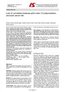
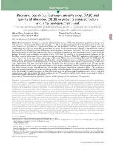
All immunohistochemical markers and AgNOR staining were obtained in all 21 microscopic slide from 21 cases (Table 1). It appears that the expression of MVP in cancer cell cytoplasm was shown as brown color (Fig. 1a,b,c). Based on MVP expressions, Table 1 classifies patients into two groups. Group 1 consists of 5 patients (18.5%) who have low MVP expression, while the 16 patients (81.5%) with strong or medium MVP expression for 16 patients are categorized as Group 2. Table 1 also indicates that 4 of 14 patient (29 percent) in clinical stage IIB show low expression of MVP, while in IIIA and IIIB stages, the same low expression is indicated by 1 of 7 patients (14%). In the same table, it appears that the AgNOR value in stage IIB is 5.69 while in stages IIIA and IIIB it is 5.38. The expression of p53 protein in nuclei as brown color is shown in Fig. 2a and indices of p53 with the values varying from 13% to 67% (mean 41%) is presented in Table 1.
(a)
(c)
and chemoradiotherapy clinical P = 0.28>0.05 (see Fig. 3d).
(a)
response,
as
(b)
(c) Fig. 2. Expression of (a) p53, (b) AgNORs, and (c) MIB-1 in cervical cancer tissue before treatment by chemoradioterapy, originally magnification10 x 40.
(b)
(a)
(b)
(c)
(d)
(d)
Fig. 1. Expression of MVP (a) weak, (b) medium, and (c) strong, in cervical cancer tissues before treatment by chemoradiotherapy, negative control from breast cancer tissue (d), originally magnification 10 x 40.
AgNORs in nuclei result in a dot and group of dots with black color (see Fig. 2b), and MIB-1 expression in nucleus as brown color one is depicted in Fig. 2c. AgNOR scores varied from 4.24 to 8.65 (mean 5.59 ± 1.09) and MIB-1 indices varied from 0.21–0.68 0.13 (see Table 1). We observed statistical correlation between the expression of MVP and AgNORs, P = 0.05 ( see Fig. 3b). There was no statistical correlation between expression of MVP and the index of p53 with P = 0.72>0.05 (see Fig. 3a), and between MVP expression and MIB-1 index with P=0.63 (see Fig. 3c). Further, there was no correlation between MVP expression
Fig. 3. (a) Index of p53 in low and strong MVP, (b) Value of AgNOR in low and strong MVP, (c) Index of MIB-1 in low and strong MVP, and (d) expression of MVP grouped chemoradiotherapy clinical response.
Theoretically, any relationship between MVP and AgNORs may be associated with degradation of nucleolin as part protein of AgNORs in preparing proliferation processes in the S phase before a cell enters the mitotic phase. An increase in AgNORs volume would indicate transcriptional activation of inactive NORs, and a reduction in number of AgNORs in nucleus may indicate association
ARTICLE IN PRESS I. Kurnia, et.al. / Atom Indonesia Vol. 40 No. 3 (2014)
processes. However, the decreases of the number of AgNORs and increases in its volume could not be explained by association processes alone, but it also results from an absolute rise in transcriptional activity [22,24]. In contrast to AgNOR values, MIB1 indices were found in all phases of cell cycle except in G0 phase (phase before entering into mitosis) [20], but it had no correlation with MVP expressions. This means that the expression of MVP is not related with the cancer cell proliferation process. The same is indicated by the expression of p53 that is not correlated with MVP. The p53 is a protein expressed in the tumor suppressor gene; it functions in cell cycle arrest if any DNA damage is found, and in initiating apoptosis. In this way, protein p53 initiates the formation of protein Bax, and also has a role in preventing apoptosis from inducing the activation of Bcl-2 protein [17,18]. The poor prognosis after radiotherapy is related with the failure of the processes of apoptosis and hypoxia. MVP and vaults have recently been linked to both major DNA double-strand break repair machineries, namely Ku70 and Ku80. Those two proteins are key proteins in non-homologue end joining, and also play a strong regulatory role in apoptosis through Bax/Bcl-2 interactions [29,30]. Our experimental results show that there is sufficient evidence to infer that the expression of MVP is not directly correlated with the proliferation of the cell. Higher percentages of high expression MVP in IIIA and IIIB than in IIB are probably related with potential metastasis and ability of cancer cell in preventing apoptosis as a part of prognosis. There is no correlation between MVP and radiation response after treatment completed. It is different with some reports that high expression of MVP will correlate with poor prognosis after radiotherapy [29,30]. This differing result is possibly due to some factors which also contribute to chemoradiotherapy response. Probably all patients included in this research were without hypoxic condition. Hypoxic condition was related to high expressions of major vault protein in cervical cancer treated with chemoradiotherapy [16,34]. This study actually has limitations, since the duration of observation of chemoradiotherapy clinical response was only three months after the treatment, and the response was only observed through pelvic control method. As high expressions of MVP tend to be found in higher clinical stages than in lower ones, we think that it will be influenced by the responses long time after treatment. It is suggested that in the next study, the response is observed for more than three months and
also to use quantitative method such as computed tomography scan or magnetic resonance imaging.
CONCLUSION High expressions of MVP are related to AgNOR degradation in S phase of cell cycle. No relationship was found between MVP and both of protein p53 and MIB-1. Expression of MVP is not associated with early clinical response of chemoradiotherapy in cervical cancer and only tends to be associated with the clinical stage of the patient.
ACKNOWLEDGMENT This research was supported by a research grant from the Center for Radiation Safety Technology and Metrology, National Nuclear Energy Agency and PKPP “Block Grant” Fiscal Year 2010. The authors thank Prof. Soehartati Gondhowiardjo MD. PhD., Endy Moegni, MD, Endang SR Harjolukito, MD, (Cipto Mangunkusumo Hospital) and Prof. Firman MD. PhD. (Hasan Sadikin Hospital) for scientific advices.
REFERENCES 1. D.M. Parkin, F. Bray, J. Ferlay et al., Cancer J. Clin. 55 (2005) 74. 2. M.F. Aziz, How to Early Detection and Prevent of Cervical Cancer, in: Prevention and Early Detection of Cancer in Indonesia, Susworo, H.R.Tjarta and H.A. Budiana (Eds.), University of Indonesia Press (1996)106. 3. S. Mufyala and A.H. Wofson, Cervical Cancer, in: Radiation Oncology, J.J. Lu and L.W. Brady (Eds.), Springer (2008) 357. 4. J.L. Benedet, H. Bender, H. Jones et al., Int. J. Gynaecol. Obstet. 70 (2000) 209. 5. S. Pecorelli, L. Zigliani and F. Odicino, Int. J. Gynaecol. Obstet. 105 (2009). 6. Anonymous, TNM Classification of Malignant Tumours, in: International Union Against Cancer, 6th ed., L.H. Sobin and Wittekind Ch (Eds.) Wiley-Liss, New York (2002) 154. 7. E.A. Pintoa, P. Ausenda, S. Giovani et al., Pathol. Res. Pract. 207 (2011) 623. 8. N.L. Kedersha, J.E. Heuser, D.C. Chugani et al., J. Cell Biol. 112 (1991) 225. 9. G.L. Scheffer, P.L. Wijngaard, M.J. Flens et al., Nat. Med. 1 (1995) 578.
ARTICLE IN PRESS I. Kurnia, et.al. / Atom Indonesia Vol. 40 No. 3 (2014)
10. V.A. Kickhoefer, A.C. Siva, N.L. Kedersha et al., J. Cell Biol. 146 (1999) 917. 11. V.A. Kickhoefer, A.G. Stephen, L. Harrington et al., J. Biol. Chem. 274 (1999) 32712. 12. G.L. Scheffer, P.L.J. Wijngaard, M.J. Flens et al., Nat. Med. 1 (1995) 578.
Asian J. Cancer 4 (2005) 202. 24. I. Kurnia, Y. Suzuki, S. Budiningsih et al., Austral. - Asian J. Cancer 8 (2009) 93. 25. R. Pearcey, M. Brundage, P Drouin et al., J. Clinical Oncology 20 (2002) 966.
13. R.J. Scheper, G.L. Scheffer, M.J. Flens et al., Role of LRP/Major Vault Protein in Multidrug Resistance, Multidrug Resistance in Cancer Cells, S Gupta, T. Tsuruo (Ed.), John Wiley & Sons Ltd., Chichester (1996) 109.
26. E.C. Halperin, L.W. Brady, C.A. Perez et al., Brachyterapy HDR and LDR, in: Perez and Brady’s Principle and Practice of Radiation Oncology, A.A. Martinez, C.G. Orton and RF Mould, (Eds.), Nucletron International BV, Netherlands (1990) 121.
14. M.A. lzquierdo, G.L. Scheffer, M.J. Flens et al., Am. J. Pathol. 148 (1996) 877.
27. C.A. Joslin, C.W. Smith, A. Mallik, Brit. J. Radiol. 45 (1972) 257.
15. L.E. Huang, R.S. Bindra, P.M. et al., J. Mol. Med. 85 (2007) 139.
28. J.H. Hong, M.S. Chen, F.J. Lin et al., Int. J. Radiat. Oncol. Biol. Phys. 22 (1991) 913.
Glazer
16. P.C. Lara, M. Lloret, B. Clavo et al., Radiother. Oncol. 89 (2008) 222. 17. R.D. Wood, Annu. Rev. Biochem. 65 (1996) 135. 18. R.S. Bindra, M.E. Crosby and P.M. Glazer, Cancer Metastasis Rev. 26 (2007) 249. 19. J. Gerdes, U. Schwab, H. Lemke et al., Int. J. Cancer 31 (1983) 13. 20. K. Oka, Y. Suzuki and T. Nakano, Cancer 89 (2000) 1526.
29. M. Lioret, P.C. Lara PC, E. Bordon et al., Gynecol. Oncol. 106 (2007) 8. 30. M. Lioret, P.C. Lara, E. Bordon et al., Int. J. Radiat. Oncol. Biol. Phys. 73 (2009) 976. 31. D. Ploton, M. Menager, P. Jeannesson et al., Histochem. J. 18 (1986) 5. 32. J. Crocker, A. David, R. Boldy et al., J. Pathol. 158 (1989) 185.
21. J.G. Gall and M.L. Pardue, Proc. Natl. Acad. Sci. 64 (1969) 600.
33. W.M. Howel, Selective Staining of Nucleolus Organizer Regions (NORs), in: The Cell Nucleus, H. Busch and L. Rothblum (Eds.), Academic Press, New York (1982) 89.
22. M. Derenzini and D. Ploton, Int. Rev. Exp. Pathol. 32 (1991) 150.
34. C.L. Pedro, L. Marta, C. Bernardino et al., Radiat. Oncol. 4 (2009) 29.
23. I. Kurnia, T. Ohno, S. Kato et al., Austral.-