Different filling defect patterns in ureteral carcinoma
CORRELATION BETWEEN FILLING DEFECT PATTERNS ON UROGRAPHY AND PATHOLOGIC STAGING OF URETERAL TRANSITIONAL CELL CARCINOMAS Yi-Lun Lee, Shu-Pin Huang,1 Min-Chen Shih,2 Yii-Her Chou,1 and Chun-Hsiung Huang1 Department of Urology, Chi-Shan Hospital, and Departments of 1Urology and 2 Radiology, Kaohsiung Medical University Chung-Ho Memorial Hospital, Kaohsiung, Taiwan.
Although a filling defect within the ureter is the most common finding with ureteral transitional cell carcinomas (TCCs), little is known about the correlation between filling defect patterns and pathologic findings. This study was conducted to address this. Between January 1995 and January 2003, 126 pathologically confirmed TCCs of the ureter were included in our study. We classified urographic filling defects into four patterns: ovoid, polypoid, infiltrating, and plaque-like. The correlation between different filling defect patterns and pathologic findings was assessed using Pearson’s Chi-squared and logistic regression methods. There were 28 (22%) ovoid filling defects, 42 (33%) polypoid filling defects, 37 (29%) infiltrating filling defects, and 19 (15%) plaque-like filling defects. Infiltrating and plaque-like filling defects were significantly associated with more advanced disease compared to ovoid and polypoid filling defects (odds ratio, 6.75; 95% confidence interval, 3.04–14.98; p < 0.0001). Our results suggest that filling defect presentations may signify different invasive behavior among TCCs. The distribution of ovoid, polypoid, infiltrating, and plaque-like filling defect patterns is significantly different between superficial and advanced ureteral TCCs. We suggest that classifying the filling defect patterns of ureteral TCCs may provide important preoperative information for planning treatment and predicting outcome.
Key Words: neoplasm, transitional cell carcinoma, ureter, urography (Kaohsiung J Med Sci 2003;19:447–52)
Transitional cell carcinoma (TCC) of the ureter is not common worldwide, representing only 1% of all genitourinary malignancies [1–3]. However, ureteral carcinoma is relatively common in Taiwan [4–7]. Urographic examinations, including intravenous pyelography (IVP), retrograde pyelography (RP), and antegrade pyelography (AP), are important tools in detecting the tumor and defining its extent and location [1,3,8]. The most common
radiographic finding in ureteral carcinoma is the presence of a filling defect within the ureter [9,10]. Although various filling defect patterns have been reported with ureteral TCCs [1,8,11], only a few studies have focused on the association between filling defect patterns and pathologic findings. Here, we report a large series of ureteral TCCs and analyze the correlation between filling defect patterns and pathologic staging.
MATERIALS AND METHODS Received: January 24, 2003 Accepted: August 12, 2003 Address correspondence and reprint requests to: Dr. Yii-Her Chou, Department of Urology, Kaohsiung Medical University Chung-Ho st Memorial Hospital, 100 Tzyou 1 Road, Kaohsiung 807, Taiwan. E-mail:
[email protected] Kaohsiung J Med Sci September 2003 • Vol 19 • No 9 © 2003 Elsevier. All rights reserved.
Between January 1995 and January 2003, patients at our institution with histopathologically confirmed TCC of the ureter and with complete radiographic and histologic data 447
Y.L. Lee, S.P. Huang, M.C. Shih, et al
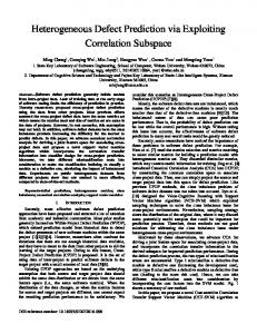
were eligible for inclusion in the study. Computed tomography was obtained in all cases and all patients underwent at least one urographic examination (IVP, RP, or AP). RP or AP was usually performed when IVP failed to localize the lesion or when patients had renal insufficiency. Urographic films were reviewed by an independent radiologist (MCS) who was blinded to clinical and pathologic data. Filling defects on urography were divided into four patterns by their shape: ovoid, polypoid, infiltrating, or plaque-like [11]. We defined ovoid as a single, rounded, nodular, exophytic lesion (Figure A); polypoid as a papillary, lobulated, exophytic lesion (Figure B); infiltrating as an extensive, straggling, sessile, broad-based lesion (Figure C); and plaque-like as a thickened-wall constriction with overhanging edges and strand-like contrast filling appearance (Figure D). Surface regularity, goblet sign, stipple sign, concomitant calcification, and pseudodiverticulum were also recorded. We used the most clearly defined images from IVP, RP, and AP to categorize the patterns of the filling defects. Pathologic staging was based on the classifications proposed by Batata et al [12]: stage A is defined as submucosal infiltration, stage B as muscular invasion, stage C as periureteral involvement, and stage D as extension to adjoining structures, regional lymph node metastases, or distant spread. Stage A was considered superficial disease because it was limited to the submucosal layer. Stages B, C, and D were considered advanced disease because invasion reached at least the muscle. Pearson’s Chi-squared and logistic regression methods were used to examine the association between various filling defect patterns and pathologic staging. Data are presented as odds ratio (OR) and 95% confidence interval (95% CI). A p value of less than 0.05 was considered statistically significant. Data analysis was performed using SPSS version 8.01 (SPSS Inc., Chicago, IL, USA).
RESULTS Of 134 histopathologically confirmed cases of TCC of the ureter between January 1995 and January 2003, eight cases were excluded due to incomplete radiographic and histologic data. A total of 126 cases were enrolled in the study. Mean age at diagnosis was 66.5 years (range, 26–85). There were more male (54%) than female patients (Table 1). Most tumors were located in the upper third of the ureter; the left ureter was more frequently involved than the right ureter. There were similar numbers of superficial and 448
A
B
C
D
Figure. Different filling defect patterns on urography: (A) ovoid filling defect; (B) polypoid filling defect; (C) infiltrating filling defect; (D) plaque-like filling defect.
advanced tumors (Table 1). Gross hematuria was the most common symptom, occurring in 70% of patients. Flank pain, urinary frequency, dysuria, lower abdominal pain, and fever were less common symptoms. Seven patients (6%) were asymptomatic and were diagnosed incidentally on ultrasonography after presentation with hydronephrosis. Filling defect patterns were determined by RP (80 cases, 63%), AP (39 cases, 31%), and IVP (7 cases, 6%). Polypoid filling defects were most common (33.3%), followed by infiltrating (29.4%), ovoid (22.2%) and plaque-like (15.1%) filling defects (Table 2). Most lesions (81.7%) manifested with an irregular surface. The goblet sign was found in 30.2% of lesions and the stipple sign in 19.8%. Seven lesions (5.6%) showed concomitant calcification. There was no pseudodiverticulum. The distribution of ovoid, polypoid, infiltrating, and plaque-like filling defects was significantly different between superficial disease and advanced disease (Chi-squared test, Kaohsiung J Med Sci September 2003 • Vol 19 • No 9
Different filling defect patterns in ureteral carcinoma
p < 0.0001) (Table 3). Infiltrating and plaque-like filling defects were significantly associated with advanced disease (OR, 6.75; 95% CI, 3.04–14.98; p < 0.0001) (Table 4). Table 1. Demographic and disease characteristics of 126 patients with ureteral transitional cell carcinoma n (%) Male Female Right side Left side Location Upper third Middle third Lower third Whole length Superficial disease (Stage A) Advanced disease (Stages B, C, D) Stage B Stage C Stage D
68 58 49 77
(54) (46) (39) (61)
50 24 49 3 60 66 26 21 19
(40) (19) (39) (2) (48) (52) (21) (17) (15)
Table 2. Radiographic presentation of filling defects in 126 ureteral transitional cell carcinomas n (%) Shape Ovoid Polypoid Infiltrating Plaque-like Surface Regular Irregular Goblet sign Stipple sign Concomitant calcification Pseudodiverticulum
28 42 37 19
(22.2) (33.3) (29.4) (15.1)
23 103 38 25 7 0
(18.3) (81.7) (30.2) (19.8) (5.6) (0)
DISCUSSION Ureteral carcinomas are mostly located in the lower third of the ureter [12–14]. In northern Taiwan, interestingly, Yang et al documented an unusually high incidence of upper urinary tract urothelial carcinoma and suggested that geographic factors or genetic predisposition might contribute to this phenomenon [4]. Our data show that the upper third of the ureter is most frequently involved in ureteral TCCs in southern Taiwan. The possible cause of this finding in southern Taiwan is undetermined. Various authors consider that the high incidence of upper urinary tract carcinomas in southern Taiwan is due to the fact that it is an area that is endemic for blackfoot disease [5–7]. Evidence suggests that the arsenic content and fluorescent substances in artesian well water are a possible etiology in blackfoot-endemic areas [15–19], but further investigation is needed to determine whether this is so. There was no significant difference in filling defect presentations among the various locations of ureteral carcinomas in our study. IVP, RP, and AP are inexpensive and are examinations which can be easily performed to detect ureteral tumors and define their extent and location [1,3,8]. IVP is the primary initial examination for ureteral lesions, but only 6% of the tumors in our study could be clearly seen with their filling defect on IVP films. RP and AP films usually provide more satisfactory delineation of the filling defect and the precise filling defect pattern. RP and AP are more reliable than IVP and clearly revealed approximately 94% of filling defects in our study. Both the goblet sign and stipple sign are specific features of TCC of the ureter [20,21]. However, in our study, only 30.2% of cases had the goblet sign and only 19.8% had the stipple sign. Wasserman et al reported that ureteral pseudodiverticula were frequently associated with uroepithelial malignancy [22], but we observed no
Table 3. Comparison of filling defect patterns with tumor stage Tumor stage Filling defect pattern
N
Superficial disease (A) n (%)
Ovoid Polypoid Infiltrating Plaque-like
28 42 37 19
20 (71) 27 (64) 9 (24) 4 (21)
Advanced disease (B, C, D) n (%) 8 15 28 15
(29) (36) (76) (79)
Chi-squared test, p < 0.0001.
Kaohsiung J Med Sci September 2003 • Vol 19 • No 9
449
Y.L. Lee, S.P. Huang, M.C. Shih, et al Table 4. Correlation between filling defect patterns and tumor stage in ureteral transitional cell carcinomas Filling defect pattern
Ovoid or polypoid Infiltrating or plaque-like
Superficial disease n (%)
Advanced disease n (%)
OR (95% CI)
47 (67) 13 (23)
23 (33) 43 (77)
1.00 6.75 (3.04–14.98)
OR = odds ratio; CI = confidence interval. Logistic regression test, p < 0.0001.
pseudodiverticulum in our study. The role of these specific signs may need to be further elucidated. The distribution of the four filling defect patterns was statistically different between superficial and advanced ureteral TCCs (p < 0.0001) (Table 3). Infiltrating and plaquelike filling defects were significantly associated with advanced TCCs, while ovoid and polypoid filling defects were associated with superficial TCCs (OR, 6.75; 95% CI, 3.04–14.98; p < 0.0001) (Table 4). These findings are compatible with the view that grossly polypoid and pedunculated urothelial tumors are less invasive than sessile or flat lesions [1]. Our results demonstrated that ureteral TCCs have different invasive behavior and can be identified through various filling defect patterns seen on urographic films. Total nephroureterectomy with excision of the bladder cuff is the standard therapy for ureteral malignancy [2,13, 14]. However, some authors advocate nephron sparing surgery (NSS), including segmental ureteral resection or transureteral endoscopic management in selected patients [23–26]. These methods are controversial due to observed recurrence rates of 12% to 50% [23–28]. To our knowledge, no study has made recommendations on how to preoperatively select patients for NSS for ureteral carcinomas. Based on our urographic and pathologic findings, we postulate that an aggressive treatment strategy is needed for infiltrating and plaque-like filling defects due to the possibility of invasive tumor behavior, whereas NSS may be appropriate for less invasive ureteral TCCs with ovoid and polypoid filling defects. Our study has some limitations due to its retrospective design. We attempted to reduce observer bias by blinding one independent radiologist to the clinical and pathologic data; the influence of observer bias should be minimal. A large-scale, prospective clinical trial is warranted to further validate our findings. In conclusion, infiltrating and plaque-like filling defect patterns are significantly associated with advanced TCCs of the ureter, while ovoid and polypoid filling defect patterns are associated with superficial TCCs of the ureter. The 450
filling defect pattern on urography may provide important preoperative information to aid treatment planning and deserves further investigation to validate its application in clinical management.
REFERENCES 1. Wong-You-Cheong JJ, Wagner BJ, Davis CJ Jr. Transitional cell carcinoma of the urinary tract: radiologic-pathologic correlation. Radiographics 1998;18:123–42. 2. Werth DD, Weigel JW, Mebust WK. Primary neoplasms of the ureter. J Urol 1981;125:628–31. 3. Leder R, Dunnick N. Transitional cell carcinoma of the pelvicalices and ureter. AJR Am J Roentgenol 1990;155:713–22. 4. Yang MH, Chen KK, Yen CC, et al. Unusually high incidence of upper urinary tract urothelial carcinoma in Taiwan. Urology 2002;59:681–7. 5. Chou YH, Huang CH. Unusual clinical presentation of upper urothelial carcinoma in Taiwan. Cancer 1999;85:1342–4. 6. Hsieh YF, Ling GC, Lu YB, et al. Incidence of tumor of the renal pelvis in Taiwan. J Formos Med Assoc 1979;78:749–53. 7. Chiang PH, Huang MS, Tsai CJ, et al. Transitional cell carcinoma of the renal pelvis and ureter in Taiwan: DNA analysis by flow cytometry. Cancer 1993;71:3988–92. 8. Schatzki SC, Fischmann J, Schatzki R. Fluoroscopic retrograde pyelography. J Urol 1971;105:554–8. 9. Babaian RJ, Johnson DE. Primary carcinoma of the ureter. J Urol 1980;123:357–9. 10. Winalski CS, Lipman JC, Tumeh SS. Ureteral neoplasms. Radiographics 1990;10:271–83. 11. Lee YL, Shih MC, Wu WJ, et al. Clinical and urographic presentation of transitional cell carcinoma of the ureter in a blackfoot disease endemic area in southern Taiwan. Kaohsiung J Med Sci 2002;18:443–9. 12. Batata MA, Whitmore WF, Hilaris BS, et al. Primary carcinoma of the ureter: a prognostic study. Cancer 1975;35:1626–32. 13. Zoretic S, Gonzales J. Primary carcinoma of ureters. Urology 1983;21:354–6. 14. Charbit L, Gendreau MC, Mee S, Cukier J. Tumors of the upper urinary tract: 10 years of experience. J Urol 1991;146:1243–6. 15. Lee SH, Lin JS, Tzai TS, et al. Prognostic factors of primary transitional cell carcinoma of the upper urinary tract. Eur Urol 1996;29:266–71. Kaohsiung J Med Sci September 2003 • Vol 19 • No 9
Different filling defect patterns in ureteral carcinoma 16. Chiou HY, Chiou ST, Hsu YH, et al. Incidence of transitional cell carcinoma and arsenic in drinking water: a follow-up study of 8,102 residents in an arseniasis-endemic area in northeastern Taiwan. Am J Epidemiol 2001;153:411–8. 17. Chen CJ, Chuang YC, Lin TM, et al. Malignant neoplasms among residents of a blackfoot disease-endemic area in Taiwan: high-arsenic artesian well water and cancers. Cancer Res 1985; 45:5895–9. 18. Chiang HS, Guo HR, Hong CL, et al. The incidence of bladder cancer in the black foot disease endemic area in Taiwan. Br J Urol 1993;71:274–8. 19. Chiou HY, Hsueh YM, Liaw KF, et al. Incidence of internal cancers and ingested inorganic arsenic: a seven-year followup study in Taiwan. Cancer Res 1995;55:1296–300. 20. Daniels RE 3rd. The goblet sign. Radiology 1999;210:737–8. 21. McLean GK, Pollack HM, Banner MP. The “stipple sign” — urographic harbinger of transitional cell neoplasms. Urol Radiol 1979;1:77–9. 22. Wasserman NF, Zhang G, Posalaky IP, et al. Ureteral pseudodiverticula: frequent association with uroepithelial
Kaohsiung J Med Sci September 2003 • Vol 19 • No 9
malignancy. AJR Am J Roentgenol 1991;157:69–72. 23. Kjaer TB, Jorgensen TM, Frederiksen P, et al. Transitional cell tumours of the upper urinary tract — radical or conservative treatment? Scand J Urol Nephrol 1981;15:235–8. 24. Elliott DS, Segura JW, Lightner D, et al. Is nephroureterectomy necessary in all cases of upper tract transitional cell carcinoma? Long-term results of conservative endourologic management of upper tract transitional cell carcinoma in individuals with a normal contralateral kidney. Urology 2001;58:174–8. 25. Tawfiek ER, Bagley DH. Upper-tract transitional cell carcinoma. Urology 1997;50:321–9. 26. Poher KS, Sheinfeld J. When is partial ureterectomy acceptable for transitional cell carcinoma of the ureter? J Endourol 2001;15: 405–9. 27. Palou J, Salvador J, Millan F, et al. Management of superficial transitional cell carcinoma in the intramural ureter: what to do? J Urol 2000;163:744–7. 28. Chen GL, Bagley DH. Ureteroscopic surgery for upper tract transitional cell carcinoma: complications and management. J Endourol 2001;15:399–404.
451