Brain Research Bulletin 124 (2016) 21–26
Contents lists available at ScienceDirect
Brain Research Bulletin journal homepage: www.elsevier.com/locate/brainresbull
Research report
Correlation between the increased release of catecholamines evoked by local anesthetics and their analgesic and adverse effects: Role of K+ channel inhibition Carmen Sircuta a,b , Alexandra Lazar a,b , Leonard Azamfirei a , Mária Baranyi b , E. Sylvester Vizi b,c,∗∗ , Zoltán Borbély b,∗ a
Intensive Care Unit, University of Medicine and Pharmacy, Tirgu Mures, Romania Department of Pharmacology, Institute of Experimental Medicine, Hungarian Academy of Sciences, Szigony str. 43, Budapest 1083, Hungary c Department of Pharmacology and Pharmacotherapy, Semmelweis University, Budapest, Hungary b
a r t i c l e
i n f o
Article history: Received 10 December 2015 Received in revised form 11 March 2016 Accepted 16 March 2016 Available online 17 March 2016 Keywords: Local anesthetics Tetrodotoxin 4-Aminopyridine Na+ and K+ channels Dopamine and noradrenaline release
a b s t r a c t Because local anesthetics are known to inhibit both sodium and potassium channels, and anesthetic properties have been attributed to the former effect, we compared their effects with those of tetrodotoxin (TTX), a selective Na+ channel inhibitor with anesthetic activity, and 4-aminopyridine (4-AP), a selective potassium channel blocker with convulsive activity, on transmitter release during rest and in response to field (axonal) stimulation using the microvolume perfusion method and isolated prefrontal cortex and spinal cord slice preparations loaded with the radioactive transmitters [3 H]dopamine ([3 H]DA) and [3 H]noradrenaline ([3 H]NA). It is also known that local anesthetics may exert analgesic effect and, rarely, some adverse effects on the central nervous system (CNS). Neurochemical evidence demonstrated that local anesthetics administered at concentrations ranging from 0.5 to 5 mM, which might have been intentionally or accidentally achieved in clinical practice (e.g., during spinal and epidural anesthesia or peripheral nerve block), led to presynaptic failures during neurochemical transmission, including inhibited transmitter release associated with axonal firing and markedly enhanced extraneuronal concentrations of transmitters due to increased resting, [Ca2+ ]o independent release. Tetrodotoxin, a toxin with selective Na+ channel-blocking properties, inhibited the stimulation-evoked release but failed to affect the resting release. In contrast, the potassium channel inhibitor 4-AP enhanced both the resting- and action potential-evoked transmitter releases. It is concluded that effects of local anesthetics on resting catecholamine release in the spinal cord may contribute to their action during neuropathic pain relief and spinal analgesia as well as to their side effects in the CNS. © 2016 The Authors. Published by Elsevier Inc. This is an open access article under the CC BY-NC-ND license (http://creativecommons.org/licenses/by-nc-nd/4.0/).
1. Introduction Local anesthetics have been widely used in clinical practice for surgical anesthesia and for short- and long-term pain management, such as for treating postoperative and neuropathic pain (Berde and Strichartz, 2010). During spinal and epidural anesthesia neurons in the dorsal horn are exposed to relatively high concentrations of local anesthetics that diffuse directly into the spinal cord (Bromage
∗ Corresponding author. ∗∗ Corresponding author at: Department of Pharmacology and Pharmacotherapy, Semmelweis University, Budapest, Hungary. E-mail addresses:
[email protected] (E.S. Vizi),
[email protected] (Z. Borbély).
et al., 1963; Berde and Strichartz, 2010) and may cause adverse effects, such as convulsions (Agarwal et al., 1992) or cauda equina syndrome (Rigler et al., 1991). Although the incidence of adverse effects of local anesthetics has significantly diminished in recent decades (Faccenda and Finucane, 2001) and the permanent toxicity is very rare (0.01–0.07%) (Berde and Strichartz, 2010; Bouwman and Morre, 2013), transient injuries do occur. The mechanisms by which local anesthetics exert adverse effects on the CNS are not well understood (Berde and Strichartz, 2010). It is generally accepted (van der Wal et al., 2015) that local anesthetics (e.g., lidocaine) have anesthetic, analgesic, antiinflammatory and antihyperalgesic properties mediated by their inhibitory effects on neuronal Na+ (Scholz, 2002; Lenkey et al., 2011), and K+ (Wolff et al., 2014) channels. Although it is
http://dx.doi.org/10.1016/j.brainresbull.2016.03.009 0361-9230/© 2016 The Authors. Published by Elsevier Inc. This is an open access article under the CC BY-NC-ND license (http://creativecommons.org/licenses/by-nc-nd/4. 0/).
22
C. Sircuta et al. / Brain Research Bulletin 124 (2016) 21–26
also known that both Na+ and K+ channels are involved in transmitter release, no study has focused on the effects of local anesthetics on chemical neurotransmission in the CNS, the frontal cortex and the spinal cord. Therefore, we studied the release of dopamine and noradrenaline. Both are transmitters in the CNS and, similar to other transmitters (e.g., glutamate), are stored in vesicles and are released by axonal stimulation-evoked exocytosis in an external Ca2+ concentration ([Ca2+ ]o )-dependent manner; thus, they serve as an example of any type of transmitter. In addition, transmitters can also be released in a [Ca2+ ]o -independent way under resting conditions, when the transmitter mainly released into the extraneuronal space results in high concentrations of catecholamines with long-lasting tonic effects on nonsynaptic receptors (Vizi et al., 2010). Therefore, the present study was designed to determine the effects of lidocaine and bupivacaine on the resting and axonal stimulation-evoked release of [3 H]dopamine ([3 H]DA) and [3 H]noradrenaline ([3 H]NA) in prefrontal cortex slices and the release of [3 H]NA in spinal cord slices, as well as to compare their actions with those of tetrodotoxin (TTX) and 4-aminopyridine (4-AP), which are selective Na+ and K+ channel inhibitors, respectively. 2. Materials and methods 2.1. Animal experiments We used Wistar (male) rats (120–150 g), and all of the studies were conducted in accordance with the principles outlined in the NIH Guidelines for the Care and Use of Laboratory Animals and were approved by the local Animal Care Committee at the Institute of Experimental Medicine, Budapest, Hungary. 2.2. Materials All chemicals were obtained from Tocris Cookson Inc. (Bristol, UK). The radioactive compounds were purchased from American Radiolabeled Chemicals, Inc. (USA) and Perkin Elmer (USA). Lidocaine HCl (MWt. 270.8) and bupivacaine HCl (MWt. 324.8) were dissolved in Krebs solution; in mM, NaCl 118, KCl 4.7, CaCl2 1.4, NaHCO3 25, KH2 PO4 1.25, MgSO4 1.25, and glucose 11.5. 2.3. Measurement of [3 H]dopamine and [3 H]noradrenaline release from prefrontal cortex and spinal cord slices The release of [3 H]DA or [3 H]NA from prefrontal cortex (Milusheva et al., 1996) and spinal cord (Nakai et al., 1999) slices was measured using the microvolume perfusion method (Milusheva et al., 1996). The animals were sacrificed under slight (isoflurane) anesthesia, their brains were quickly removed, and the prefrontal cortex or spinal cord was dissected. Catecholaminergic terminals efficiently accumulate radiolabeled NA or DA due to the activities of transporters (Moron et al., 2002) located in the prefrontal cortex and spinal cord. Prefrontal cortex slices (400 m thick and 15.8 ± 0.52 mg in weight, n = 30) and spinal cord sections at the L6 to S1 level (400 m thick and 13.9 ± 2.9 mg in weight, n = 48) were prepared with a McIlwain tissue chopper from rats weighing 120–150 g. The slices were loaded with levo-[(7,8)3 H]DA (60Ci/mmol and 3 Ci/ml; Amersham) or levo-[7-3 H]-NA (15Ci/mmol and 5 Ci/ml) for 45 min at 37 ◦ C in Krebs solution containing 0.3 mM ascorbic acid and 0.03 mM Na2 EDTA, and they were aerated with a 95% O2 and 5% CO2 gas mixture. After 45 min of loading, the tissues were transferred to an organ bath with a 0.5 ml capacity, and they were superfused with Krebs solution at a steady rate of 0.5 ml/min. The effluent was not collected for the first 60 min. Subsequently, fractions were collected every 3 min. The radioactivity remaining in the tissue was measured after perfusion
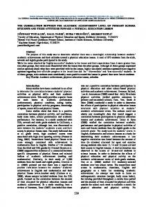
(Milusheva et al., 1996), and we calculated the amount (neuronal uptake) of radioactivity at the beginning of the experiments (the sum of radioactivity released during 19 collection periods and the radioactivity remaining after perfusion). In the cerebral cortex samples, 75.2 ± 5.7% (the mean ± SEM of 30 observations) of the total radioactivity was accounted for DA, and 85.6 ± 4.1% (n = 18) was attributed to NA, as measured by high-pressure liquid chromatography combined with scintillation spectrometry. For the spinal cord slices 83.6 ± 5.8% (n = 16) of the radioactivity was released as NA. The remaining radioactivity was due to metabolites (Milusheva et al., 1996), indicating that the radioactivity measured in the fractions can be safely attributed to labeled transmitter release into the perfusion fluid. The level of radioactivity in the samples (by [3 H]DA or [3 H]NA and 3 H metabolites) was determined using a liquid scintillation counter (Packard 1900 Tri-Carb liquid scintillation spectrometer), and the counts were converted to absolute activity using external standards. The tritium release was expressed in Bq/g and as a percentage of the amount of radioactivity in the tissue at the time the release was measured (fractional release, FR%). FRR1 and FRS1 were taken as internal standards (controls) and are indicated in the figures. The release of tritium in response to the drug treatment or field (axonal) stimulation was compared with the basal release level and was expressed as the increase in the fractional release of tritium. 2.4. Nerve stimulation A 2 Hz electrical field stimulation (supramaximal, 2 ms impulse duration) was used with a Grass S88 stimulator (Grass Medical Instrument, Quincy, MA, USA) through a pair of platinum ring electrodes. A total of 360 or 240 supramaximal shocks were delivered, as indicated. 2.5. Statistics Statistical analysis of the data was performed with a two-tailed Student t-test and analysis of variance (ANOVA) with Dunnett’s test. The results are presented as the mean ± SEM. The error bars in the figures represent the SEM and were only plotted when they exceeded the symbol size. 3. Results 3.1. Release of [3 H]DA from cerebral cortex slice preparations In 30 experiments, the prefrontal cortex (PFC) slices contained 220.2 ± 9.5 kBq/g radioactivity following 45 min of loading with [3 H]DA and subsequent washing. Over a 3 min collection period at rest, the average release from the second resting samples of the PFC slices was 1.9 ± 0.1 kBq/g (1.00 ± 0.03% (n = 30) of the total radioactive content). The change in resting release over a 30 min period was always compared with the control value (FRR1 ) determined during the 3 min collection period (FRR2 /FRR1 = 0.86 ± 0.05, n = 6). In response to electrical field stimulation (2 Hz, 2 ms, and 360 shocks), the release was 14.0 ± 0.7 kBq/g (n = 30), which was 6.31 ± 0.23% of the radioactivity present in the PFC tissue at the time of stimulation. When the stimulation (FRS1 = 5.83 ± 0.93%) was repeated 30 min after the first stimulation (S2 ), the level of radioactivity released was 5.32 ± 1.33% (FRS2 ), resulting in an FRS2 /FRS1 ratio of 1.04 ± 0.07 (n = 6, Fig. 1a). When voltage-dependent Na+ influx (i.e., axonal conduction) was inhibited by the application of TTX (1 M) between the first and second stimulation, the evoked [3 H]DA release in response to field stimulation was significantly blocked with FRS2 /FRS1 = 0.12 ± 0.03 (Fig. 1b). This finding indicates the
C. Sircuta et al. / Brain Research Bulletin 124 (2016) 21–26
23
Fig. 1. The release of [3 H]dopamine ([3 H]DA) from rat frontal cortex slices. The slices were stimulated two times (S1 and S2 ), as indicated (supramaximal voltage, 2 ms impulse duration, 360 shocks, 2 Hz). The vertical lines represent the SEM. The fractional release (FR% = the percentage of radioactivity in the tissue at the time when the release was measured) is plotted vs. the fractions collected in 3 min. (a) Control experiments. Note that the radioactivity release in response to field stimulations (repeated after 30 min [S1 and S2 ] (FRS2 /FRS1 = 0.87 ± 0.07) and during rest [R1 and R2 ] (FRR2 /FRR1 = 1.04 ± 0.05) was fairly constant (n = 6). (b) Effect of tetrodotoxin (TTX, 1 M). TTX was added to and maintained in the Krebs perfusion solution as indicated. Note that the stimulation-evoked release of radioactivity was blocked (n = 6).
neuronal origin of the released radioactivity. The resting release was not affected (FRR2 /FRR1 = 1.03 ± 0.008). Epidural anesthetics are administered at concentrations of 1.5–2% (lidocaine, 55.2 mM; bupivacaine, 46.2 mM); anesthetics are administered at 1.5–5% for spinal anesthesia and at 1–2% for major nerve blocks. After epidural injection, the spread of the solution into the tissues of the epidural space is unpredictable (Hogan, 2002), and neurons are then exposed to relatively high concentrations (e.g., 5–50 mM) of local anesthetics. Therefore, we administered anesthetics at concentrations ranging from 0.1 mM to 5 mM to test their effects on neurochemical transmission in the CNS. Lidocaine, at a concentration of 5 mM (Fig. 2c), enhanced the resting release of [3 H]DA by more than six-fold, and 1.5 mM bupivacaine (Fig. 2d) increased the release by more than nine-fold. Note that the stimulation-evoked release was fully inhibited (Fig. 2).
When CaCl2 was removed and EGTA (1 mM), a Ca2+ chelator, was added to the Krebs solution, 5 mM lidocaine enhanced the resting release of [3 H]DA (FRR2 /FRR1 = 5.94 ± 0.34 (n = 4)) (Fig. 3) to approximately the same extent as that observed in the normal solution (FRR2 /FRR1 = 6.84 ± 0.10) (Fig. 2c). Under the control condition of the removal of Ca2+ from the Krebs solution (1 mM EGTA was also added), the axonal stimulation failed to release [3 H]DA, but the resting release was not affected (data not shown). significantly increased both the resting 4-AP (FRR2 /FRR1 = 1.75 ± 0.13, p < 0.05) and axonal firing-associated (FRS2 /FRS1 = 2.52 ± 0.21, p < 0.01) [3 H]DA release (Fig. 4a and b). Our observations are in agreement with previous studies that have shown that 4-AP increases the release of dopamine in the brain (King et al., 2012), the release of acetylcholine (Vizi et al., 1977) and noradrenaline in the heart (Sugimori et al., 1987), and glutamate from synaptosomes (Tapia and Sitges, 1982).
Fig. 2. Effects of lidocaine and bupivacaine on the resting and stimulation-evoked release of [3 H]dopamine ([3 H]DA). The slices were stimulated (2 Hz, 360 shocks) once or twice (S1 and/or S2 ), as indicated. Lidocaine or bupivacaine was added to the solution, as indicated. (a–c) Effect of various concentrations of lidocaine (0.5, 1.5 and 5 mM, n = 6–6) on the resting and stimulation-evoked release of [3 H]dopamine ([3 H]DA) expressed as the fractional release (FR%). At 5 mM lidocaine, while the resting release was increased by more than six-fold (FRR2 /FRR1 = 6.84 ± 0.10), the stimulation-evoked release (S1 = 15.2 ±1.8 kBq/g, FRS1 = 6.46 ± 1.04%, n = 6) was fully inhibited (S2 = 0). The asterisks indicate the level of significance, p < 0.01. (d) The effects of bupivacaine (1.5 mM) on the resting and stimulation (2 Hz, 360 shocks)-evoked release of [3 H]dopamine ([3 H]DA). Bupivacaine (1.5 mM) was added to and maintained in the Krebs solution, as indicated. Note the large increase in [3 H]DA release not associated with axonal firing. There was a complete presynaptic deficit in the transmitter release in response to field stimulation (FRR2 /FRR1 = 9.40 ± 0.45, S1 = 13710 ± 2684 Bq/g, FRS1 = 6.47 ± 0.35%, S2 = 0, n = 6). The asterisks indicate the level of significance, p < 0.01.
24
C. Sircuta et al. / Brain Research Bulletin 124 (2016) 21–26
Tetrodotoxin (1 M) fully inhibited the stimulation-evoked release (data not shown).
4. Discussion
Fig. 3. The [Ca2+ ]o -independent release of [3 H]dopamine ([3 H]DA) in response to lidocaine (5 mM) from frontal cortex slices. Lidocaine was added to the Ca2+ -free (CaCl2 was omitted and 1 mM EGTA was added) Krebs perfusion solution. The slices were stimulated (2 Hz, 360 shocks) once (S1 = 12.2 ± 0.6 kBq/g, FRS1 = 5.50 ± 0.44%, n = 6), as indicated. The asterisks indicate the level of significance, p < 0.01.
3.2. Release of [3 H]NA from cerebral cortex and spinal cord slice preparations After the cerebral cortex slices were loaded with [3 H]NA, the average tissue uptake of radioactivity was 435.0 ± 69.8 kBq/g (n = 6). During the collection period (3 min), the fractional release of radioactivity was 0.89 ± 0.15% (1.9 ± 0.2 kBq/g) at rest. Axonal stimulation (S1 ) led to the release of 10.9 ± 0.2 kBq/g of radioactivity (Fig. 5a). The effect of lidocaine on the resting release depended on the concentration. The FRR2 /FRR1 values were 0.85 ± 0.02 (p > 0.05, n = 5), 1.87 ± 0.10 (p < 0.05, n = 5) and 2.53 ± 0.20 (p < 0.01, n = 6) at concentrations of 0.1, 1.5 and 3 mM, respectively. At a concentration of 3 mM, lidocaine significantly enhanced the resting release and reduced the stimulation-evoked release of [3 H]NA (Fig. 5b). In clinical practice, solutions of 0.5% lidocaine (18.4 mM) and bupivacaine (15.4 mM) that are locally injected into spinal space might result in relatively high local concentrations of these drugs. Therefore, we also studied the effect of lidocaine on [3 H]NA release from the spinal cord slices (Fig. 6a). After 45 min of loading, the tissue contained 283.6 ± 50.4 kBq/g of radioactivity (n = 14). At rest, 0.7 ± 0.05% of the radioactive content was released during the 3 min collection period. Electrical field stimulation released [3 H]NA from the preparations (FRS1 = 7.4 ± 0.9 kBq/g, n = 6) and the FRS2 /FRS1 was 0.91 ± 0.08 (n = 6). At a concentration of 5 mM, lidocaine enhanced the resting release and completely blocked the stimulation-evoked release (Fig. 6a). 4-Aminopyridine (300 M) enhanced both the resting and stimulation-evoked release (Fig. 6b).
We have shown that local anesthetics (lidocaine and bupivacaine) at concentrations > 0.5 mM produced a partial or complete dissociation of chemical neurotransmission, i.e., reduced or inhibited neuronal stimulation-evoked, [Ca2+ ]o -dependent release and increased [Ca2+ ]o -independent resting release of [3 H]DA and [3 H]NA from frontal cortex slices and [3 H]NA release from spinal cord slices. To the best of our knowledge, our study is the first to provide neurochemical evidence that local anesthetics enhance transmitter release at rest (Fig. 2b–d). The selective K+ channel inhibitor 4-AP also enhanced the resting transmitter release, which is independent of the axonal activity and induces clonic and tonic seizures in patients (King et al., 2012). However, while local anesthetics partially or fully inhibit it, 4-AP potentiates stimulation-evoked release from prefrontal cortex (Fig. 4) and spinal cord preparations (Fig. 6b). Therefore, we suggest that the rare occurrence of adverse effects to local anesthetics is due to their effects on chemical transmission influenced by combined inhibitory effects on Na+ and K+ channels inhibiting the stimulation–evoked (an effect on Na+ channels) and enhancing the resting release of catecholamines (an effect on K+ channels). This conclusion is strongly supported by the findings that diazoxide, a K+ channel opener reduces both the resting and stimulation-evoked release of noradrenaline (Takata et al., 1992; Oe et al., 1999); in addition, pinacidil, a drug with a similar effect on K+ channels, has been shown to inhibit the stimulation-induced release of endogenous dopamine and noradrenaline (Soares-da-Silva and Fernandes, 1990). Tetrodotoxin, a selective Na+ channel blocker (Narahashi, 1972; Kohane et al., 1998) with local anesthetic activity (Padera et al., 2006), fully inhibited the [Ca2+ ]o -dependent release of transmitters evoked by axonal stimulation in both preparations. TTX is similar to local anesthetics in this respect, it is even able to potentiate the action of local anesthetics (Kohane et al., 1998; Padera et al., 2006), but it failed to affect the [Ca2+ ]o -independent release of [3 H]DA at rest and does not cause adverse CNS effects after systemic administration (Marcil et al., 2006). TTX has no inhibitory effect on K+ permeability. Voltage-gated sodium channels are the pharmacological targets of a variety of drugs, such as analgesics, local anesthetics, antiarrhythmics and antiepileptics (Lenkey et al., 2010, 2011). Presynaptic potassium channels play a key role in cellular physiology by regulating the efflux of K+ ions and setting the resting membrane potential (Dodson and Forsythe, 2004; Kim and Kang, 2015). It is known that local anesthetics are also able to inhibit K+ channels (Scholz, 2002; Wolff et al., 2014) in neocortical
Fig. 4. Effect of 4-aminopyridine (4-AP) on the release of [3 H]dopamine ([3 H]DA) from the rat prefrontal cortex slices. The tissues were stimulated twice (2 Hz, 360 shocks). The asterisks indicate p < 0.01. (a) 4-Aminopyridine was added at a concentration of 100 M. Two stimulations were applied as indicated (S1 = 15.9 ± 1.1 kBq/g, FRS1 = 5.96 ± 0.63%, n = 5). (b) 4-Aminopyridine at a concentration of 300 M increased both the resting (FRR2 /FRR1 = 2.68 ± 0.03, p < 0.01) and stimulation-evoked release (FRS2 /FRS1 = 3.50 ± 0.20, p < 0.01) of [3 H]dopamine (n = 6). (S1 15.6 ± 0.7 kBq/g, FRS1 = 6.44 ± 0.07%).
C. Sircuta et al. / Brain Research Bulletin 124 (2016) 21–26
25
Fig. 5. Effect of lidocaine (3 mM) on the resting and stimulation-evoked release of [3 H]noradrenaline ([3 H]NA) from rat prefrontal cortex slices. The slices were stimulated twice (2 Hz, 360 shocks) (S1 and S2 ), as indicated. R1 and R2 are also indicated. (a) Control experiments. Resting (FRR2 /FRR1 = 0.93 ± 0.06) and stimulation-evoked (FRS2 /FRS1 = 0.89 ± 0.12) release were relatively constant (n = 6). (b) The effect of lidocaine (3 mM) on the resting (FRR2 /FRR1 = 2.53 ± 0.20 compared with the control (0.93 ± 0.06) p < 0.01)) and stimulation-evoked release of [3 H]noradrenaline (S1 = 16.2 ± 2.3 kBq/g (n = 5). (FRS2 /FRS1 = 0.54 ± 0.07, compared with the control (0.89 ± 0.12) p < 0.01). Notably, lidocaine enhanced the resting release, but reduced the stimulation-evoked release.
Fig. 6. Effects of lidocaine (5 mM) and 4-aminopyridine (300 M) on the release of [3 H]noradrenaline ([3 H]NA) from in vitro rat spinal cord slice preparations. The slices were stimulated (3 Hz, 240 shocks) twice (S1 and S2 ) as indicated. Fractions were collected every 3 min. The release is expressed as the% fractional release (FR) n = 6–6. The asterisks indicate p < 0.01. (a) Lidocaine (5 mM) significantly enhanced the resting release (asterisks indicate p < 0.01) of [3 H]NA (FRR2 /FRR1 = 5.22 ± 0.20, p < 0.001). Note that field stimulation (S2 ) failed to enhance the release to the same extent. S1 = 6.3 ± 0.9 and S2 = 3.2 ± 0.8 kBq/g. (b) Effect of 4-aminopyridine (300 M) on the release of [3 H]NA. 4-AP significantly increased the resting release, R1 = 1.6 ± 0.3 kBq/g, FRR2 /FRR1 = 5.02 ± 0.17, p < 0.001 and markedly potentiated the stimulation-evoked release of [3 H]NA. S1 = 6.9 ± 0.9 kBq/g, S2 = 17.4 ± 2.5 (n = 4).
(Andreasen and Hablitz, 1993) and dorsal horn (Olschewski et al., 1998) neurons. Since a variety of K+ currents are known to be involved in the repolarization of the neuron action potential, any effect on K+ channels may influence transmitter release. It is notable that locally applied anesthetics (0.5–5% w/v, which is 15.4–154 mM for bupivacaine) that penetrate the tissue can easily result in local concentrations of >0.5 mM that are required to develop the local anesthetic effect (Schmidtmayer and Ulbricht, 1980); these concentrations are also sufficiently high to affect transmitter release, as observed in this study. Subarachnoid injection of a local anesthetic (lidocaine, 75 mg) produces anesthesia lasting from 60 to 115 min (Moore et al., 1987). It has been shown that the volume (42–81 ml) of lumbosacral cerebrospinal fluid is the primary determinant of sensory block extent and duration (Higuchi et al., 2004). Accordingly, 75 mg of lidocaine administered into the lumbosacral cerebrospinal fluid (∼60 ml) might produce a 4–5 mM concentration, high enough to affect transmitter release. Continuous spinal anesthesia even with a relatively lower concentration that is increasingly administered over a long time period to treat postoperative neuropathic pain (Berde and Strichartz, 2010) also increases the chance of neurotoxicity. It has been shown that dopaminergic (Cobacho et al., 2014) and noradrenergic (Takano and Yaksh, 1992) mechanisms are involved in the antiallodynic action of neuropathic pain; therefore, the release of NA in response to a spinal administration of local anesthetics is very likely involved in the pain-relief action. This conclusion is supported by the fact that the descending noradrenergic system originating from brainstem nuclei A5, A6 and A7 exerts
analgesia by inhibiting glutamate release from the spinal nociceptive primary afferent fiber (Kamisaki et al., 1993), which is an effect that can be blocked by ␣2 -adrenoceptor antagonists (Takano and Yaksh, 1992). Indeed, clinically, intrathecal and epidural administration of ␣2 -adrenoceptor agonists (e.g., clonidine), drugs that are able to mimic the effect of NA with an agonist effect on these ␣2 -adrenoceptors, have been used for spinal analgesia for the treatment of intractable pain (Rauck et al., 1993). Local anesthetics increase the release of [3 H]NA or [3 H]DA at rest, resulting in a high ambient concentration of catecholamines and their toxic aldehyde metabolites (3,4-dihydroxyphenylacetaldehyde (DOPAL) and 3,4dihydroxyphenylglycolaldehyde (DOPEGAL)) (Burke et al., 2004); these effects might also be involved in neurotoxic effects. In clinical practice, an overdose, accidental rapid injection or local administration of a local anesthetic may produce adverse effects in the nervous system. An interesting problem that remains to be studied is how patients suffering from different somadendritic and axo-somatic channelopathies (Brager and Johnston, 2014) or being treated with noradrenaline uptake blockers may respond to local anesthetics. Nevertheless, although neurotoxicity from local anesthetics occurs very rarely, it may limit their clinical utility, and, at a minimum, caution should be exercised in selecting the volume, total dose and concentration of a specific anesthetic.
Declaration of interest None declared.
26
C. Sircuta et al. / Brain Research Bulletin 124 (2016) 21–26
Acknowledgments The authors thank Ms. Kate Windisch and Ms. Judit Oszi for their technical help and Ms. Judit Csek for her administrative work. We also thank Dr. Árpád Mike for valuable discussion of the results. References Agarwal, R., Gutlove, D.P., Lockhart, C.H., 1992. Seizures occurring in pediatric patients receiving continuous infusion of bupivacaine. Anesth. Analg. 75 (2), 284–286. Andreasen, M., Hablitz, J.J., 1993. Local anesthetics block transient outward potassium currents in rat neocortical neurons. J. Neurophysiol. 69 (6), 1966–1975. Berde, Strichartz, 2010. Local Anesthetics in Miller’s Anesthesia. In: Miller, R.D. (Ed.), pp. 913–939. Bouwman, N.A., Morre, H.H., 2013. Lidocaine-induced seizure during carotid endarterectomy. Clin. Neurophysiol. 124 (7), 1481–1483. Brager, D.H., Johnston, D., 2014. Channelopathies and dendritic dysfunction in fragile X syndrome. Brain Res. Bull. 103, 11–17. Bromage, P.R., Joyal, A.C., Binney, J.C., 1963. Local anesthetic drugs: penetration from the spinal extradural space into the neuraxis. Science 140 (3565), 392–394. Burke, W.J., Li, S.W., Chung, H.D., Ruggiero, D.A., Kristal, B.S., Johnson, E.M., Lampe, P., Kumar, V.B., Franko, M., Williams, E.A., Zahm, D.S., 2004. Neurotoxicity of MAO metabolites of catecholamine neurotransmitters: role in neurodegenerative diseases. Neurotoxicology 25 (1–2), 101–115. Cobacho, N., de la Calle, J.L., Paino, C.L., 2014. Dopaminergic modulation of neuropathic pain: analgesia in rats by a D2-type receptor agonist. Brain Res. Bull. 106, 62–71. Dodson, P.D., Forsythe, I.D., 2004. Presynaptic K+ channels: electrifying regulators of synaptic terminal excitability. Trends Neurosci. 27 (4), 210–217. Faccenda, K.A., Finucane, B.T., 2001. Complications of regional anaesthesia incidence and prevention. Drug Saf. 24 (6), 413–442. Higuchi, H., Hirata, J., Adachi, Y., Kazama, T., 2004. Influence of lumbosacral cerebrospinal fluid density, velocity, and volume on extent and duration of plain bupivacaine spinal anesthesia. Anesthesiology 100 (1), 106–114. Hogan, Q., 2002. Distribution of solution in the epidural space: examination by cryomicrotome section. Reg. Anesth. Pain Med. 27 (2), 150–156. Kamisaki, Y., Hamada, T., Maeda, K., Ishimura, M., Itoh, T., 1993. Presynaptic alpha 2 adrenoceptors inhibit glutamate release from rat spinal cord synaptosomes. J. Neurochem. 60 (2), 522–526. Kim, D., Kang, D., 2015. Role of K(2)p channels in stimulus-secretion coupling. Pflugers Arch. 467 (5), 1001–1011. King, A.M., Menke, N.B., Katz, K.D., Pizon, A.F., 2012. 4-Aminopyridine toxicity: a case report and review of the literature. J. Med. Toxicol. 8 (3), 314–321. Kohane, D.S., Yieh, J., Lu, N.T., Langer, R., Strichartz, G.R., Berde, C.B., 1998. A re-examination of tetrodotoxin for prolonged duration local anesthesia. Anesthesiology 89 (1), 119–131. Lenkey, N., Karoly, R., Lukacs, P., Vizi, E.S., Sunesen, M., Fodor, L., Mike, A., 2010. Classification of drugs based on properties of sodium channel inhibition: a comparative automated patch-clamp study. PLoS One 5 (12), e15568. Lenkey, N., Karoly, R., Epresi, N., Vizi, E., Mike, A., 2011. Binding of sodium channel inhibitors to hyperpolarized and depolarized conformations of the channel. Neuropharmacology 60 (1), 191–200. Marcil, J., Walczak, J.S., Guindon, J., Ngoc, A.H., Lu, S., Beaulieu, P., 2006. Antinociceptive effects of tetrodotoxin (TTX) in rodents. Br. J. Anaesth. 96 (6), 761–768. Milusheva, E.A., Doda, M., Baranyi, M., Vizi, E.S., 1996. Effect of hypoxia and glucose deprivation on ATP level, adenylate energy charge and [Ca2+]o-dependent and independent release of [3H]dopamine in rat striatal slices. Neurochem. Int. 28 (5–6), 501–507.
Moore, D.C., Chadwick, H.S., Ready, L.B., 1987. Epinephrine prolongs lidocaine spinal: pain in the operative site the most accurate method of determining local anesthetic duration. Anesthesiology 67 (3), 416–418. Moron, J.A., Brockington, A., Wise, R.A., Rocha, B.A., Hope, B.T., 2002. Dopamine uptake through the norepinephrine transporter in brain regions with low levels of the dopamine transporter: evidence from knock-out mouse lines. J. Neurosci. 22 (2), 389–395. Nakai, T., Milusheva, E., Baranyi, M., Uchihashi, Y., Satoh, T., Vizi, E.S., 1999. Excessive release of [3H]noradrenaline and glutamate in response to simulation of ischemic conditions in rat spinal cord slice preparation: effect of NMDA and AMPA receptor antagonists. Eur. J. Pharmacol. 366 (2–3), 143–150. Narahashi, T., 1972. Mechanism of action of tetrodotoxin and saxitoxin on excitable membranes. Fed. Proc. 31 (3), 1124–1132. Oe, K., Sperlagh, B., Santha, E., Matko, I., Nagashima, H., Foldes, F.F., Vizi, E.S., 1999. Modulation of norepinephrine release by ATP-dependent K(+)-channel activators and inhibitors in guinea-pig and human isolated right atrium. Cardiovasc. Res. 43 (1), 125–134. Olschewski, A., Hempelmann, G., Vogel, W., Safronov, B.V., 1998. Blockade of Na+ and K+ currents by local anesthetics in the dorsal horn neurons of the spinal cord. Anesthesiology 88 (1), 172–179. Padera, R.F., Tse, J.Y., Bellas, E., Kohane, D.S., 2006. Tetrodotoxin for prolonged local anesthesia with minimal myotoxicity. Muscle Nerve 34 (6), 747–753. Rauck, R.L., Eisenach, J.C., Jackson, K., Young, L.D., Southern, J., 1993. Epidural clonidine treatment for refractory reflex sympathetic dystrophy. Anesthesiology 79 (6), 1163–1169 (discussion 1127A). Rigler, M.L., Drasner, K., Krejcie, T.C., Yelich, S.J., Scholnick, F.T., DeFontes, J., Bohner, D., 1991. Cauda equina syndrome after continuous spinal anesthesia. Anesth. Analg. 72 (3), 275–281. Schmidtmayer, J., Ulbricht, W., 1980. Interaction of lidocaine and benzocaine in blocking sodium channels. Pflugers Arch. 387 (1), 47–54. Scholz, A., 2002. Mechanisms of (local) anaesthetics on voltage-gated sodium and other ion channels. Br. J. Anaesth. 89 (1), 52–61. Soares-da-Silva, P., Fernandes, M.H., 1990. Inhibition by the putative potassium channel opener pinacidil of the electrically-evoked release of endogenous dopamine and noradrenaline in the rat vas deferens. Naunyn Schmiedebergs Arch. Pharmacol. 342 (4), 415–421. Sugimori, T., Nagashima, H., Vizi, E.S., Harsing Jr., L.G., Chaudhry, I., Lalezari, I., Duncalf, D., Goldiner, P.L., Foldes, F.F., 1987. Effect of mono- and diaminopyridines on release of [3H]norepinephrine from isolated guinea-pig atrium. Neuropharmacology 26 (6), 621–626. Takano, Y., Yaksh, T.L., 1992. Characterization of the pharmacology of intrathecally administered alpha-2 agonists and antagonists in rats. J. Pharmacol. Exp. Ther. 261 (2), 764–772. Takata, Y., Shimada, F., Kato, H., 1992. Differential effects of diazoxide, cromakalim and pinacidil on adrenergic neurotransmission and 86Rb+ efflux in rat brain cortical slices. J. Pharmacol. Exp. Ther. 263 (3), 1293–1301. Tapia, R., Sitges, M., 1982. Effect of 4-aminopyridine on transmitter release in synaptosomes. Brain Res. 250 (2), 291–299. Vizi, E.S., van Dijk, J., Foldes, F.F., 1977. The effect of 4-aminopyridine on acetylcholine release. J. Neural Transm. 41 (4), 265–274. Vizi, E.S., Fekete, A., Karoly, R., Mike, A., 2010. Non-synaptic receptors and transporters involved in brain functions and targets of drug treatment. Br. J. Pharmacol. 160 (4), 785–809. van der Wal, S.E., van den Heuvel, S.A., Radema, S.A., van Berkum, B.F., Vaneker, M., Steegers, M.A., Scheffer, G.J., Vissers, K.C., 2015. The in vitro mechanisms and in vivo efficacy of intravenous lidocaine on the neuroinflammatory response in acute and chronic pain. Eur. J. Pain, http://dx.doi.org/10.1002/ejp.794. Wolff, M., Schnobel-Ehehalt, R., Muhling, J., Weigand, M.A., Olschewski, A., 2014. Mechanisms of lidocaine’s action on subtypes of spinal dorsal horn neurons subject to the diverse roles of Na(+) and K(+) channels in action potential generation. Anesth. Analg. 119 (2), 463–470.