AJNR Am J Neuroradiol 26:1832–1839, August 2005
Correlation of Apparent Diffusion Coefficient with Neuropsychological Testing in Temporal Lobe Epilepsy Yvonne W. Lui, Annette O. Nusbaum, William B. Barr, Glyn Johnson, James S. Babb, Darren Orbach, Alice Kim, Georgia Laliotis, and Orrin Devinsky
BACKGROUND AND PURPOSE: Patients with nonlesional temporal lobe epilepsy have long been known to have abnormalities of memory. Recently, these patients have been shown to have increased diffusivity in the hippocampus. We hypothesized that in these patients, a negative correlation would exist between diffusivity measures of the mesial temporal lobe and performance on neuropsychological tests. METHODS: Twenty presurgical patients with temporal lobe epilepsy and 20 age- and sex-matched healthy controls underwent MR imaging of the brain. Apparent diffusion coefficient region of interest measures were taken in both hippocampi and parahippocampal gyri by 2 independent observers. Mean whole brain diffusivity was calculated. All patients completed neuropsychological testing. Electroencephalogram and pathology results were collected. Patients and controls were compared with respect to each apparent diffusion coefficient measure. In patients, apparent diffusion coefficients ipsilateral and contralateral to the seizure focus were compared. Associations were assessed between diffusivity measures and neuropsychological scores. RESULTS: Eleven patients had right-sided seizure foci and 9 had left-sided seizure foci. Patients demonstrated higher apparent diffusion coefficient values than controls over the whole brain, in the hippocampi, and in the parahippocampal gyri (P < .05). Patients demonstrated higher apparent diffusion coefficient within the ipsilateral hippocampus (1.19 ⴞ 0.22 ⴛ 10ⴚ3 s/mm2) and parahippocampal gyrus (1.02 ⴞ 0.12 ⴛ 10ⴚ3 s/mm2) compared with the contralateral side (1.02 ⴞ 0.16 ⴛ 10ⴚ3 s/mm2 and 0.96 ⴞ 0.09 ⴛ 10ⴚ3 s/mm2, respectively) (P < .05). Negative correlations were seen between hippocampal apparent diffusion coefficients and multiple memory tests (P < .05). CONCLUSION: Quantitative diffusion measurements in the hippocampus correlate with memory dysfunction in patients with temporal lobe epilepsy. In nonlesional mesial temporal lobe epilepsy, patients suffer from seizures that originate in the limbic structures of the mesial temporal lobe (1). In patients with long-standing disease, these structures undergo histopathological changes. Hippocampal sclerosis is the most common pathologic finding in adult patients
with temporal lobe epilepsy (2), and ultrastructural changes include neuronal loss and extracellular disorganization (3, 4). It is believed that these changes are reflected in a variety of MR imaging abnormalities. Investigators have documented volume loss (5, 6), hyperintensity on T2-weighted images with quantitative abnormalities by using T2 relaxometry (7–10), alterations of MR spectra (11–13), and abnormalities of diffusion (13–21). The hippocampus is a critical structure for anterograde declarative memory (22); however, the mechanisms underlying encoding, reinforcement, and retrieval of memories are not well understood. Patients with chronic temporal lobe epilepsy demonstrate deficits in verbal and nonverbal memory that correlate with decreased neuronal attenuation within the hippocampi and hippocampal atrophy (3, 23, 24). Studies using MR imaging– based volume measurements con-
Received August 14, 2004; accepted after revision January 31, 2005. From the Departments of Radiology (Y.W.L., A.O.N., G.J., J.S.B., D.O., A.K.), Neuropsychology (W.B.B.), and Neurology (G.L., O.D.), NYU Medical Center, 530 First Avenue, New York, NY 10016. Abstract was presented at the 89th annual meeting of the Radiological Society of North America, December 2003. Address correspondence to Yvonne W. Lui, Department of Radiology, Section of Neuroradiology, NYU Medical Center, 530 First Avenue, New York, NY 10016 (email:
[email protected]).
© American Society of Neuroradiology 1832
AJNR: 26, August 2005
firm this relationship (8, 24 –26). Quantified MR imaging abnormalities using T2 relaxometry and spectroscopy have also been shown to correlate with memory loss (8, 27, 28). In particular, abnormalities of the left hippocampus have been correlated with verbal memory deficits (8, 24, 26, 27), and abnormalities of the right hippocampus, with visuospatial memory deficits (25, 27). Multiple investigators have shown that patients with temporal lobe epilepsy demonstrate increased apparent diffusion coefficient in the hippocampus (17, 18, 20, 21). Differences in tissue water diffusivity are brought out by using diffusion-weighted imaging via the application of strong pulsed-gradient fields. Tissue water diffusivity is dependent on a variety of factors, including the number and organization of neurons as well as the amount of extracellular water. Most recently, Briellmann et al (20) showed a correlation between language localization using functional MR imaging and fractional anisotropy measures within the hippocampus in patients with temporal lobe epilepsy. They described 9 patients, 2 of whom had atypical language localization and abnormal left temporal fractional anisotropy and 7 of whom had typical language localization and normal left temporal diffusion tensor measures (20). These findings suggest that patients with temporal lobe epilepsy have white matter tract abnormalities within the mesial temporal lobe. Thus, diffusion abnormalities may well reflect microstructural abnormalities in patients with temporal lobe epilepsy. The purpose of our study was to determine if abnormal diffusion correlates with a patient’s clinical picture and memory function. In particular, we hypothesized that there is a relationship between mean diffusivity measurements in patients with temporal lobe epilepsy and their performance on neuropsychological testing.
Methods Patients We conducted an institutional review board–approved retrospective study of patients with a clinical diagnosis of nonlesional temporal lobe epilepsy who were evaluated at our institution as presurgical candidates between June 1, 1999, and February 28, 2003. Only English-speaking adult patients (ⱖ18 years old at the time of MR imaging and neuropsychological testing) who had both MR imaging and neuropsychological test results available were included. These patients were compared with age- and sex-matched controls, namely individuals who presented with a chief complaint of headache with no previous medical history, no other symptoms or signs, and no abnormalities on MR imaging. MR Imaging Technique Routine MR imaging of the brain using a standard head coil was performed at 1.5T and included 3D T1-weighted gradientecho imaging (magnetization-prepared rapid acquisition gradient-echo) (TR/TE, 1170/4.38), fast spin-echo 3-mm coronal T2-weighted images (TR/TE, 4000/106; number of excitations, 3), 5-mm coronal fluid-attenuated inversion-recovery images (TR/TE, 9000/111), and diffusion-weighted images. All coronal images were obtained along a plane perpendicular to the long
TEMPORAL LOBE EPILEPSY
1833
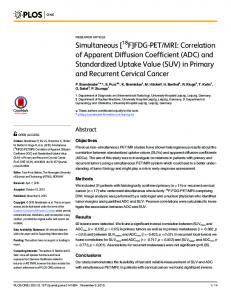
FIG 1. Axial diffusion-weighted image with b ⫽ 0 seconds mm–2 obtained in a 30-year-old patient through the mesial temporal lobe structures. Placement of oval and round regions of interest is demonstrated in the hippocampi and parahippocampal gyri bilaterally.
axis of the hippocampus. Diffusion-weighted images were acquired in the axial plane with an echo-planar imaging sequence using b values of 0, 500, and 1000 seconds mm–2. Apparent diffusion coefficients were calculated with diffusion weighting in 3 orthogonal directions and averaged. Mean diffusivity region of interest measurements were obtained independently by 2 observers (YL and DO) who were blinded to the clinical information as well as to other MR images. Both observers were neuroradiology fellows: one (YL) with approximately 3 years of experience in epilepsy imaging. Region of interest measurements were obtained in the bilateral hippocampi and parahippocampal gyri. Regions of interest were placed using the images obtained at b ⫽ 0 seconds mm–2. An oval region of interest with an anteroposterior diameter of 5 pixels and a side-to-side diameter of 3 pixels was used for the hippocampal measurements, and a circular region of interest with a diameter of 5 pixels was used for the parahippocampal measurements, with a pixel size of 1.875 ⫻ 1.875 mm (Fig 1). Two sets of measurements using the same technique were obtained by 1 observer (YL) to provide a measure of intraobserver variability. In obtaining the second set of region of interest measurements, YL was blinded to all previously obtained results. The mean whole brain diffusivity was also calculated using a thresholding analysis to exclude cerebrospinal fluid. This measure is observer-independent and, therefore, only calculated once. MR imaging studies were reviewed using all sequences except diffusion-weighted images. In particular, coronal T1weighted, coronal T2-weighted, and coronal fluid-attenuated inversion recovery images obtained through the temporal lobes were evaluated for hippocampal atrophy and for abnormal hippocampal signal intensity. The studies were interpreted retrospectively by 2 independent readers (AN and YL): a neuroradiologist experienced in epilepsy imaging (AN) and a neuroradiology fellow with approximately 3 years of experience in epilepsy imaging (YL). Both observers were blinded to the
1834
LUI
clinical information as well as the results of the region of interest measurements for mean diffusivity. Clinical Assessment All patients completed neuropsychological testing as part of a routine presurgical work-up, and testing was standardized as follows: The California Verbal Learning Test was used for verbal memory assessment (29). Each patient began the test by being shown 16 words. These words were then removed from the patient’s view, and the patient was immediately asked to recall as many of the 16 words as possible. This was repeated 5 times. A total score was calculated by summing the total number of words recalled over 5 trials (CVTOT) with a maximal score of 80 (16 ⫻ 5). After a 30-minute delay, the patient was again asked to recall the same 16 words for a measure of delayed recall (DR) with a maximum score of 16. To measure recognition, the patient was shown a word and asked to determine if that word was a member of the original set of 16 words previously shown to him or her or whether it was a new word (REC). The Logical Memory subtest from the Wechsler Memory Scale: Revised was used to assess a patient’s ability to recall meaningful verbal material (30). A story was read to the patient, and the patient was asked to recall the story immediately (LM1) and then following a 30-minute delay (LM2). Visuospatial memory evaluation included the Brief Visuospatial Memory Test which, as the nonverbal analog to the California Verbal Learning Test, also involves 5 trials of being shown multiple simple images for a measure of immediate recall (BV1) followed by a measure of delayed recall at 30 minutes (BV2) and a measure of recognition (BV3) (31). In addition, delayed recall of the Rey-Osterrieth Complex Figure (RD) was evaluated with a maximal score of 36 (32). A chart review was also conducted and clinical data obtained, including age at first seizure (including febrile seizures), age at onset of chronic seizure disorder, and frequency of seizures. In addition, electroencephalography and surgical pathology results were collected where available. Statistical Analysis For the purposes of statistical analysis, the gold standard used to determine laterality of disease was video electroencephalography, unless a patient’s results of video electroencephalography and preoperative subdural electrode recording were discordant. In the latter case, the results of the subdural electrode recording were used as the gold standard for laterality. Five main statistical analyses were performed. The first compared MR imaging interpretation of routine images not including diffusion with the gold standard for laterality of disease. Cohen’s kappa was used to assess agreement between MR imaging and the gold standard assessments of laterality as well as the agreement between the MR imaging assessments provided by the 2 readers. Statistical significance was assessed by using exact tests; separate tests were conducted for each reader in the case of assessing agreement between MR imaging and the gold standard. In addition, interobserver and intraobserver variability in the interpretation of routine MR images was also measured. Generalized estimating equations based on a binary logistic regression model were used to evaluate whether diagnostic accuracy (concordance between MR imaging and the gold standard assessments) exhibited significant variation between readers or depended on the nature of the gold standard assessment (leftversus-right laterality). The binary indicator of agreement between the MR imaging and gold standard assessments of laterality constituted the dependent variable, and the logistic regression model included the gold standard assessment and reader identification and fixed classification factors. The second main statistical analysis compared patients with controls with respect to regional apparent diffusion coefficient
AJNR: 26, August 2005 measures and whole brain apparent diffusion coefficient. Mixed-model analysis of variance was used to compare patients with controls with respect to regional apparent diffusion coefficient measures while adjusting for differences between regions, sides, and readers. The apparent diffusion coefficient measures in the left and right hippocampus and parahippocampus of each subject constituted the dependent variable. The model included subject type (patient versus control), brain region, side, and reader as fixed classification factors as well as terms representing all 2-factor interactions among these factors and the 3-factor interactions between subject type, region, and side (to assess whether differences between patients and controls varied within and between brain regions). A Mann-Whitney test was used to compare patients and controls with respect to whole brain apparent diffusion coefficient, a measure which is observer-independent. The third statistical analysis involved comparing, in patients, regional apparent diffusion coefficient measures ipsilateral and contralateral to the seizure focus as determined by the gold standard. Mixed-model analysis of variance was used, adjusting for differences between regions (hippocampus, parahippocampus) and readers. The apparent diffusion coefficient measures in the ipsilateral and contralateral hippocampus and parahippocampus of each patient constituted the dependent variable. The model included brain region, side (ipsilateral versus contralateral) and reader as fixed classification factors as well as terms representing all 2-factor interactions among these factors. The fourth statistical analysis was performed to determine if a correlation exists between apparent diffusion coefficient measures and performance on neuropsychological tests. Mixedmodel analysis of variance was again used while evaluating and adjusting for differences between regions (hippocampus, parahippocampus), laterality (GS determination of left versus right), sides (ipsilateral versus contralateral), and readers. The apparent diffusion coefficient measures in the ipsilateral and contralateral hippocampus and parahippocampus of each subject constituted the dependent variable. The model included brain region, laterality, side, and reader as fixed classification factors as well as terms representing all 2-factor interactions among these factors. The final statistical analysis used Spearman rank correlation coefficients to assess the relationship between the clinical characteristics and neuropsychological test results of patients with temporal lobe epilepsy.
Results Eight male and 12 female patients with temporal lobe epilepsy were included in the study, with an average age of 34 years (age range, 18 – 48 years; median, 31 years). The control group consisted of 8 men and 12 women with an average age of 34 years (age range, 19 – 47 years; median, 31 years). The average age of patients at the time of first seizure was 9 years (age range, 6 months–38 years; median, 2 years), and the average age at the onset of chronic seizure disorder was 18 years (age range, 6 months–38 years; median, 19 years) with an average duration of disease of 15 years at the time of the study (range, 4 –26 years; median, 13 years). Nine patients presented with febrile seizures (average age, 2 years). One patient presented with seizures secondary to meningitis at 6 months. Seizure frequency ranged from ⬍1 to 252 seizures/month with a median of 4 seizures/month. Two patients had a prior history of status epilepticus more than 6 years before imaging. No patients reported having a seizure in the 48 hours
AJNR: 26, August 2005
TEMPORAL LOBE EPILEPSY
1835
FIG 2. Bar graph shows that patients with temporal lobe epilepsy demonstrate higher apparent diffusion coefficient (ADC) values compared with controls over the whole brain, in the hippocampi, and in the parahippocampal gyri. The differences were statistically significant (P ⬍ .05). There were no significant differences comparing right- and left-sided diffusivity measures; therefore, the right and left regional measures are depicted together. FIG 3. Bar graph shows that in patients with temporal lobe epilepsy, mean diffusivity is higher in the hippocampus and parahippocampal gyrus ipsilateral to the seizure focus compared with the contralateral side. These differences are statistically significant (P ⬍ .05).
immediately preceding MR imaging, and no patients were in status epilepticus at the time of imaging. Seizure focus was determined by video electroencephalography. In 3 patients, video electroencephalography and preoperative subdural electrode recording results differed, and for these patients, subdural electrode recordings were used to determine the seizure focus. Eleven patients had right-sided seizure foci, and 9 patients had left-sided seizure foci. Seventeen of 20 patients underwent surgical excision of the affected temporal lobe, and all specimens confirmed gliosis within the mesial temporal lobe structures. Three patients declined surgery. In the evaluation of routine MR imaging sequences not including diffusion (T1-weighted, T2-weighted, and fluid-attenuated inversion recovery), the 2 observers had completely concordant findings for 17 of 20 patients: right hippocampal atrophy and abnormal signal intensity (6), left hippocampal atrophy and abnormal signal intensity (6), left hippocampal atrophy without abnormal signal intensity (4), and bilateral hippocampal atrophy without abnormal signal intensity (1). Of the 3 patients with discordant findings between observers, 2 patients were thought by both readers to have right-sided hippocampal atrophy as well as abnormal signal intensity; however, AN also reported the left mesial temporal lobe to be abnormal, whereas YL did not. The finding in the third discordant patient were considered normal by AN and were thought to represent right-sided hippocampal atrophy by YL. This assessment of MR images for laterality exhibited a level of concordance with the gold standard assessment that was significantly higher (P ⬍ .005) than could be ascribed to chance alone: Cohen’s kappa was 0.61 for reader 1, 0.80 for reader 2, and 0.71 overall. Furthermore, agreement between the MR imaging assessments of the 2 readers (Cohen’s kappa ⫽ 0.79) was significantly higher (P ⬍ .006) than could be expected on the basis of chance.
In comparing patients with controls, we found that patients demonstrated statistically significantly higher apparent diffusion coefficient values than controls over the whole brain, in the hippocampus, and in the parahippocampal gyrus. All apparent diffusion coefficients were measured in units of 10⫺3 mm2 s–1. The average whole brain apparent diffusion coefficient in patients was 1.05 ⫾ 0.07 compared with 0.98 ⫾ 0.03 in controls (P ⬍ .05) (Fig 2). In the analysis of regional apparent diffusion coefficient measures, comparing patients with controls, we found a highly significant difference between patients and controls (P ⬍ .0001) but no significant difference between readers (P ⬎ .074), and there was no significant variation across the left and right sides of the same brain region. More specifically, the mean apparent diffusion coefficient in each region was significantly higher for patients than for controls, with the magnitude of the difference between patients and controls being greatest in the hippocampus (Fig 2). The correlation between the regional apparent diffusion coefficient measures provided by the 2 readers for the same location (eg, left hippocampus) of the same patient was 0.881. The correlation between the 2 apparent diffusion coefficient measures provided by the observer who obtained 2 sets of data (YL) for the same location of the same patient was 0.805. Hence, the level of agreement between 2 different readers was actually observed to be slightly higher than the agreement between the replicate assessments of a single reader. In addition, there was no statistical evidence for systematic difference between the apparent diffusion coefficient assessments generated by the readers in any brain location. In patients, regional apparent diffusion coefficient measures were statistically significantly higher in the hippocampus and parahippocampal gyrus ipsilateral to the seizure focus compared with the contralateral side. The ipsilateral versus contralateral difference in mean apparent diffusion coefficient levels was signif-
1836
LUI
AJNR: 26, August 2005
icant in the hippocampus (P ⬍ .0001) and parahippocampus (P ⫽ .008), with the difference being greater in the hippocampus (Fig 3). There was no statistically significant difference between measurements taken in the corresponding right and left regions of interest in healthy controls. An interobserver correlation of 0.847 was found between the regional apparent diffusion coefficient measures provided by the 2 readers for the same location (eg, ipsilateral hippocampus) in the same patient. The correlation between the 2 apparent diffusion coefficient measures provided by reader 2 for the same location in the same patient was 0.712. Again, the level of agreement between 2 different readers was actually observed to be slightly higher than the agreement between the replicate assessments of a single reader. Statistically significant negative correlations were seen between apparent diffusion coefficient measurements in the left hippocampus and the following neuropsychological tests: DR (r ⫽ ⫺0.3, P ⬍ .05), REC (r ⫽ ⫺0.4, P ⬍ .05), and RD (r ⫽ ⫺0.4, P ⬍ .05) (Fig 4A–C). Significant negative correlations were also demonstrated between apparent diffusion coefficient measurements in the right hippocampus and REC (r ⫽ ⫺0.4) (Fig 4A–D). A trend that did not reach statistical significance was seen toward a negative correlation between right hippocampal apparent diffusion coefficient and RD (r ⫽ ⫺0.23, P ⫽ .08) (Fig 4E). Apparent diffusion coefficient measures in the right parahippocampal gyrus, however, demonstrated a positive correlation with BV2, which was statistically significant (r ⫽ 0.5, P ⬍ .05), and a trend toward a positive correlation with BV1, which did not reach statistical significance (r ⫽ 0.3, P ⫽ .09) (Fig 5). These correlations were only seen using non-Bonferroni adjusted significance levels. No correlations were found between clinical characteristics and neuropsychological assessment scores.
Discussion Our findings demonstrate that increased apparent diffusion coefficient measurements in the hippocampal structures correlate with impaired memory in patients with temporal lobe epilepsy. Specifically, we found that patients with greater diffusion abnormalities in the hippocampus, particularly the left hippocampus, have decreased verbal and visuospatial memory function. Similar trends were seen in the relationship between the right hippocampal diffusivity and memory function, though not as many correlations involving the right hippocampus reached statistical significance. Several previous studies have tried to correlate MR imaging findings with memory dysfunction in patients with temporal lobe epilepsy. In particular, investigators have documented a correlation between left hippocampal MR volumetric analysis and performance on verbal memory tasks (8, 24). Baxendale et al (25) found a correlation between right hippocampal volume and recall of a complex figure. In a study of 20
preoperative patients before temporal lobectomy, Loring et al (26) showed that patients who had asymmetric WADA test results also tended to have asymmetric hippocampal volumes. Prolonged T2 relaxation times have also been demonstrated to have a negative correlation with patients’ scores on verbal memory tasks (8). Wendel et al (28) showed that signal intensity abnormalities on T2weighted images in the left hippocampi of patients with temporal lobe epilepsy could be quantified and were predictive of postoperative verbal memory. In a study of 22 children with temporal lobe epilepsy, Gadian et al (27) found an association between abnormalities of MR spectroscopy in the left temporal lobe and a loss of verbal IQ, and similarly, abnormalities in the right temporal lobe were associated with a loss of nonverbal function. To our knowledge, there are no published studies that evaluate the relationship between mean diffusivity measures and clinical memory testing in patients with temporal lobe epilepsy. We found abnormalities of left hippocampal mean diffusivity to be negatively correlated not only with verbal memory tests but also with the Rey-Osterreith Complex Figure test, a visuospatial memory test. Similarly, right hippocampal diffusivity demonstrated negative correlation with one verbal memory test and additionally showed a trend toward a negative correlation with the Rey-Osterreith Complex Figure test. This finding suggests that verbal and nonverbal memory are incompletely lateralized and is supported by studies of healthy subjects showing bilateral functional MR imaging localization of visuospatial memory (33) and delayed right hippocampal activation in verbal memory retrieval, in addition to left-sided activation (34). The correlations we found were only statistically significant using non-Bonferroni adjusted significance levels. We believe that these associations are real; however, review of the scatterplots generated (Figs 4 and 5) shows a wide dispersion of data points. Greater statistical power would be necessary to unequivocally show hippocampal apparent diffusion coefficient measures to be independent predictors of memory function. Several studies have shown that patients with temporal lobe epilepsy have increased apparent diffusion coefficient measurements in mesial temporal lobe structures. This abnormality is seen bilaterally in patients with unilateral temporal lobe epilepsy, the maximal apparent diffusion coefficient being ipsilateral to the seizure focus (15, 17). Most recently, Londono et al (18) showed that when used in conjunction with visual inspection of MR images, apparent diffusion coefficient measures improved the detection of abnormal hippocampi but, when used alone, were not predictive of laterality. Our findings agree with prior results in that our patients demonstrated bilateral abnormal increased mean diffusivity region of interest measures within the hippocampus compared with controls. Additionally, apparent diffusion coefficient measures in patients within the hippocampus ipsilateral to the sei-
AJNR: 26, August 2005
TEMPORAL LOBE EPILEPSY
1837
FIG 4. Scatterplots demonstrate negative correlations between mean diffusivity measures in the hippocampus of patients with mesial temporal sclerosis and multiple neuropsychological tests assessing verbal memory (delayed recall [DR] and new word [REC]) and visuospatial memory (RD). Each data point corresponds to the average apparent diffusion coefficient (ADC) measure in the specified region for a single patient. Error bars indicate ⫾1 standard deviation derived from multiple readings and multiple readers. Correlation coefficients are calculated using a mixed-model analysis. Solid lines and solid data points indicate statistically significant findings (P ⬍ .05) using non-Bonferroni adjusted significance levels. Dotted line and hollow data points indicate that the relationship does not reach statistical significance. A. Left hippocampal ADC correlation with DR. B. Left hippocampal ADC correlation with REC. C. Left hippocampal ADC correlation with RD. D. Right hippocampal ADC correlation with REC. E. Right hippocampal ADC correlation with RD (P ⫽ .08). FIG 5. Scatterplot demonstrates a positive correlation between right parahippocampal apparent diffusion coefficient (ADC) measures and visuospatial memory test scores. Data points and error bars represent average ADC and standard deviation based on multiple readers and multiple readings. The correlation coefficient is calculated by using mixed-model analysis. Non-Bonferroni adjusted statistically significant positive correlation (P ⬍ .05) is seen between right parahippocampal ADC and BV2 (measure of delayed recall at 30 minutes). The relationship between right parahippocampal ADC and BV1 (measure of immediate recall) does not reach statistical significance (P ⫽ .09).
1838
LUI
zure focus were statistically significantly higher than those of the contralateral side. These findings may reflect underlying microstructural abnormalities including neuronal loss, gliosis, and an increase in the amount of extracellular fluid (18, 20, 21). We took mean diffusivity region of interest measures not only in the hippocampi but also in the parahippocampal gyri and over the whole brain. Because the limbic structures are tightly connected, abnormalities are not frequently isolated to a single area. Patients who have undergone excision of nonhippocampal mesial temporal lobe structures including the amygdala and entorhinal cortex demonstrate a high rate of seizure relief (35). This finding suggests that limbic structures other than the hippocampus have a role in the pathophysiology of seizures. Bernasconi et al (36, 37) have shown that patients with temporal lobe epilepsy demonstrate entorhinal cortex atrophy and that this can occur even in the absence of hippocampal atrophy. Our results demonstrate increased mean diffusivity region of interest measures bilaterally in the parahippocampal gyri in all our patients with temporal lobe epilepsy. This finding supports the notion that there are abnormalities of surrounding interconnected structures also involved in the storage of memories. In addition, our study demonstrates whole brain apparent diffusion coefficient to be increased in patients with temporal lobe epilepsy, possibly suggesting that these patients have widespread central nervous system abnormalities. For example, long-standing seizures may result in complex neuronal reorganization. However, because diffusion region of interest analysis was not performed in nonlimbic structures, it is unclear whether the increase in whole brain apparent diffusion coefficient in patients reflects higher apparent diffusion coefficient values only within limbic structures or whether there are concurrent diffusion abnormalities in nonlimbic parts of the brain. An unexpected observation supporting the notion that the brains of patients with temporal lobe epilepsy undergo diffuse complex change was the positive correlation we found between right parahippocampal apparent diffusion coefficient and BV2 in the subgroup of patients with left-sided seizure foci. This finding is not readily explained but suggests that the brain undergoes complex reactionary and perhaps compensatory changes and reorganization in the face of long-standing seizures. Briellmann et al (20) described 2 patients with temporal lobe epilepsy with atypical language localization on functional MR imaging who also had abnormal left temporal diffusion tensor measures, suggesting white matter reorganization. Further work using diffusion tensor imaging and tractography may elucidate some of these changes to better advantage. The main limitation of our study was small sample size. As discussed previously, the correlations we found between left hippocampal apparent diffusion coefficient and DR, REC, and RD and the correlation between right hippocampal apparent diffusion coefficient and REC were only statisti-
AJNR: 26, August 2005
cally significant using non-Bonferroni adjusted significance levels. A second limitation was the accuracy and precision of our region of interest measurements. Region of interest analysis has come under some scrutiny in the literature, and there is controversy as to how reproducible the results are (38). Yoo et al (17) suggested in their study that apparent diffusion coefficient measures in the temporal lobe of patients with hippocampal atrophy may be falsely elevated secondary to partial volume averaging of cerebrospinal fluid. In their study, large regions of interest that encompassed cerebrospinal fluid within sulci as well as within the temporal horn were used. We attempted to minimize this problem by using relatively small and uniform regions of interest, which were placed within gray matter structures, and special care was taken to exclude the temporal horn. Using this technique, we found good interobserver and intraobserver correlation; in fact, there was slightly less variability between measurements obtained by 2 observers than between 2 measurements obtained by the same reader. The section thickness of the diffusion sequence was 5 mm; therefore, some volume averaging into cerebrospinal fluid structures may still have been present. However, volume averaging would affect the control group as well as the patient group, making the differences found still meaningful. Additionally, our regions of interest were placed on axial images. Hippocampal anatomy is usually examined in the coronal plane. Unfortunately, our diffusion-weighted images are acquired axially and are of low spatial resolution, not allowing meaningful multiplanar analysis. On the other hand, this simple method uses a routine axial diffusion sequence, which is part of routine brain protocol of our institution, and allows relatively reproducible region of interest analysis of the mesial temporal lobe. In conclusion, the relationship between apparent diffusion coefficient values and memory performance is complex. MR diffusion abnormalities affect not only the mesial temporal lobe structures but possibly the whole brain in patients with temporal lobe epilepsy. Our preliminary findings suggest that diffusion abnormalities within the brain are related to a patient’s cognitive function and that a negative correlation exists between hippocampal apparent diffusion coefficients and both verbal and visuospatial memory scores, particularly on the left. Further study is needed to elucidate the mechanism by which diffusion abnormalities occur in these patients and to determine whether tissue water diffusivity in the hippocampus can be used as an indicator of cognitive function in patients with temporal lobe epilepsy.
References 1. Commission on Classification and Terminology of the International League Against Epilepsy. Proposal for the classification of epilepsy and epileptic syndromes. Epilepsia 1989;30:389 –399 2. Babb TL. Pathology of the temporal lobe: hippocampal sclerosis.
AJNR: 26, August 2005
3. 4. 5. 6.
7. 8.
9. 10.
11. 12. 13. 14. 15. 16.
17.
18. 19.
In: Lu ¨ders HO, Comari YG, eds. Epilepsy Surgery. Philadelphia, PA: Lippincott Williams & Wilkins; 2001;901–906 Blumcke I, Thom M, Wiestler OD. Ammon’s horn sclerosis: a maldevelopmental disorder associated with temporal lobe epilepsy. Brain Pathol 2002;12:199 –211 Thom M, Sisodiya SM, Beckett A, et al. Cytoarchitectural abnormalities in hippocampal sclerosis. J Neuropathol Exp Neurol 2002;61:510 –519 Jack CR Jr, Sharbrough FW, Twomey CK, et al. Temporal lobe seizures: lateralization with MR volume measurements of the hippocampal formation. Radiology 1990;175:423– 429 Luby M, Spencer DD, Kim JH, deLanerolle N, McCarthy G. Hippocampal MRI volumetrics and temporal lobe substrates in medial temporal lobe epilepsy. Magn Reson Imaging 1995;13:1065–1071 Triulzi F, Franceschi M, Fazio F, Del Maschio A. Nonrefractory temporal lobe epilepsy: 1.5T MR imaging. Radiology 1988;166:181–185 Kalviainen R, Partanen K, Aikia M, et al. MRI-based hippocampal volumetry and T2 relaxatometry: correlation to verbal memory performance in newly diagnosed epilepsy patients with left sided temporal lobe focus. Neurology 1997;48:286 –287 Bernasconi A, Bernasconi N, Caramanos Z, et al. T2 relaxometry can lateralize mesial temporal lobe epilepsy in patients with normal MRI. Neuroimage 2000;12:739 –746 Mackay CE, Webb JA, Eldridge PR, Chadwick DW, Whitehouse GH, Robert N. Quantitative magnetic resonance imaging in consecutive patients evaluated for surgical treatment of temporal lobe epilepsy. Magn Reson Imaging 2000;18:1187–1199 Connelly A, Jackson GD, Duncan JS, King MD, Gadian DG. Magnetic resonance spectroscopy in temporal lobe epilepsy. Neurology 1994;44:1411–1417 Capizzano AA, Vermathen P, Laxer KD, et al. Multisection proton MR spectroscopy for mesial temporal lobe epilepsy. AJNR Am J Neuroradiol 2002;23:1359 –1368 Kantarci K, Shin C, Britton JW, So EL, Cascino GD, Jack CR Jr. Comparative diagnostic utility of 1H MRS and DWI in evaluation of temporal lobe epilepsy. Neurology 2002;58:1745–1753 Hugg JW, Butterworth EJ, Kuzniecky RI. Diffusion mapping applied to mesial temporal lobe epilepsy: preliminary observations. Neurology 1999;52:173–176 Wieshmann UC, Clark CA, Symms MR, Barker GJ, Birnie KD, Shorvon SD. Water diffusion in the human hippocampus in epilepsy. Magn Reson Imaging 1999;17:29 –36 Sener RN. Diffusion MRI: apparent diffusion coefficient (ADC) values in the normal brain and a classification of brain disorders based on ADC values. Comput Med Imaging Graph 2001;25:299 –326 Yoo SY, Chang K-H, Song IC, et al. Apparent diffusion coefficient value of the hippocampus in patients with hippocampal sclerosis and in healthy volunteers. AJNR Am J Neuroradiol 2002;23:809 – 812 Londono A, Castillo M, Lee YZ, Smith JK. Apparent diffusion coefficient measurements in the hippocampi in patients with temporal lobe seizures. AJNR Am J Neuroradiol 2003;24:1582–1586 Duzel E, Kaufmann J, Guderian S, et al. Measures of hippocampal
TEMPORAL LOBE EPILEPSY
20. 21. 22. 23. 24.
25. 26. 27. 28. 29. 30. 31. 32. 33. 34. 35. 36. 37. 38.
1839
volumes: diffusion and 1H MRS metabolic abnormalities in temporal lobe epilepsy provide partially complementary information. Eur J Neurol 2004;11:195–205 Briellmann RS, Mitchell LA, Waites AB, et al. Correlation between language organization and diffusion tensor abnormalities in refractory partial epilepsy. Epilepsia 2003;44:1541–1545 Assaf BA, Mohamed FB, Abou-Khaled KH, et al. Diffusion tensor imaging of the hippocampal formation in temporal lobe epilepsy. AJNR Am J Neuroradiol 2003;24:1857–1862 Burgess N, Maguire EA, O’Keefe J. The human hippocampus and spatial and episodic memory. Neuron 2002;35:625– 641 Sass KJ, Spencer DD, Kim J, Westerveld M, Novelly RA, Lencz T. Verbal memory impairment correlates with hippocampal pyramidal cell density. Neurology 1990;40:1694 –1697 Lencz T, McCarthy G, Bronen RA, et al. Quantitative magnetic resonance imaging in temporal lobe epilepsy: relationship to neuropathology and neuropsychological function. Ann Neurol 1992;31:629 –37 Baxendale SA, van Paesschen W, Thompson PJ, et al. The relationship between quantitative MRI and neuropsychological functioning in temporal lobe epilepsy. Epilepsia 1998;39:158 – 66 Loring DW, Murro AM, Meador KJ, et al. Wada memory testing and hippocampal volume measurements in the evaluation for temporal lobectomy. Neurology 1993;43:1789 –1793 Gadian DG, Isaacs EB, Gross JH, et al. Lateralization of brain function in childhood revealed by magnetic resonance spectroscopy. Neurology 1996;46:974 –977 Wendel JD, Trenerry MR, Xu YC, et al. The relationship between quantitative T2 relaxometry and memory in nonlesional temporal lobe epilepsy. Epilepsia 2001;42:863– 868 Delis DC, Kramer JH, Kaplan E, et al. California Verbal Learning Test: Adult Version. San Antonio, TX: The Psychological Corporation; 1987 Wechsler D. Wechsler Memory Scale: Revised. San Antonio, TX: The Psychological Corporation; 1987 Benedict RHB. Brief Visuospatial Memory Test: Revised. Lutz, FL: Psychological Assessment Resources, Inc.; 1997 Osterrieth PA. Le test de copie d’une figure complexe. Archives de Psychologie 1944;30:206 –356 Angioi-Duprez K, Braun M, Jonveaux T, Picard L, George JL. Exploration of visual memory by functional MRI. J Fr Ophtalmol 2000;23:19 –26 Dupont S, Samson Y, Le Bihan D, Baulac M. Anatomy of verbal memory: a functional MRI study. Surg Radiol Anat 2002;24:57– 63 Zentner J, Hufnagel A, Wolf HK, et al. Surgical treatment of TLE: clinical, radiological, and histopathological findings in 178 patients. J Neurol Neurosurg Psychiatry 1995;58:666 – 673 Bernasconi N, Bernasconi A, Andermann F, Dubeau F, Feindel W, Reutens DC. Entorhinal cortex in temporal lobe epilepsy: a quantitative MRI study. Neurology 1999;52:1870 –1876 Bernasconi N, Bernasconi A, Caramanos Z, et al. Entorhinal cortex atrophy in epilepsy patients exhibiting normal hippocampal volumes. Neurology 2001;56:1335–1339 Zimmerman RD. Is there a role for diffusion-weighted imaging in patients with brain tumors or is the “bloom off the rose”? [Editorial] AJNR Am J Neuroradiol 2001;22:1013–1014