Cortical feed-forward networks for binding different streams of sensory information Bjo¨rn M Kampa1,2, Johannes J Letzkus1 & Greg J Stuart1 Different streams of sensory information are transmitted to the cortex where they are merged into a percept in a process often termed ‘binding.’ Using recordings from triplets of rat cortical layer 2/3 and layer 5 pyramidal neurons, we show that specific subnetworks within layer 5 receive input from different layer 2/3 subnetworks. This cortical microarchitecture may represent a mechanism that enables the main output of the cortex (layer 5) to bind different features of a sensory stimulus. Over the last 50 years, the idea of the ‘functional column’ has provided a dominant influence on our understanding of mammalian cortical circuits1,2. More recent studies have indicated that neurons within a column are further organized into subnetworks. The over-representation of reciprocal connections and triplet patterns in networks of layer 5 (L5) pyramidal neurons3,4 indicates that their connections are clustered in subnetworks. Similarly, synaptically connected layer 2/3 (L2/3) pyramidal neurons in visual cortex5 and within barrels (but not septa) of the barrel cortex6 form subnetworks that receive common inputs from within L2/3 and from layer 4. How these subnetworks in L2/3 and L5 interact with one another is currently unknown. To address this, we investigated communication between subnetworks in L2/3 and L5 using triple whole-cell recordings from pyramidal neurons in brain slices of rat somatosensory cortex (see Supplementary Methods online). To investigate whether L5 subnetworks receive common inputs from L2/3 pyramidal neurons, we recorded from pairs of L5 pyramidal neurons and sequentially from different presynaptic L2/3 pyramidal
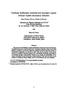
neurons (Fig. 1a; all experiments were carried out in accordance with the guidelines approved by the Animal Ethics Committee of the Australian National University). The probability that both L5 neurons received input from the same L2/3 neuron was significantly higher if the L5 neurons were also connected to each other. Both L5 neurons received synaptic input from the same L2/3 neuron in 22.1% of recordings when the L5 neurons were synaptically connected (total of 68 pairs with 15 double and 12 single connections), whereas this was the case in only 2.1% of recordings when the L5 neurons were not connected to each other (total of 340 pairs with 7 double and 100 single connections). From this data it can be calculated that, compared with random connectivity, the probability that a L2/3 neuron makes a synaptic connection with two L5 neurons is fourfold higher (4.4 ± 1.0; P ¼ 0.001) if the L5 neurons are synaptically connected with each other (Fig. 1b, left), whereas the probability that two L5 neurons receive input from the same L2/3 neuron is reduced, although not significantly (0.69 ± 0.26; P ¼ 0.1), if they are not synaptically connected (Fig. 1b, right). These findings show that individual
a L2/3
L5 pair connected
L5 pair not connected
L5 Stimulate:
C
Figure 1 L2/3 neurons target the same L5 subnetwork. (a) Examples of triple recordings from connected (left panels) or unconnected L5 pairs (right panels). Inset shows recording scheme. Simultaneous recordings were made from two L5 neurons (blue and green) and one L2/3 neuron (orange). Black traces indicate presynaptic current injection used to stimulate action potentials. Scale bars: 100 ms, 1 mV for excitatory postsynaptic potential traces and 50 mV for action potential traces. (b) Numbers of connections between L2/3 neurons and one (single) or both (double) L5 neurons for connected (left panel) or unconnected (right panel) L5 pairs, shown relative to the expected counts for random network connectivity. Note the increase in double connections between L2/3 neurons and connected pairs of L5 neurons. Error bars represent s.d. (see Supplementary Methods).
b
L5 pair connected Counts relative to random
© 2006 Nature Publishing Group http://www.nature.com/natureneuroscience
B R I E F C O M M U N I C AT I O N S
L5 pair not connected
6 5 4 3 2 1 0 Single
Double
Single
Double
L2-L5 connection
1The John Curtin School of Medical Research, Australian National University, Mills Road, ACT 0200, Canberra, Australia. 2Present address: Brain Research Institute, University of Zu¨rich, Winterthurerstrasse 190, 8057 Zu¨rich, Switzerland. Correspondence should be addressed to B.M.K. (
[email protected]).
Received 27 June; accepted 18 October; published online 12 November 2006; doi:10.1038/nn1798
NATURE NEUROSCIENCE ADVANCE ONLINE PUBLICATION
1
B R I E F C O M M U N I C AT I O N S
a
L2/3 pair connected
L2/3 pair not connected
L2/3
L5
b
Counts relative to random
© 2006 Nature Publishing Group http://www.nature.com/natureneuroscience
Stimulate:
6
L2/3 pair connected
L2/3 pair not connected
5 4 3 2 1 0 Single
Double Single L2-L5 connection
Double
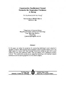
L2/3 pyramidal neurons preferentially target L5 pyramidal neurons in the same L5 subnetwork. To investigate whether L2/3 inputs onto L5 neurons originate from the same or different L2/3 subnetworks, we recorded from pairs of L2/3 pyramidal neurons and studied the connectivity of these neurons with L5 pyramidal neurons (Fig. 2a). The probability that the L5 neuron received input from both L2/3 neurons was significantly higher if the two L2/3 neurons were not connected to each other. Both L2/3 neurons connected to the same L5 neuron in 7.4% of recordings when the L2/3 neurons were not connected (total of 148 pairs with 11 double and 30 single connections), whereas this was the case in only 1.9% of recordings when the L2/3 neurons were connected (total of 106 pairs with 2 double and 35 single connections). Compared with random connectivity, the probability that the L5 neuron received input from both L2/3 neurons is therefore threefold higher (3.3 ± 1.0; P ¼ 0.006) if the L2/3 neurons are not connected with each other (Fig. 2b, right), whereas this probability is halved (0.5 ± 0.3; P ¼ 0.05) if the L2/3 neurons are connected (Fig. 2b, left). These findings indicate that L5 pyramidal neurons receive input preferentially from L2/3 pyramidal neurons located in different subnetworks. The resulting connectivity scheme (Supplementary Figure 1 online) includes connections between subnetworks in different layers, but not in a strict feed-forward manner. Recent work has indicated that subnetworks in L2/3 receive common inputs from L4 (ref. 5), and other studies have described the existence of specific subnetworks in L5 (ref. 3). We now show that L5 subnetworks share common inputs from individual L2/3 pyramidal neurons (Fig. 1). Moreover, we show that the output of individual L2/3 subnetworks is spread across different L5 subnetworks (Fig. 2), rather than simply being propagated from one subnetwork to the next. This enables individual L5 pyramidal neurons to integrate and bind information coming from different L2/3 subnetworks, which may encode different features of a stimulus. Consistent with this notion, several previous studies have indicated that neurons that code for the same orientation are connected to each other7–10. Furthermore, there is evidence that L5 neurons possess larger and more complex receptive fields than L2/3 or L4 neurons11,12.
2
Figure 2 L5 neurons integrate inputs from different L2/3 subnetworks. (a) Examples of triple recordings from connected (left panels) or unconnected L2/3 pairs (right panels). Inset shows recording scheme. Simultaneous recordings were made from two L2/3 neurons (orange and red) and one L5 neuron (blue). Black traces indicate presynaptic current injection used to stimulate action potentials. Scale bars: 100 ms, 0.2 mV for excitatory postsynaptic potential traces and 50 mV for action potential traces. (b) Numbers of connections between one (single) or both (double) L2/3 neurons and the L5 neuron for connected (left panel) or unconnected (right) L2/3 pairs, shown relative to the expected counts for random network connectivity. Note the increase in double connections between unconnected L2/3 pairs and L5 neurons. Error bars represent s.d. (see Supplementary Methods).
Connections between cortical subnetworks could evolve through Hebbian synaptic plasticity. Layer 2/3 neurons that receive similar inputs from L4 would be expected to be active at similar times, and would therefore be likely to connect to each other (‘neurons that fire together wire together’). Synchronous activity in different L2/3 subnetworks that project to the same L5 subnetwork (Supplementary Figure 1), may be sufficiently powerful to trigger dendritic spikes and burst firing in L5 neurons13. Recent work indicates that burst firing and dendritic spikes in L5 neurons can lead to the induction of spike timing–dependent synaptic plasticity at both L2/3 to L5 and L5 to L5 connections14,15. In this way, Hebbian plasticity could lead to the described cortical connectivity scheme, and thereby may have an important role in binding information in the cortex. In summary, we present data on the microarchitecture of cortical subnetworks, and propose that the convergence of information from different L2/3 subnetworks onto specific L5 subnetworks may represent a mechanism by which the main output pathway of the cortex, L5 pyramidal neurons, can bind different streams of sensory input. Note: Supplementary information is available on the Nature Neuroscience website.
ACKNOWLEDGMENTS We thank C. Stricker and J. Bekkers for help with the analysis and F. Helmchen, C. Gee and W. Schweer for comments on the manuscript. AUTHOR CONTRIBUTIONS B.M.K. designed the experiments and did the calculations; B.M.K. and J.J.L. performed the experiments and data analysis; B.M.K., J.J.L. and G.J.S. jointly discussed the results and wrote the manuscript. COMPETING INTERESTS STATEMENT The authors declare that they have no competing financial interests. Published online at http://www.nature.com/natureneuroscience Reprints and permissions information is available online at http://npg.nature.com/ reprintsandpermissions/
1. Hubel, D.H. & Wiesel, T.N. J. Physiol. (Lond.) 160, 106–154 (1962). 2. Mountcastle, V.B. J. Neurophysiol. 20, 408–434 (1957). 3. Song, S., Sjostrom, P.J., Reigl, M., Nelson, S. & Chklovskii, D.B. PLoS Biol. [online] 3, e68 (2005) (doi:10.1371/journal.pbio.0030068). 4. Wang, Y. et al. Nat. Neurosci. 9, 534–542 (2006). 5. Yoshimura, Y., Dantzker, J.L. & Callaway, E.M. Nature 433, 868–873 (2005). 6. Shepherd, G.M. & Svoboda, K. J. Neurosci. 25, 5670–5679 (2005). 7. Bosking, W.H., Zhang, Y., Schofield, B. & Fitzpatrick, D. J. Neurosci. 17, 2112–2127 (1997). 8. Marino, J. et al. Nat. Neurosci. 8, 194–201 (2005). 9. Sincich, L.C. & Blasdel, G.G. J. Neurosci. 21, 4416–4426 (2001). 10. Weliky, M., Kandler, K., Fitzpatrick, D. & Katz, L.C. Neuron 15, 541–552 (1995). 11. Berman, N., Payne, B.R., Labar, D.R. & Murphy, E.H. J. Neurophysiol. 48, 1362–1377 (1982). 12. Martinez, L.M. et al. Nat. Neurosci. 8, 372–379 (2005). 13. Williams, S.R. & Stuart, G.J. Science 295, 1907–1910 (2002). 14. Kampa, B.M., Letzkus, J.J. & Stuart, G.J. J. Physiol. (Lond.) 574, 283–290 (2006). 15. Letzkus, J.J., Kampa, B.M. & Stuart, G.J. J. Neurosci. 26, 10420–10429 (2006).
ADVANCE ONLINE PUBLICATION NATURE NEUROSCIENCE
Kampa et al. (2006), Supplementary Figure
Supplementary Figure. Integrative feed-forward networks in the cortex. Neurons in L2/3 form local sub-networks defined by lateral connections that receive common inputs from L4 (grey connections, as shown by5). Neurons in L5 also form highly interconnected sub-networks (grey connections, as shown by3, 4). L2/3 networks project to different L5 sub-networks that in turn integrate inputs from different L2/3 sub-networks (black connections). This circuitry allows the binding of different streams of information from L2/3 (shown in blue and yellow) in layer 5 of the cortex (green).
Cortical feed-forward networks for binding different streams of sensory information
Björn M. Kampa, Johannes J. Letzkus and Greg J. Stuart
Online Methods
Electrophysiology All animal work was carried out in accordance with the guidelines approved by the Animal Ethics Committee of the Australian National University. Sagittal slices (300 µm) were obtained from hindlimb and forelimb areas of somatosensory cortex from 3-5 weeks old rats and were perfused with oxygenated extracellular solution containing 125 mM NaCl, 3mM KCl, 1.25 mM NaH2PO4, 25 mM NaHCO3, 25 mM glucose, 2 mM CaCl2, 1 mM MgCl2 (pH 7.4 with 5% CO2) at 35 ± 1 ºC. Whole-cell current-clamp recordings were made from the soma of L2/3 and thick tufted L5 pyramidal neurons (mainly located in deeper areas of layer 5) using current-clamp amplifiers (Dagan Corporation). Pipette solution contained 130 mM K-gluconate, 20 mM KCl, 10 mM HEPES, 4 mM MgATP, 0.3 mM NaGTP and 10 mM Na2Phosphocreatine (pH 7.4 with KOH; 290 mOsm with sucrose). Data were acquired with AxoGraph 4.9 (Axon Instruments, USA), stored on a Macintosh computer and analysed in IGOR (Wavemetrics, USA).
Triple Recordings Pyramidal neurons were identified by their shape, location and action potential firing pattern in response to somatic current injection. During triple recordings from two L5 and one L2/3 neuron, we located the mid-point between the two L5 somata and followed the apical dendrites of L5 neurons at this location to L2/3. We then targeted L2/3 neurons in this area. The same procedure was used in cases when there was a connection between the two L5 neurons and when there was no connection. Similarly, during triple recordings from two L2/3 and one L5 neuron, we targeted L5 neurons
directly below the mid-point between the recorded L2/3 neurons irrespective of whether the L2/3 neurons were connected or not. Pairs of neurons within L2/3 or L5 were separated by less than 100 µm. We did not observe a dependence of L5 connection probability on the distance between L5 somata, as also reported previously for L5 somata within 100 µm of each other1. There was also no significant difference in the distance between the somata of L5 neurons in experiments with a L5 connection (42 µm ± 4 µm; range = 20-80 µm) compared to experiments without a L5 connection (46 µm ± 2 µm; range = 10-100 µm; P = 0.39). Likewise, the distance between L5 somata was similar when the L2/3 neuron connected both L5 neurons (46 µm ± 5 µm; range = 20-100 µm) compared to when the L2/3 neuron only contacted one L5 neuron (45 µm ± 2 µm; range = 10-100 µm; P = 0.97). These data suggest that the observed connectivity pattern does not arise from systematic differences in the relative location of L2/3 or L5 somata.
Assessment of synaptic connections Synaptic connectivity was assessed using trains of 5 unitary EPSPs (at 20 Hz) evoked by APs generated by brief (2 ms) somatic current injections in the presynaptic neuron. A synaptic connection was scored as present if the mean EPSP amplitude (after averaging at least 10 sweeps) was larger than 3 times the root-mean-square (RMS) of the baseline noise. EPSP peak was measured as the difference in amplitude between a 20 ms baseline window and a 2 ms window around the maximum amplitude within 40 ms after stimulation of the presynaptic neuron. The average RMS noise level was calculated in the same way by sliding the baseline and amplitude windows 50-100 ms before or after the train of action potentials in the presynaptic neuron. In 100 randomly chosen traces from paired recordings without a detected synaptic connection, the ratio of the maximum amplitude within the 40 ms time window after stimulation of the potential presynaptic neuron divided by the RMS of the baseline noise was always less than 3, and on average was significantly smaller than in traces with detected connections (connections: 26.4 ± 34.9 versus no connection: 1.0 ± 1.4; P < 0.0001). Consistent with the idea that detected EPSPs were monosynaptic, the onset latency (at 5% of EPSP peak) was on average 1.87 ± 0.56 ms (range: 0.6 to 3.4 ms) after the peak of the presynaptic action potential (see2).
Calculation of connection probability The probability that a L2/3 pyramidal neuron makes a synaptic connection with a L5 pyramidal neuron (PL2/3-L5) assuming random network connectivity was calculated as follows. In triple recordings with either one L2/3 neuron and two L5 neurons or two L2/3 neurons and one L5 neuron there were always two possibilities for a connection between a L2/3 neuron and a L5 neuron. The probability of a double connection (Pdouble) is Pdouble = (PL 2 / 3− L 5 ) . 2
(1)
Whereas the probability of a single connection (Psingle) is
Psingle = 2 × (PL 2 / 3− L 5 × (1 − PL 2 / 3− L 5 )) .
(2)
The Factor two occurs as there are two pairs where a connection can be measured. Hence, the obtained number of triple recordings in which at least one L2/3 to L5 connection was observed divided by the total number of experiments is
(connections trials ) = (PL 2 / 3− L 5 )2 + 2 × PL 2 / 3− L 5 × (1 − PL 2 / 3− L 5 ) .
(3)
Equation 3 can be rearranged to give:
(PL 2 / 3− L5 )2 − 2 × PL 2 / 3− L 5 + (connections trials ) = 0
(4)
The solution to this quadratic equation has two roots, of which only one is meaningful because PL2/3-L5 cannot be greater than 1: PL 2 / 3− L 5 = 1 − 1 − (connections trials )
(5)
To account for possible differences in connectivity because of variability in slice quality or slicing angle between experiments we separated the data set into groups of connected or unconnected L5 pairs and connected or unconnected L2/3 pairs and
calculated PL2/3-L5 for each of these 4 data sets. The probability that a L2/3 pyramidal neuron made a synaptic connection with a L5 pyramidal neuron was found to be similar in all 4 cases (PL2/3-L5 = 0.22 in the case of connected L5 neurons and 0.17 for unconnected L5 neurons; PL2/3-L5 = 0.19 in the case of connected L2/3 neurons and 0.15 for unconnected L2/3 neurons; P = 0.37; Chi-Squared Test for Independency). We further tested for differences in EPSP rise time which has been shown to indicate synaptic distance from the soma3,4. There was no significant difference between the rise time of EPSPs in these four groups (2.56 ± 0.93 ms in the case of connected L5 neurons and 2.64 ± 1.06 ms for unconnected L5 neurons; 2.90 ± 1.29 ms in the case of connected L2/3 neurons and 2.67 ± 1.22 ms for unconnected L2/3 neurons; P = 0.56; single factor ANOVA). Together, these data suggest that the observed connectivity pattern is not caused by systematic differences in slice quality or slicing angle.
Calculation of statistical significance Assuming random network connectivity, the expected number of single and double connections between single L2/3 neurons and L5 pairs (Fig. 1B), and between L2/3 pairs and single L5 neurons (Fig. 2B), was calculated from the average connection probability for the 4 different cases as described above and compared to the observed number of single or double connections. To compute statistical significance we generated bootstrap distributions for each connectivity data set with N observations by drawing 1,000 trials of N samples each from the connectivity data set with replacement. We counted the number of single, double or no connections in each trial and computed the standard deviation and cumulative probability distribution for all 1,000 trials. Values are given as mean ± s.d. and statistical significance as obtained from the cumulative probability distribution.
References: 1. Song, S., Sjostrom, P. J., Reigl, M., Nelson, S. & Chklovskii, D. B. PLoS Biol 3, e68 (2005). 2. Markram, H., Lubke, J., Frotscher, M., Roth, A. & Sakmann, B. J Physiol 500, 409-440 (1997). 3. Sjostrom, P.J. & Hausser, M. Neuron 51(2), 227-38 (2006) 4. Letzkus, J.J., Kampa, B.M. & Stuart, G.J. J Neurosci 26(41), 10420-9 (2006)