[Cell Cycle 5:20, 2314-2318, 15 October 2006]; ©2006 Landes Bioscience
Extra View
Coupling Cell Cycle Exit, Neuronal Differentiation and Migration in Cortical Neurogenesis Laurent Nguyen1,* Arnaud Besson2 James M. Roberts2 François Guillemot1,*
Abstract
Hughes Medical Institute; Fred Hutchinson Cancer Research Centre; Division of Basic Sciences; Seattle, Washington USA
Introduction
ON
*Correspondence to: Laurent Nguyen; Division of Molecular Neurobiology; National Institute for Medical Research; The Ridgeway, Mill Hill; London NW7 1AA, UK; Tel.: +44.208.816.2741; Fax: +44.208.816.2109; Email: loinuk@ gmail.com/Francois Guillemot; Division of Molecular Neurobiology; National Institute for Medical Research; The Ridgeway, Mill Hill; London, NW7 1AA UK; Tel.: +44.208.816.2740; Fax: +44.208.816.2109; Email:
[email protected]. ac.uk
RIB
2Howard
IST
of Molecular Neurobiology; National Institute for Medical Research; London, UK
OT D
1Division
UT E
.
The generation of new neurons in the cerebral cortex requires that progenitor cells leave the cell cycle and activate specific programs of differentiation and migration. Genetic studies have identified some of the molecules controlling these cellular events, but how the different aspects of neurogenesis are integrated into a coherent develop‑ mental program remains unclear. One possible mechanism implicates multifunctional proteins that regulate, both cell cycle exit and cell differentiation.1 A prime example is the cyclin‑dependent kinase inhibitor ��� p27Kip1, which has recently been shown to function beyond cell cycle regulation and promote both neuronal differentiation and migration of newborn cortical neurons, through distinct and separable mechanisms. p27 ���Kip1 is there‑ fore part of a machinery that couples the multiple events of neurogenesis in the cerebral cortex.
Original manuscript submitted: 09/05/06 Manuscript accepted: 09/07/06
Key words
IEN
p27Kip1, radial migration, RhoA, neuronal differentiation, Ngn2
CE
.D
Previously published online as a Cell Cycle E-publication: http://www.landesbioscience.com/journals/cc/abstract.php?id=3381
The cerebral cortex is a highly specialized brain region derived from the dorsal telencephalon, which is responsible for higher order cognitive functions in mammals. Two major types of neurons populate the cortex, glutamatergic projection neurons which are excitatory and are produced locally by cortical progenitors �����������2 and GABAergic interneurons which are inhibitory and are born outside the cortex in the ganglionic eminences (GE).3 In mammals, the cerebral cortex is organized into six horizontal cellular layers and is regionally subdivided into specialized areas.4 Each layer contains neurons that share common features including gene expression, birth date, morphologies and patterns of connectivity.5 The laminar organization of the cerebral cortex arises as a consequence of the sequential birth �����6 and orderly migration ���������2 of neurons during histogenesis. There is a systematic progression in the laminar destination of neurons produced in the cortical progenitor compartment, with later‑born neurons migrating past earlier‑born neurons and settling in more superficial layers, resulting in an inside‑first and outside‑last neurogenic gradient.7 This process must involve the coordination of the timing of cell cycle exit of progenitors with their laminar fate determination, which is established around the S‑phase of their last cell division.8 Unfortunately, the nature of the factors that specify laminar fate and drive migrating cortical neurons to their appropriate laminar position remain poorly characterized. Complex relationships are believed to take place between cell cycle components and factors regulating neural development.1 Cyclin‑dependent kinase inhibitors (CKIs) play a major role in controlling cell cycle progression and are subdivided into two families, the Cip/Kip family that includes p21Cip1, p27Kip1 and p57Kip2 and the INK4 family composed of p15Ink4b, p16Ink4a, p18Ink4c and p19Ink4d.9 CKIs promote cell cycle exit during cell cycle progression at the G1 restriction point, by associating with specific cyclins and Cdks, preventing them from binding to ATP, and hence blocking their catalytic activity.10 p27Kip1 is the most important CKI for cerebral cortex development. Genetic disruption of the p27Kip1 gene causes a general rise in cell proliferation, reflected in an increased brain size in p27Kip1 knockout ������������������ mice (p27‑/‑).11 A detailed analysis of ��� p27Kip1 function in the embryonic cortex indicates that its expression levels in cortical progenitors determine two cell cycle parameters, the cell cycle length and the probability of cell cycle ���� exit12 and 13 ‑/‑ hence the birth date of cortical projection neurons. Accordingly, p27 ����������������� cortices show an enlargement of upper cortical layers resulting from the reduction in neuronal production during mid‑corticogenesis followed by an increase in production of late‑born neurons.14 Conversely, overexpression of p27Kip1 in cortical progenitors results in a reduced number
SC
Acknowledgements
©
20
06
LA
ND
ES
BIO
Our work was supported by a grant from the European Commission Research and Technological Development programme to F.G. and institutional funds from the Medical Research Council. L.N. was supported by an EMBO Long-term fellowship and a Medical Research Council career development fellowship, A.B. is a Leukemia & Lymphoma Society Special Fellow and J.M.R. is an investigator of the Howard Hughes Medical Institute.
2314
Cell Cycle
2006; Vol. 5 Issue 20
Multiple Roles of p27Kip1 in Cortical Neurogenesis
of upper layer neurons.15 The situation appears to be more complex in the macaque where ��� p27Kip1 expression levels in progenitors differ dramatically between cortical areas, thus implicating ��� p27Kip1 in areal differences in neuronal production.12 In addition to its well‑documented role in the control of cell proliferation, p27 ���Kip1 has been shown to influence other developmental processes in the nervous system and other tissues, including cell fate choices and differentiation.16‑21 Studies performed in Xenopus have led to the conclusion that ��� p27Xic1—which has Kip1 homology to the mammalian ��� p27 —promotes the differentiation of Müller glial cells in the ������ retina19 and is required in combination with the proneural protein X‑NGNR1 for the formation of primary neurons.17 Structural analysis of p27 ���Xic1 have ��������������������������� uncovered overlapping but separable domains in the amino‑terminus region that are independently responsible for driving progenitors out of the cell cycle and controlling their fate determination.17,19 Recent experiments performed in vitro have highlighted an additional role for ��� p27Kip1 in 22,23 regulating cell migration. Fibroblasts isolated from p27‑/‑ mice exhibit reduced cell motility and increased numbers of stress fibres and focal adhesions that are characteristic features of Rho activity.22 Indeed, ��� p27Kip1 �������������������������������������������������� promotes cell migration by preventing RhoA activation and the subsequent activation of the Rho‑kinases ROCK1 and ROCK2, which in turn allows a dynamic remodelling of the actin cytoskeleton.22 In the embryonic cerebral cortex, p27 ���Kip1 expression is not restricted to the progenitor zones but extends to post‑mitotic compartments where neurons migrate and differentiate,24 suggesting that ��� p27Kip1 may influence several aspects of cortical neurogenesis independently of its cell cycle function. Indeed, two recent studies provide evidences that ��� p27Kip1 acts as a modular protein that independently regulates and couples pathways controlling the differentiation and migration of cortical projection neurons.25,26 This extra‑view discusses the diverse functions of ��� p27Kip1 in the developing cerebral cortex, focusing on the molecular mechanisms by which p27 ���Kip1 couples the differentiation and migration of cortical projection neurons.
p27Kip1 Promotes Neuronal differentiation in the Cerebral Cortex During corticogenesis, dorsal progenitors initiate genetic programs that commit them to progressively more restricted cell lineages.27 Thereafter, these cells must receive appropriate cues to exit the cell cycle and terminally differentiate into functional neurons. Given its functions in regulation of the cell cycle in cortical progenitors and terminal neuronal differentiation in several mammalian cell lines,20,21 p27Kip1 ������������������������������������������������������������� is a good candidate to couple several events contributing to neurogenesis in the cerebral cortex. To address whether p27 ���Kip1 regulates neuronal differentiation in addition to cell cycle exit in the cerebral cortex, we analysed the rate of neuronal differentiation in cortices of p27‑/‑ embryos by performing bromodeoxyuridine (BrdU) birth‑dating experiments. Our analysis revealed a significant reduction in the number of newly born cells expressing post‑mitotic neuronal markers.25 Additional experimental support for a neuronal differentiation activity of ��� p27Kip1 arose from siRNA mediated knock‑down experiments performed by in utero electroporation in the cerebral cortex.25 While deletion of p27Kip1 impaired neuronal differentiation, the overexpression of p27Kip1 or a mutant version of ��� p27Kip1 that no longer binds to cyclins and CDKs and does not induce cell cycle exit (p27ck‑ 22), promoted the differentiation of cortical progenitors into neurons. Interestingly, www.landesbioscience.com
overexpression of other Cip/Kip genes, p21Cip1 or p57Kip2, did not affect neuronal differentiation. Although overexpression of p27ck‑ in cortical progenitors promoted their differentiation into neurons, the cortices of mice in which the coding sequence of ��� p27Kip1 ��������� has been replaced by p27 ���ck‑ (��� p27CK‑ mice) did not show any overt defects in neuronal differentiation.25 This observation led us to hypothesize that the suppression of the cell cycle regulatory function of p27 ���Kip1 in cortical progenitors does not abolish its neurogenic activity, and therefore that ��� p27Kip1 ������������������������������������������������ regulates cell cycle exit and neuronal differentiation through distinct molecular mechanisms. To further explore this hypothesis, we investigated the molecular mechanism underlying the activity of ��� p27Kip1 in neuronal differentiation. Several classes of transcription factors have been implicated in the fate specification and progressive differentiation of cortical progenitors into projection neurons.27 Among them, proneural basic Helix‑Loop‑Helix (bHLH) proteins, that include Neurogenins 1 and 2 (Ngn1/2) and Mash1, have a prominent role.28 These factors promote the selection of neuronal precursors from neuroepithelial cells and drive their differentiation into specific subsets of neurons.29 Ngn2 is the main proneural factor expressed by cortical progenitors �����������30 and it regulates both their differentiation into projection neurons and their laminar destination.31 Interestingly, Ngn2 and ��� p27Kip1 25,32 are extensively coexpressed in cortical VZ and SVZ cells. The finding that the regulation of primary neurogenesis by ��� p27Xic1 in Xenopus relies on the stabilization of X‑NGNR1, an homologue of mammalian Ngn2 ����17 raised the possibility that p27 ���Kip1 promotes the differentiation of cortical progenitors by regulating Ngn2. Indeed, our results support such a mechanism, as coexpression of Ngn2 rescued the defect in neuronal differentiation induced by p27 ���Kip1 siRNA electroporation. Strikingly, there was a significant reduction in the number of VZ/SVZ cells expressing Ngn2 in p27 ���Kip1 ��������� knockout CK‑ cortices that was not observed in ��� p27 cortices. These observations suggest that p27 ���Kip1 promotes ������������������������������������������������ the differentiation of cortical progenitor into neurons by up‑regulating Ngn2 expression. Importantly, we found that ��� p27Kip1 stabilises Ngn2 protein in cortical progenitors by a mechanism that depends on the integrity of its N‑terminal half but does not require interactions with cyclin and ���� CDKs25 (see Fig. 1). The molecular mechanism by which ��� p27Kip1 operate to regulate the stability of Ngn2 proteins is still unclear but may depend on the ability of ��� p27Kip1 to interact and sequestrate specific ubiquitin 33 ligases that may target Ngn2 to the proteasome for degradation, or to mask some residues that are important for Ngn2 ubiquitination.34 The data discussed here outline a previously unrecognized function of p27 ���Kip1 in corticogenesis that corresponds to an evolutionary conserved role in neuronal differentiation. Although stabilization of Ngn2 appears to play a significant role in neuronal differentiation, it is however not excluded that p27 ���Kip1 also influences this process by regulating the expression of other factors or by extending the cell cycle duration and providing a time window for the accumulation of cell fate determinants in VZ cells. Indeed, the regulation of cell cycle kinetics, and in particular of the G1 checkpoint is one of the fundamental mechanisms underlying determination of cell fate,35 and over expression of ��� p27Kip1 in cortical progenitors induces a premature lengthening of their cell cycle duration mostly resulting from an extension of G1 duration.12,36 Thus, it is reasonable to postulate that p27Kip1 may�������������������������������������������������������� ����������������������������������������������������������� also ������������������������������������������������������� promotes neuronal differentiation by allowing the accumulation of specific neuronal determinant factors during G1.37
Cell Cycle
2315
Multiple Roles of p27Kip1 in Cortical Neurogenesis
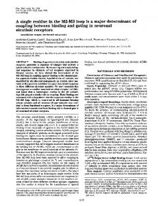
Figure 1. p27Kip1 couples multiple signalling pathways that underlie cortical neurogenesis. (A) ��� p27Kip1 promotes cell cycle exit by associating with specific Cdk/cylins complexes through a N‑terminal binding domain and hence block‑ ing their catalytic activity and preventing G1‑S phase transition (molecular pathway in green). By stabilising Ngn2 in the nucleus of cortical progenitors, p27Kip1 regulates neuronal differentiation, an activity that resides in its N‑terminal half (molecular pathway in purple). Ngn2 may act in positive feedback loop to promote the transcription of p27 ���Kip1 in cortical progeni‑ 56 tors as demonstrated for Ngn1 in P19 cells. p27 ���Kip1 promotes the radial migration of cortical neurons by blocking the RhoA signalling pathway, an activity residing in its C‑terminal domain (molecular pathway in blue). Ngn2 promotes migration by activating transcription of target genes regulating radial migration.42 Cdk5 phosphorylates ��� p27Kip1 and hence regulates its stability and cytoplasmic distribution.26 In this pathway, cofilin (an actin‑ binding protein with actin‑severing activity) has been proposed to be a downstream target of the Rho‑kinase pathway mediating p27 ���Kip1 activity on the actin cytoskeleton (molecular pathway in grey). Black arrows represent nontranscriptional interactions and white arrows represent transcriptional interactions. (B) Diagram illustrating the functional roles played by ��� p27Kip1 during cortical histogenesis. The drawing illustrates a cortical cell at different steps of its maturation. The accumulation of p27 ���Kip1 in the nucleus of a VZ cell (yellow) induces its exit from the cell cycle by inhibiting the catalytic activity of Cdks. As this cell moves towards the SVZ, ��� p27Kip1 stabilizes ����������� Ngn2 protein that accumulates until reaching a threshold level that triggers neuronal differentiation. The newly differentiate neuron poses in the SVZ before initiating its migration to the cortical plate, a step that involves inhibition of RhoA activity by the cytoplasmic fraction of p27 ���Kip1 (yellow colour outside the nucleus). Given that ��� p27Kip1 expression is maintained in the nucleus of neurons that have reached their final destination in the corti‑ cal plate,25 it is possible that ��� p27Kip1 performs other yet uncharacterized functions in mature neurons.
p27Kip1 Regulates Radial Migration in the Cerebral Cortex The extraordinary degree of organization of the cerebral cortex is the result of elaborate patterns of migratory movements during corticogenesis. Cell migration in the cortex can broadly be divided into two categories: radial and tangential migrations.3 Glutamatergic projection neurons are born in the VZ and SVZ of the cortex and migrate radially from these progenitor layers to the cortical plate, whereas GABAergic interneurons are generated ventrally in the GE and navigate over long distances using multiple tangential migratory routes to integrate into the cortex.3 Dynamic remodelling of the actin and microtubule cytoskeletons provides the driving force for cell migration in all tissues. ��� p27Kip1 has been shown to regulates the actin cytoskeleton dynamics in several in vitro models.23,38‑41 The finding that migrating cortical projection neurons express p27 ���Kip1 25 suggested that p27 Kip1 ��� ����������������������������������������� may also contributes to the cytoskeletal changes that underlie radial migration in the cerebral cortex. To address this possibility, we examined whether neuronal migration is affected in p27‑/‑ cerebral cortices.25 Birth‑dating analysis indeed revealed an aberrant distribution of newly born cells throughout the cortex of p27‑/‑ embryos with a reduced number of neurons reaching the cortical plate. In contrast, ��� p27CK‑ cortices, that express a cell cycle mutant form of ��� p27Kip1, did no not present any significant migration defect.25 Radial migration was also impaired when ��� p27Kip1 2316
was knocked down by electroporation of siRNAs in cortical VZ cells.25 Conversely, overexpression of p27Kip1 or the cell cycle mutant form p27ck‑, but not other Cip/Kip genes, accelerated the migration of newly born neurons away from the VZ/SVZ, resulting in an increased number of projection neurons accumulating in the cortical plate. Altogether, these results indicate that p27Kip1 is required for the proper radial migration of cortical neurons and that this activity is independent of its cell cycle regulatory function. There is compelling evidence that the radial migration of projection neurons in the cortex is controlled by Ngn2 through the transcriptional regulation of genes important for cell migration42 as well as an uncharacterized mechanism that requires the phosphorylation of a tyrosine���������������������������������� residue ��������������������������������� in its C‑terminal domain.43 Given the ability of p27 ���Kip1 to stabilize Ngn2 in cortical progenitors,25 it was thus conceivable that the migratory property of ��� p27Kip1 reflected its capacity to regulate Ngn2 expression in cortical cells. To address this possibility, we attempted to rescue the migration defect of cortical VZ induced by p27 ���Kip1 knock‑down, by coelectroporating Ngn2 with siRNAs directed against p27 ���Kip1. To our surprise and in striking contrast with the differentiation phenotype, the radial migration defect caused by p27 ���Kip1 knock‑down was not corrected by overexpression of Ngn2. These results therefore demonstrate that, although Ngn2 is epistatic to p27 ���Kip1 for the differentiation Kip1 of cortical neurons, p27 ��� regulates neuron migration through a distinct mechanism that does not involve Ngn2. Small GTPase of the Rho family play an important regulatory role in the the organisation of the actin cytoskeleton,44 and cortical neuron migration requires the inhibition of the activity of the small GTPase RhoA.43,45 Moreover, ��� p27Kip1 has been shown to promote migration of fibroblasts by blocking the activation of the Rho‑kinase pathway, an activity that involves an interaction of its C‑terminal half with RhoA.22 Considering that most cortical VZ and SVZ
Cell Cycle
2006; Vol. 5 Issue 20
Multiple Roles of p27Kip1 in Cortical Neurogenesis
cells express RhoA,25 it was thus possible that p27 ���Kip1 regulates the cytoskeletal change contributing to radial migration in the cortex by interacting with RhoA. Indeed, we found that coelectroporating a dominant negative version of RhoA with p27 ���Kip1 siRNAs rescued the neuronal migration defect caused by p27 ���Kip1 knock‑down.25 Moreover, the migration‑promoting activity of p27 ���Kip1 lies in the C‑terminal half of protein, suggesting that the mechanism by which p27Kip1 promotes the migration of fibroblasts, involving the inactivation of the Rho‑kinase signalling pathway, also operates for the radial migration of cortical neurons �������25 (see Fig. 1). In the cerebral cortex, the atypical cyclin‑dependant kinase Cdk5 associated with its coactivator p35 promotes radial migration through phosphorylation of several targets that influence microtubule stability, dynein motor complex activity and actin cytoskeleton dynamics.2 A recent study supports a mechanism by which Cdk5 regulates the migration of cortical neurons through phosphorylation of p27 ���Kip1.26 In this work, Kawauchi and collaborators have identified a novel molecular pathway that links Cdk5 to the actin cytoskeleton. The authors propose that phosphorylation of p27 ���Kip1 by Cdk5 at Serine 10 allows the translocation and accumulation of p27Kip1 in the ��������� cytoplasm46 where it mediates its migratory func26 tion. The role of Cdk5 in phosphorylating and stabilizing p27 ���Kip is based on biochemical evidence obtained in cell culture, and evaluation of its importance in the migratory‑promoting activity of p27Kip1 in vivo awaits analysis of ��� p27Kip1 knock‑in ������������������������� mice harbouring a mutation of serine 10 (��� p27S10A).47 The authors also propose that the p27 ���Kip1‑mediated block in the Rho‑kinase pathway promotes actin reorganisation by activating the actin‑binding protein cofilin.26 However, the extent to which cofilin mediates p27 ���Kip1 activity in actin cytoskeleton remodelling remains to be assessed, as overexpression of a constitutively active form of cofilin prevents radial migration rather than promoting it.26 In contrast with its activity in the cerebral cortex, ��� p27Kip1 inhibits the migration of sarcoma cells in culture.48 In this system, ��� p27Kip1 impairs cell migration by altering microtubule dynamics through cytoplasmic binding to the microtubule (MT)‑destabilising protein stathmin and inhibition of its activity.48 This discrepancy may reflect differences in ��� p27Kip1 activity ��������������������������������������� depending on the cellular and molecular contexts. In contrast with the amoeboid‑like mode of cell migration used by sarcoma cells,49 highly polarised cells such as migrating neurons require a stable MT network to maintain cell polarity and to couple nucleus and centrosome through bridges of stabilized MT during nucleokinesis.50 Stathmin is expressed in cortical neurons,51,52 and ��� p27Kip1 may also promote neuronal migration by blocking the activity of stathmin and that of other related MT‑destabilising proteins.53
Concluding Remarks From the identification of p27 ���Kip1 as a cell cycle inhibitor almost 15 years ago,54 a more complex picture of its biology has emerged with the recent findings that p27 ���Kip1 regulates multiple cellular processes that are critical for histogenesis of various tissues. In the cerebral cortex, p27 ���Kip1 ������������������������������������������������ regulates and couples cell cycle exit, neuronal differentiation and cell migration through the regulation of distinct molecular pathways.25,26 p27Kip1 is therefore an essential regulator of cortical development that orchestrates the major steps by which a progenitor cell becomes a mature projection neuron. By identifying novel functions of p27 ���Kip1 in the cerebral cortex, we have shed some light on how distinct cellular events are regulated and coupled during neurogenesis. Based on recent findings demonstrating that www.landesbioscience.com
other known regulators display unexpected activities in the cerebral cortex,43,55 it is however likely that important aspects of the whole story are still missing, and that p27 ���Kip1 is only one element of a complex machinery that couples multiple events contributing to neurogenesis in the cerebral cortex. References 1. Ohnuma S, Philpott A, Harris WA. Cell cycle and cell fate in the nervous system. Curr Opin Neurobiol 2001; 11:66‑73. 2. Gupta A, Tsai LH, Wynshaw‑Boris A. Life is a journey: A genetic look at neocortical development. Nat Rev Genet 2002; 3:342‑55. 3. Marin O, Rubenstein JL. A long, remarkable journey: Tangential migration in the telencephalon. Nat Rev Neurosci 2001; 2:780‑90. 4. Rash BG, Grove EA. Area and layer patterning in the developing cerebral cortex. Curr Opin Neurobiol 2006; 16:25‑34. 5. Hevner RF, Daza RA, Rubenstein JL, Stunnenberg H, Olavarria JF, Englund C. Beyond laminar fate: Toward a molecular classification of cortical projection/pyramidal neurons. Dev Neurosci 2003; 25:139‑51. 6. Caviness Jr VS. Neocortical histogenesis in normal and reeler mice: A developmental study based upon [3H]thymidine autoradiography. Brain Res 1982; 256:293‑302. 7. Sidman RL, Rakic P. Neuronal migration, with special reference to developing human brain: A review. Brain Res 1973; 62:1‑35. 8. McConnell SK, Kaznowski CE. Cell cycle dependence of laminar determination in developing neocortex. Science 1991; 254:282‑5. 9. Elledge SJ, Harper JW. Cdk inhibitors: On the threshold of checkpoints and development. Curr Opin Cell Biol 1994; 6:847‑52. 10. Sherr CJ, Roberts JM. CDK inhibitors: Positive and negative regulators of G1‑phase progression. Genes Dev 1999; 13:1501‑12. 11. Fero ML, Rivkin M, Tasch M, Porter P, Carow CE, Firpo E, Polyak K, Tsai LH, Broudy V, Perlmutter RM, Kaushansky K, Roberts JM. A syndrome of multiorgan hyperplasia with features of gigantism, tumorigenesis, and female sterility in p27(Kip1)‑deficient mice. Cell 1996; 85:733‑44. 12. Lukaszewicz A, Savatier P, Cortay V, Giroud P, Huissoud C, Berland M, Kennedy H, Dehay C. G1 phase regulation, area‑specific cell cycle control, and cytoarchitectonics in the primate cortex. Neuron 2005; 47:353‑64. 13. Caviness Jr VS, Goto T, Tarui T, Takahashi T, Bhide PG, Nowakowski RS. Cell output, cell cycle duration and neuronal specification: A model of integrated mechanisms of the neocortical proliferative process. Cereb Cortex 2003; 13:592‑8. 14. Goto T, Mitsuhashi T, Takahashi T. Altered patterns of neuron production in the p27 knockout mouse. Dev Neurosci 2004; 26:208‑17. 15. Tarui T, Takahashi T, Nowakowski RS, Hayes NL, Bhide PG, Caviness VS. Overexpression of p27 Kip 1, probability of cell cycle exit, and laminar destination of neocortical neurons. Cereb Cortex 2005; 15:1343‑55. 16. Munoz‑Alonso MJ, Acosta JC, Richard C, Delgado MD, Sedivy J, Leon J. p21Cip1 and p27Kip1 induce distinct cell cycle effects and differentiation programs in myeloid leukemia cells. J Biol Chem 2005; 280:18120‑9. 17. Vernon AE, Devine C, Philpott A. The cdk inhibitor p27Xic1 is required for differentiation of primary neurones in Xenopus. Development 2003; 130:85‑92. 18. Vernon AE, Philpott A. A single cdk inhibitor, p27Xic1, functions beyond cell cycle regulation to promote muscle differentiation in Xenopus. Development 2003; 130:71‑83. 19. Ohnuma S, Philpott A, Wang K, Holt CE, Harris WA. p27Xic1, a Cdk inhibitor, promotes the determination of glial cells in Xenopus retina. Cell 1999; 99:499‑510. 20. Baldassarre G, Barone MV, Belletti B, Sandomenico C, Bruni P, Spiezia S, Boccia A, Vento MT, Romano A, Pepe S, Fusco A, Viglietto G. Key role of the cyclin‑dependent kinase inhibitor p27kip1 for embryonal carcinoma cell survival and differentiation. Oncogene 1999; 18:6241‑51. 21. Sasaki K, Tamura S, Tachibana H, Sugita M, Gao Y, Furuyama J, Kakishita E, Sakai T, Tamaoki T, Hashimoto‑Tamaoki T. Expression and role of p27(kip1) in neuronal differentiation of embryonal carcinoma cells. Brain Res Mol Brain Res 2000; 77:209‑21. 22. Besson A, Gurian‑West M, Schmidt A, Hall A, Roberts JM. p27Kip1 modulates cell migration through the regulation of RhoA activation. Genes Dev 2004; 18:862‑76. 23. McAllister SS, Becker‑Hapak M, Pintucci G, Pagano M, Dowdy SF. Novel p27(kip1) C‑terminal scatter domain mediates Rac‑dependent cell migration independent of cell cycle arrest functions. Mol Cell Biol 2003; 23:216‑28. 24. van Lookeren Campagne M, Gill R. Tumor‑suppressor p53 is expressed in proliferating and newly formed neurons of the embryonic and postnatal rat brain: Comparison with expression of the cell cycle regulators p21Waf1/Cip1, p27Kip1, p57Kip2, p16Ink4a, cyclin G1, and the proto‑oncogene Bax. J Comp Neurol 1998; 397:181‑98. 25. Nguyen L, Besson A, Heng JI, Schuurmans C, Teboul L, Parras C, Philpott A, Roberts JM, Guillemot F. p27kip1 independently promotes neuronal differentiation and migration in the cerebral cortex. Genes Dev 2006; 20:1511‑24. 26. Kawauchi T, Chihama K, Nabeshima Y, Hoshino M. Cdk5 phosphorylates and stabilizes p27(kip1) contributing to actin organization and cortical neuronal migration. Nat Cell Biol 2006; 8:17‑26. 27. Schuurmans C, Guillemot F. Molecular mechanisms underlying cell fate specification in the developing telencephalon. Curr Opin Neurobiol 2002; 12:26‑34. 28. Bertrand N, Castro DS, Guillemot F. Proneural genes and the specification of neural cell types. Nat Rev Neurosci 2002; 3:517‑30.
Cell Cycle
2317
Multiple Roles of p27Kip1 in Cortical Neurogenesis 29. Parras CM, Schuurmans C, Scardigli R, Kim J, Anderson DJ, Guillemot F. Divergent functions of the proneural genes Mash1 and Ngn2 in the specification of neuronal subtype identity. Genes Dev 2002; 16:324‑38. 30. Fode C, Ma Q, Casarosa S, Ang SL, Anderson DJ, Guillemot F. A role for neural determination genes in specifying the dorsoventral identity of telencephalic neurons. Genes Dev 2000; 14:67‑80. 31. Schuurmans C, Armant O, Nieto M, Stenman JM, Britz O, Klenin N, Seibt J, Brown C, Tang H, Cunningham JM, Dyck R, Walsh C, Campbell K, Polleux F, Guillemot F. Sequential phases of neocortical fate specification involve Neurogenin‑dependent and ‑independent pathways. EMBO J 2004; 23:2892‑902. 32. Kawaguchi A, Ogawa M, Saito K, Matsuzaki F, Okano H, Miyata T. Differential expression of Pax6 and Ngn2 between pair‑generated cortical neurons. J Neurosci Res 2004; 78:784‑95. 33. Laman H, Funes JM, Ye H, Henderson S, Galinanes‑Garcia L, Hara E, Knowles P, McDonald N, Boshoff C. Transforming activity of Fbxo7 is mediated specifically through regulation of cyclin D/cdk6. Embo J 2005; 24:3104‑16. 34. Reynaud EG, Leibovitch MP, Tintignac LA, Pelpel K, Guillier M, Leibovitch SA. Stabilization of MyoD by direct binding to p57(Kip2). J Biol Chem 2000; 275:18767‑76. 35. Pardee AB. G1 events and regulation of cell proliferation. Science 1989; 246:603‑8. 36. Mitsuhashi T, Aoki Y, Eksioglu YZ, Takahashi T, Bhide PG, Reeves SA, Caviness Jr VS. Overexpression of p27Kip1 lengthens the G1 phase in a mouse model that targets inducible gene expression to central nervous system progenitor cells. Proc Natl Acad Sci USA 2001; 98:6435‑40. 37. Iacopetti P, Michelini M, Stuckmann I, Oback B, Aaku‑Saraste E, Huttner WB. Expression of the antiproliferative gene TIS21 at the onset of neurogenesis identifies single neuroepithelial cells that switch from proliferative to neuron‑generating division. Proc Natl Acad Sci USA 1999; 96:4639‑44. 38. Sun J, Marx SO, Chen HJ, Poon M, Marks AR, Rabbani LE. Role for p27(Kip1) in vascular smooth muscle cell migration. Circulation 2001; 103:2967‑72. 39. Goukassian D, Diez‑Juan A, Asahara T, Schratzberger P, Silver M, Murayama T, Isner JM, Andres V. Overexpression of p27(Kip1) by doxycycline‑regulated adenoviral vectors inhibits endothelial cell proliferation and migration and impairs angiogenesis. Faseb J 2001; 15:1877‑85. 40. Diez‑Juan A, Andres V. Coordinate control of proliferation and migration by the p27Kip1/ cyclin‑dependent kinase/retinoblastoma pathway in vascular smooth muscle cells and fibroblasts. Circ Res 2003; 92:402‑10. 41. Supriatno, Harada K, Kawaguchi S, Yoshida H, Sato M. Effect of p27Kip1 on the ability of invasion and metastasis of an oral cancer cell line. Oncol Rep 2003; 10:527‑32. 42. Ge W, He F, Kim KJ, Blanchi B, Coskun V, Nguyen L, Wu X, Zhao J, Heng JI, Martinowich K, Tao J, Wu H, Castro D, Sobeih MM, Corfas G, Gleeson JG, Greenberg ME, Guillemot F, Sun YE. Coupling of cell migration with neurogenesis by proneural bHLH factors. Proc Natl Acad Sci USA 2006; 103:1319‑24. 43. Hand R, Bortone D, Mattar P, Nguyen L, Heng JI, Guerrier S, Boutt E, Peters E, Barnes AP, Parras C, Schuurmans C, Guillemot F, Polleux F. Phosphorylation of Neurogenin2 specifies the migration properties and the dendritic morphology of pyramidal neurons in the neocortex. Neuron 2005; 48:45‑62. 44. Hall A. Rho GTPases and the actin cytoskeleton. Science 1998; 279:509‑14. 45. Kholmanskikh SS, Dobrin JS, Wynshaw‑Boris A, Letourneau PC, Ross ME. Disregulated RhoGTPases and actin cytoskeleton contribute to the migration defect in Lis1‑deficient neurons. J Neurosci 2003; 23:8673‑81. 46. Rodier G, Montagnoli A, Di Marcotullio L, Coulombe P, Draetta GF, Pagano M, Meloche S. p27 cytoplasmic localization is regulated by phosphorylation on Ser10 and is not a prerequisite for its proteolysis. Embo J 2001; 20:6672‑82. 47. Besson A, Gurian‑West M, Chen X, Kelly‑Spratt KS, Kemp CJ, Roberts JM. A pathway in quiescent cells that controls p27Kip1 stability, subcellular localization, and tumor suppression. Genes Dev 2006; 20:47‑64. 48. Baldassarre G, Belletti B, Nicoloso MS, Schiappacassi M, Vecchione A, Spessotto P, Morrione A, Canzonieri V, Colombatti A. p27(Kip1)‑stathmin interaction influences sarcoma cell migration and invasion. Cancer Cell 2005; 7:51‑63. 49. Sahai E, Marshall CJ. Differing modes of tumour cell invasion have distinct requirements for Rho/ROCK signalling and extracellular proteolysis. Nat Cell Biol 2003; 5:711‑9. 50. Tsai LH, Gleeson JG. Nucleokinesis in neuronal migration. Neuron 2005; 46:383‑8. 51. Gavet O, El Messari S, Ozon S, Sobel A. Regulation and subcellular localization of the microtubule‑destabilizing stathmin family phosphoproteins in cortical neurons. J Neurosci Res 2002; 68:535‑50. 52. Ozon S, Byk T, Sobel A. SCLIP: A novel SCG10‑like protein of the stathmin family expressed in the nervous system. J Neurochem 1998; 70:2386‑96. 53. Tararuk T, Ostman N, Li W, Bjorkblom B, Padzik A, Zdrojewska J, Hongisto V, Herdegen T, Konopka W, Courtney MJ, Coffey ET. JNK1 phosphorylation of SCG10 determines microtubule dynamics and axodendritic length. J Cell Biol 2006; 173:265‑77. 54. Polyak K, Lee MH, Erdjument‑Bromage H, Koff A, Roberts JM, Tempst P, Massague J. Cloning of p27Kip1, a cyclin‑dependent kinase inhibitor and a potential mediator of extracellular antimitogenic signals. Cell 1994; 78:59‑66. 55. Ferguson KL, McClellan KA, Vanderluit JL, McIntosh WC, Schuurmans C, Polleux F, Slack RS. A cell‑autonomous requirement for the cell cycle regulatory protein, Rb, in neuronal migration. Embo J 2005. 56. Farah MH, Olson JM, Sucic HB, Hume RI, Tapscott SJ, Turner DL. Generation of neurons by transient expression of neural bHLH proteins in mammalian cells. Development 2000; 127:693‑702.
2318
Cell Cycle
2006; Vol. 5 Issue 20