Creating an Immune-Privileged Site Using Retinal Progenitor Cells and Biodegradable Polymers. TAT FONG NG,a ERIN LAVIK,b HIROSHI KEINO,a ANDREW ...
TECHNOLOGY DEVELOPMENT Creating an Immune-Privileged Site Using Retinal Progenitor Cells and Biodegradable Polymers TAT FONG NG,a ERIN LAVIK,b HIROSHI KEINO,a ANDREW W. TAYLOR,a ROBERT S. LANGER,c MICHAEL J. YOUNGa a
Schepens Eye Research Institute, Department of Ophthalmology, Harvard Medical School, Boston, Massachusetts, USA; bDepartment of Biomedical Engineering, Yale University, New Haven, Connecticut, USA; cDepartment of Chemical and Biomedical Engineering, Massachusetts Institute of Technology, Cambridge, Massachusetts, USA
Key Words. Stem cells • Polymer • Transplantation • Immunology
ABSTRACT We describe the creation of local immune privilege (IP) using retinal progenitor cells (RPCs) and biodegradable polymers. Murine RPCs were seeded on poly(lactic-coglycolic acid) polymers to generate composite grafts. Composites or RPCs alone were transplanted into allogeneic kidney capsules. Grafts survived at all time points, differentiating into neurons and astrocytes. Upon treatment with interferon ␥ (IFN␥), major histocompatibility complex antigens were upregulated. Although 10% of IFN␥-treated RPC grafts survived 14 days, 66% of the IFN␥-treated composites survived in part by producing im-
mune suppressive factors transforming growth factor-2, Fas ligand, and indoleamine 2,3-dioxygenase. The composites were assayed for delayed-type hypersensitivity (DTH) by seeding composites with antigen-presenting cells incubated with ovalbumin. This resulted in suppression of ovalbumin-specific DTH, indicating that composite grafts consisting of biodegradable polymers and central nervous system progenitor cells can be used to generate local IP. This technology may be used to promote the survival of nonprivileged grafts (e.g., pancreas, liver, or skin). STEM CELLS 2007;25:1552–1559
Disclosure of potential conflicts of interest is found at the end of this article.
INTRODUCTION Foreign tissue such as skin, liver, and kidney is typically rejected when transplanted to a conventional site (e.g., skin or kidney capsule) in allogeneic recipients. However, some exceptions exist when foreign tissues are transplanted to sites such as the brain, eyes, and testis. These compartments are termed immune-privileged (IP) sites. The anterior chamber in the mouse eye has been studied extensively for its property as an IP site. It has been shown that minor histoincompatible tumor cells transplanted into the anterior chamber grow progressively and do not result in immune rejection [1]. These allogeneic tumors induced a deviant systemic immune response known as anterior chamber associated immune deviation (ACAID). This is an active process that suppresses the host’s systemic immune response. Similarly, grafts composed of cells from IP tissues survive for extended periods in allogeneic recipients [2, 3]. Stem cells and progenitor cells have been shown to possess some IP properties, including low immunogenicity [4] and the production of immunosuppressive factors [5, 6]. Retinal progenitor cells (RPCs) are multipotent cells that can differentiate into cells of the retinal lineage, including retinal neurons and glia [7–9]. The commitment of these cells to the retinal lineage makes RPCs attractive candidates for clinical applications designed to treat degenerative retinal diseases. In addition, progenitor cells in general have been shown to possess low immunogenicity [10 –12], which may
protect them from T-cell recognition, allowing them to survive in allogeneic hosts for extended periods of time. It has also been shown that progenitor cells are capable of producing immunosuppressive factors including transforming growth factor (TGF)-2 [13]. Polymer substrates combined with progenitor cells have been used to generate a number of tissue equivalents including bones, blood vessels, and cartilages. We have shown that poly(lactic-coglycolic acid) (PLGA) polymers can bind to and retain growth factors (e.g., epidermal growth factor), which in turn supports the survival of the progenitor cells after transplantation [9]. We thus hypothesize that biodegradable polymer scaffolds not only provide a substrate for the growth of RPCs, but may also provide a “sink” for the accumulation of immunosuppressive factors produced by these progenitor cells. These properties may facilitate RPC survival in allogeneic recipients by suppressing the immune response against donor alloantigens. In this study, we evaluate the survival of RPCs seeded onto biodegradable PLGA polymers before and after challenge with interferon ␥ (IFN␥) treatment. These constructs were grafted into the kidney subcapsular space and assessed for the properties of IP. We propose that RPC/biodegradable polymers actively generate a local IP site. The results of these experiments prompted us to look further into the mechanism underlying altered immunity in the local microenvironment. This has implications for the use of such constructs in regenerative medicine.
Correspondence: Michael J. Young, Ph.D., Schepens Eye Research Institute, Department of Ophthalmology, Harvard Medical School, 20 Staniford Street, Boston, Massachusetts 02114, USA. Telephone: 617-912-7419; Fax: 617-912-0100; e-mail: michael.young@schepens. harvard.edu Received December 11, 2006; accepted for publication February 12, 2007. ©AlphaMed Press 1066-5099/2007/$30.00/0 doi: 10.1634/stemcells.2006-0780
STEM CELLS 2007;25:1552–1559 www.StemCells.com
Ng, Lavik, Keino et al.
MATERIALS
1553
AND
METHODS
Scaffold Preparation Poly(lactic-coglycolic acid) with a lactic to glycolic acid ratio of 50:50 and a number average molecular weight of ⬃35,000 g/mol, noted here as PLGA 504, was obtained from Boehringer Ingelheim GmbH (Ingelheim, Germany, http://www.boehringer-ingelheim.com). All solvents were from Sigma-Aldrich (St. Louis, http:// www.sigmaaldrich.com) (ACS Grade).
Phase-Inversion Membrane Formation Solutions were prepared with concentrations of 20% (wt/vol) polymer (PLGA 504) in dimethylsulfoxide. We added 5% (vol/vol) glycerol to the solution to promote formation of larger and less asymmetric pore structures. We added 400 l of the complete solution to a glass microscope slide and allowed it to spread evenly over the entire surface. The glass slide was then immersed in 18 M-Ohm water (Milli-Q system; Millipore, Billerica, MA, http:// www.millipore.com) at room temperature, and the solvent transfer process was initiated. Slides were removed from the water once the transfer process was complete, approximately 10 minutes after immersion. The completion of the transfer was indicated by the absence of solution at the glass-slide interface. Samples that completed the transfer were easily removed from the glass slides without any apparent sticky residue. Membranes were then dried with blotting paper and lyophilized overnight to remove residual water and solvent.
Cell Maintenance and Seeding Retinal progenitor cells and fibroblasts were isolated from the retina (RPC) and skin (fibroblast) of P1 enhanced green fluorescent protein (eGFP) C57BL/6 and maintained in epidermal growth factorenriched neural basal medium. Scaffolds were soaked in 70% ethanol for 24 hours and rinsed three times in Hanks’ balanced saline solution (HBSS) with 3⫻ penicillin/streptomycin (Sigma-Aldrich) prior to receiving cells. Two hundred microliters containing approximately 107 cells per milliliter RPC cells were seeded on the polymer (⬃1 cm2) in a 6-well culture plate. Cells were permitted to attach to the polymer for 2 hours, after which time the polymer was submerged in culture medium and grown for 7 days under routine conditions of 95% air, 5% CO2 at 37°C. After 7 days in culture, one group received fresh medium and the other groups received IFN␥ at a concentration of 33 ng/ml and were grown for an additional 7 days prior to transplantation.
lows: specific swelling ⫽ ([24-hour numerical values of right ear ⫺ 0 hour numerical values of right ear] ⫺ [24 hour numerical values of left ear ⫺ 0 hour numerical values of left ear]) ⫻ 10⫺3 mm. Ear-swelling responses at 24 hours after ear injection are presented as group mean ⫾ SEM.
Immune Deviation Peritoneal exudates containing cells were collected from BALB/c mice (same strain as recipients) 3 days after thioglycolate injection into the abdomen. Transplantation ready RPC/polymer and IFN␥treated RPC/polymer composites were incubated with peritoneal exudate cells (PEC) for 18 hours. Ovalbumin (OVA) (2 mg/ml) was added to the culture 1 hour before transplantation. A positive control group that contained only PEC/polymer was also prepared. The polymer composites were then transplanted into the kidney capsule of the above groups of BALB/c mice. All mice also received an injection of OVA (2 mg/ml in HBSS) prepared in complete Freund’s adjuvant 7 days post-transplantation. OVA in HBSS (20 mg/ml) was injected into the right pinnae of all four mice described above and into a negative group of BALB/c mice 14 days after transplantation. Both ear pinnae were measured immediately before injection as well as 24 and 48 hours later. DH was calculated based on the method described above.
Immunohistochemistry At appropriate time points, kidneys were harvested, and the portion bearing the polymer was dissected, fixed overnight in 4% paraformaldehyde, cryoprotected overnight at 4°C in 20% sucrose, and cryosectioned for 16-m thickness. A panel of antibodies was used to detect antigens expressed by progenitor cells before and after transplantation. These included anti-major histocompatibility complex (MHC) class I (H2-Kb; BD Pharmingen, San Diego, http:// www.bdbiosciences.com/index_us.shtml; used at a dilution of 1:100 in phosphate-buffered saline); anti-MHC class II (I-Ad; BD Pharmingen; 1:100); anti-F4/80 (Caltag Laboratories, Burlingame, CA, http://www.caltag.com; 1:100); anti-CD3e (BD Pharmingen; 1:200); anti-neurofilament high (Sigma-Aldrich; 1:1,000); anti-glial fibrillary acidic protein (GFAP; Sigma-Aldrich; 1:1,000); antiTGF-2 (Santa Cruz Biotechnology Inc., Santa Cruz, CA, http:// www.scbt.com; 1:100); anti-Fas ligand (FasL) (Santa Cruz Biotechnology Inc.; 1:100); and anti-indoleamine 2,3-dioxygenase (IDO) (Santa Cruz Biotechnology Inc.; 1:100).
Western Blot Analysis
Implantation of the progenitor cells/polymer composite graft beneath the kidney capsule was performed as described [3]. Recipient mice received general anesthesia using a mixture of 150 mg/kg ketamine (Phoenix Pharmaceutical, St. Joseph, MO) and 6 mg/kg xylazine (Phoenix Pharmaceuticals Inc., Belmont, CA, http://www. phoenixpeptide.com) before the surgery. A 1.5-cm-long opening was made in the back of the animals, parallel to the spinal cord. The peritoneum was opened, and the kidney was extruded. A small pouch was made in the kidney capsule, after which time the composite graft was inserted into the pouch. The kidney was then returned to its original position, and the wound was closed using a surgical stapler.
Progenitor cells and IFN␥-treated progenitor cells were lysed in cell lysis buffer (Cell Signaling Technology, Beverly, MA, http:// www.cellsignal.com), after which time 50 g of protein from each sample was loaded, run in 4%–12% 2-[bis(2-hydroxyethyl)amino]2-(hydroxymethyl)propane-1,3-diol (BisTris) gradient gel, and transferred to a polyvinylidene difluoride membrane (Invitrogen, Carlsbad, CA, http://www.invitrogen.com). A SeeBlue Prestained marker (Invitrogen) was added as the molecular weight ladder. The blot was then incubated in primary antibodies consisting of either anti-FasL (1:200) or anti-IDO (1:200) overnight at 4°C. After several washes, the blot was incubated in secondary antibodies conjugated with horseradish peroxidase (Chemicon, Temecula, CA, http://www.chemicon.com; 1:5,000) for 1 hour and then incubated with developing substrate (SuperSignal; Cell Signaling Technology) and subjected to chemiluminescence detection.
Delayed Hypersensitivity
TGF- Production Measurement
An ear-swelling assay was used to measure delayed hypersensitivity (DH) using a minimum of five animals per group. Irradiated (2000R) splenocytes (1 ⫻ 106 cells per 10 l) from C57BL/6 donors were injected into the right pinnae of recipient mice. Positive control mice were immunized subcutaneously with 1 ⫻ 107 C57BL/6 splenocytes 1 week before ear challenge. Negative control mice received only an ear pinnae challenge. Both ear pinnae were measured immediately before injection and 24 hours later with a low-pressure engineer’s micrometer (Mitsutoyo, Kawasaki, Japan, http://www.mitutoyo.co.jp/eng). Ear swelling was expressed as fol-
A mink lung cell bioassay was used to measure TGF- production secreted into culture supernatant of progenitor cells [14]. Briefly, 1 ⫻ 105 mink lung cells were incubated with culture supernatant in a 96-well plate for 96 hours. In parallel, serial dilution of TGF-2 standard that ranged from 1 ng to 10 g was also incubated with mink lung cells in the same culture plate for 96 hours. At the appropriate time point, 10 l of the CCK-8 solution (Dojindo Molecular Technologies Inc., Gaithersburg, MD, http://www.dojindo. com) were added to each well, and the absorbance was measured at 450 nm after a 1-hour incubation. The TGF-2 content in the
Surgical Procedures
www.StemCells.com
Creating an Immune-Privileged Site
1554
naı¨ve BALB/c mice (Fig. 3A, 3B). These data demonstrated a lack of DH in animals that received RPC or composite grafts.
IFN␥-Treated Retinal Progenitor Cells Survived as Composite Grafts
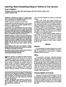
Figure 1. Photomicrographs of the retinal progenitor cell (RPC) grafts in the kidney subcapsular space. (A): Scanning electron micrograph of the polymer scaffold surface depicting its highly porous surface. (B): Green fluorescent protein (GFP) RPCs seeded on the polymer scaffold prior to transplantation. (C): A piece of polymer transplanted to the kidney capsule of a BALB/c mouse at day 28. (D): An accepted GFP RPC graft in BALB/c mice at day 28. (E, F): Accepted GFP retinal progenitor cells/polymer composite grafts at day 42 in syngeneic C57BL/6 (E) and BALB/c mice (F) showing that the majority of the scaffold was covered with GFP fluorescence. Arrows point to pores.
culture supernatants would be derived from the standard curve plotted by the absorbance of the TGF-2 standard.
RESULTS Retinal Progenitor Cells Survive in the Allogeneic Kidney Capsule and Fail to Induce Delayed Hypersensitivity RPCs seeded onto polymers attached to the substrates, where they sent processes into the pores and migrated into the channels (Fig. 1A). Polymers containing RPCs (Fig. 1B) or RPCs alone were then transplanted into the subcapsular space of the kidney in BALB/c (allogeneic) and C57BL/6 (syngeneic) mice. In control experiments, unseeded polymers were transplanted into the kidney subcapsular space of C57BL/6 and BALB/c mice. Although no neovascularization was seen in the unseeded polymer (Fig. 1C), all RPC grafts and composite grafts induced neovascularization at the transplantation site (Fig. 1E, 1F). All grafts survived in both syngeneic and allogeneic conditions up to 42 days post-transplantation (Table 1). The presence of delayed hypersensitivity to allogeneic antigens was tested to determine whether composite grafts sensitized the recipients to these allo-antigens. BALB/c recipients who received RPC or composite grafts (green fluorescent protein [GFP]-C57BL/6) were ear challenged with allogeneic C57BL/6 spleen cells 14 days after transplantation. The resulting ear thickness was not significantly different from that detected in similarly challenged
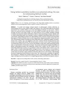
The survival of RPCs and composite grafts may be attributed to low expression of MHC class I and class II antigens. Here, we used IFN␥ treatment to upregulate MHC class I and class II expression in RPCs [13]. Expression of MHC class I and class II in untreated RPCs grown on polymers was very low, but these transplantation antigens were upregulated following 7 days of exposure to IFN␥ (Fig. 2I–2P). The treatment did not have any toxic effects on the progenitor cells and did not alter the expression of nestin, Ki-67, or neurofilament (data not shown). IFN␥treated RPCs or IFN␥-treated composite grafts were transplanted into the kidney subcapsular space in BALB/c (allogeneic) and C57BL/6 (syngeneic) mice. In control experiments, eGFP-C57BL/6 fibroblasts grown on the scaffold were transplanted into the kidney subcapsular space of C57BL/6 and BALB/c mice. Fibroblasts were found to express high levels of MHC class I. The fibroblast composite grafts (Fig. 2A) and IFN␥-treated RPCs survived on day 7 (Fig. 2B) but had all disappeared by day 14. In contrast, more than two-thirds of IFN␥-treated RPC/polymer grafts survived in allogeneic conditions for at least 42 days, the longest time point tested (Table 1 and Fig. 2C, 2D). Typically, tissues or cells expressing high levels of transplantation antigens, such as MHC Class I or II, trigger the host immune response that leads to their destruction. However, when delivered as a polymer construct, and in spite of the increased expression of MHC class I and II, the composite grafts were not rejected.
Delayed Hypersensitivity To study the recipient’s systemic immune response to the allogeneic transplant, the DH of recipients that received the composite grafts was measured and compared in groups as well as in individuals within the experimental group. A positive DH was observed in recipients receiving either IFN␥-treated RPCs (Fig. 3A) or the fibroblast/polymer grafts (Fig. 3B). A detectable DH response was observed in animals that received IFN␥-treated composite grafts (Fig. 3C). A positive DH correlated with the disappearance or diminished size of GFP fluorescence in the grafts (Fig. 3C), leading to the speculation that the emergence of DH due to the upregulation of MHC class I and class II resulted in the immune destruction of the progenitor cells.
Differentiation of IFN␥-Treated RPC The emergence of DH may be correlated with the infiltration of immune cells in the graft. F4/80⫹ macrophages were found in composite grafts in both syngeneic and allogeneic recipients on day 7 and at all time points until day 42 (Fig. 2Q–2T). CD3⫹ T cells were first found on day 14 and were present at all time points in allogeneic recipients but not syngeneic recipients (Fig. 2U–2X). In order to determine whether the upregulation of MHC antigens influenced the differentiation of progenitor cells, immunohistochemistry was used to stain the RPC/composite grafts at different time points with neuronal-specific and astroglial-specific markers. The upregulation of MHC class I and II did not affect the differentiation of progenitor cells. Similar to the untreated cells, IFN␥-treated progenitor cells were able to differentiate into neurofilament-high positive neurons as well as GFAP positive astrocytes (Fig. 2E–2H).
Ng, Lavik, Keino et al.
1555
Table 1. Survival of progenitor cells or fibroblasts in kidney capsule Survival (days)
7 14 28 42
RPC/polymer
IFN-RPC/polymer
Fibroblast/polymer
RPC BALB/c
IFN-RPC BALB/c
B6
BALB/c
B6
BALB/c
B6
BALB/c
3/3 5/5 5/5 ND
3/3 1/10 ND ND
5/5 5/5 5/5 8/8
5/5 5/5 5/5 8/8
5/5 5/5 5/5 3/4
5/5 7/8 4/5 6/9
5/5 0/5 0/5 ND
0/5 0/5 0/5 ND
Abbreviations: IFN, interferon; ND, not determined; RPC, retinal progenitor cell.
Figure 2. Photomicrographs of IFN␥treated RPCs. (A): A rejected GFP fibroblast/polymer composite graft at day 14 showing an absence of GFP fluorescence. (B): A rejected IFN␥-treated RPC graft in the kidney subcapsular space in a BALB/c mouse. (C, D): An accepted GFP RPC/polymer composite graft at (C) day 14 and (D) day 42. Scale bar: 500 m. (E–H): GFP fluorescence in differentiated RPCs expressing either NFh or GFAP (red). (I–P): GFP fluorescence and MHC expression in untreated and IFN␥-treated RPCs seeded on polymer at day 14. (I, K, M, O): GFP fluorescence; (J, L): H2-Kb immunostaining; (N, P): I-Ab; (Q–T): GFP fluorescence and F4/80 positive macrophages in the transplant. (Q, S): GFP fluorescence; (R, T): F4/80. (U–X): GFP fluorescence and F4/80positive macrophages in the transplant. (U, W): GFP fluorescence; (V, X): CD3. Scale bar: 100 m. Abbreviations: GFAP, glial fibrillary acidic protein; GFP, green fluorescent protein; IFN␥, interferon ␥; MHC, major histocompatibility complex; NFh, neurofilament high; RPC, retinal progenitor cell.
Expression of Immunosuppressive Factors by Some RPCs We speculate that the progenitor cells may employ one or more immunosuppressive factors to alter the recipient’s immune response. In the ocular environment, TGF-2 and FasL are important for maintenance of an environment devoid of inflammation [15, 16]. IDO is an important factor to maintain the immune privilege in the placenta [17]. The tryptophanmediated degradation may be involved in inhibiting infiltrating allogeneic T cells. Interestingly, IDO is found in the eye www.StemCells.com
[18] and may be a factor produced by the RPCs. In the current study, we tested whether RPC expressed TGF-, FasL, or IDO. Culture mediums collected from both nontreated and IFN␥-treated progenitor cells on days 7, 9, and 14 were used for measurement of TGF-. TGF- was detected in the culture medium of IFN␥-treated as well as untreated composite graft in vitro at day 7 and day 14 (Fig. 4A). Progenitor cells treated with IFN␥ were lysed and tested for the presence of FasL or IDO by Western blot. The level of FasL was increased after IFN␥ treatment, whereas the
1556
Creating an Immune-Privileged Site
Figure 3. Assessment of donor-specific delayed hypersensitivity (DH). The procedures of DH were performed as in the following: Ear pinnae of BALB/c mice bearing the transplant in the subcapsular space of the kidney were challenged with C57BL/6 spleen cells. Positive control BALB/c mice were immunized subcutaneously with C57BL/6 spleen cells (prime). Negative control mice (naı¨ve) were only ear challenged with spleen cells. Ear swelling responses were assessed at 24 and 48 hours. (A): BALB/c mice were ear challenged after RPC grafts (or IFN␥-treated RPC grafts) and were in residence beneath the kidney capsule for 14 days. Positive DH was observed in recipients receiving IFN␥-treated RPC. Error bars indicate SE. ⴱ, significantly higher mean ear swelling response than in naı¨ve mice (p ⬍ .05). (B): No positive DH was observed in BALB/c mice that received RPC/polymer composite grafts beneath the kidney. Positive DH was observed in BALB/c recipients of fibroblasts/polymer or IFN␥-treated RPC/polymer composite grafts were ear challenged 14 days after progenitor cells/polymer composite grafts were implanted beneath the kidney capsule. Error bars indicate SE. ⴱ, significantly higher mean ear swelling response than in naı¨ve animals (p ⬍ .05). (C): Each BALB/c mouse that had received the IFN␥-treated RPC/polymer composite graft was evaluated and the area of green fluorescent protein fluorescence-covered scaffold measured. Three ⫻ SD was considered to be positive. Abbreviations: h, hours; hrs, hours; IFN␥, interferon ␥; RPC, retinal progenitor cell.
level of IDO was decreased in IFN␥-treated RPCs (Fig. 4B). Immunostaining of RPCs using anti-TGF-2 antibodies revealed a high amount of progenitor cells expressing TGF-2 (Fig. 4C), with only a few TGF-2-positive cells in the deteriorating transplant. Immunostaining indicated that progenitor cells in the surviving grafts also expressed FasL (Fig. 4D) and IDO (Fig. 4E, 4F). These results demonstrated that RPCs and IFN␥-treated RPCs produced TGF- and FasL, which may modify the microenvironment to be immune-privileged. We also evaluated the distribution of immunosuppressive factors within the polymer composite grafts. In a graft in which the majority of the progenitor cells were located on the superficial surface of the polymer, it was possible to determine the distribution of TGF-2 within the polymer itself. TGF-2 expression was found to be highest at the graft/polymer interface, with positive staining also found within the polymer (Fig. 5A– 5D). This suggests that the polymer is indeed able to adsorb immunosuppressive factors and thereby generate local immune privilege.
Induction of Immune Privilege to Foreign Antigens by RPC We tested the hypothesis that the immune response to an antigen (OVA) injected into allogeneic recipients could be induced using RPC/polymer composite grafts. PECs were incubated
with composite grafts for 18 hours and then with OVA for an additional hour. In parallel experiments, PECs were also incubated with IFN␥-treated composite grafts as described. PECs that were seeded onto the polymer scaffold and then pulsed with OVA served as the positive control. All polymer composite grafts were then transplanted into BALB/c recipients. Animals that received polymers seeded with PECs alone showed a positive DH response to OVA (Fig. 6A). In contrast, those animals that received treated or untreated composite grafts showed a suppressed DH response to OVA, demonstrating that the presentation of an unrelated antigen (OVA) in the microenvironment of RPC/polymer composite graft induced immune suppression to that antigen. The results suggest that progenitor cells placed on the polymer scaffold can manipulate transplanted antigens.
DISCUSSION In this study, we demonstrate that the use of a PLGA scaffold promotes the survival of transplanted progenitor cells. RPCs or RPC composite grafts survived for extended periods of time in the allogeneic kidney subcapsular space. IFN␥-treated RPCs with upregulation of MHC class I and II did not survive in the allogeneic kidney capsule. Seeding the RPCs onto the biode-
Ng, Lavik, Keino et al.
1557
Figure 5. Staining of TGF-2 on the polymer scaffold. (A): Expression of GFP. (B): TGF-2 in graft with superficial distribution of progenitor cells, thereby allowing for evaluation of the staining for immunosuppressive factors adsorbed to the polymer scaffold. TGF-2 is highest at the surface of the polymer scaffold, with positive staining also found within the polymer itself. (C, D): Control staining in polymer composite grafts shows that positive staining in the polymer is not caused by nonspecific binding of antibody. Abbreviations: GFP, green fluorescent protein; TGF, transforming growth factor.
Figure 4. Production of immunosuppressive factors by progenitor cells. (A): TGF- was measured in the culture supernatant by the mink lung cell bioassay. (B): Western blot analysis showed that progenitor cells with or without IFN␥ treatment made FasL. FasL expression was upregulated after IFN␥ treatment. -Actin was used as an internal control. K-562 was human chronic myelogenous leukemia cells (Santa Cruz Biotechnology Inc.) and served as positive control. Kidney cell lysate served as negative control. (C): IFN␥-treated RPC/polymer composite grafts express TGF-2 14 days after transplantation into the kidney capsule. (D): IFN␥-treated RPC/polymer composite grafts were shown to express FasL 14 days after transplantation. (E): IFN␥-treated RPC/polymer composite grafts express IDO 14 days after transplantation. (F): A higher magnification showing that the IDO expression (red) was in the cytoplasm of the green fluorescent protein (GFP) positive RPCs and the nucleus is blue (4,6-diamidino-2-phenylindole) (inset in [E]). (G, H): GFP fluorescence and isotype control (normal goat IgG) in IFN␥-treated RPCs 14 days after transplantation into kidney capsule. Abbreviations: FasL, Fas ligand; IFN␥, interferon ␥; IDO, indoleamine 2,3-dioxygenase; RPC, retinal progenitor cell; TGF, transforming growth factor.
gradable polymer scaffold results in the prolonged survival of these IFN␥-treated RPCs. Immunosuppressive factors including TGF-, FasL, and IDO are produced by RPCs. Thus, we hypothesize that the scaffold may act as a sink to accumulate these factors and transform the incoming antigen-specific bone marrow-derived cells into “ACAID” type cells that suppress the DH response. Importantly, we show that this deviant immune rewww.StemCells.com
sponse can also be used to prevent the response directed at other antigens, thereby allowing one to engineer a graft of potentially “immunogenic” cells or tissue that is protected from rejection. We have previously shown that injection of RPCs as a single cell suspension imposes a variety of stressors on the graft, leading to significant cell death and poor graft survival [7, 9]. Seeding progenitor cells on biodegradable polymer scaffolds prior to transplantation reduces this effect by decreasing trauma at the time of grafting and providing a substrate for cell attachment. The scaffolds employed here were specifically designed to be porous, with numerous channels to allow for anchoring the cells to the substrate. It has been shown that polymer scaffolds also prevent the infiltration of fibroblasts, thereby preventing the formation of scar tissue at the graft site [19]. Progenitor grafts composed of allogeneic cells can survive in the kidney capsule for a period of 4 weeks [4]. However, in that study, the progenitor cells were rejected after challenging the recipients with allogeneic spleen cells derived from the same source [20]. We presented evidence that neural progenitor cells (NPCs) upregulated MHC class I and were recognized by cytotoxic T cells causing graft rejection. In the present study, we demonstrate that IFN␥-treated RPCs upregulated expression of MHC class I and II, making them targets for recognition and rejection in an allogeneic kidney capsule. However, when the IFN␥-treated RPCs were grown on biodegradable polymers, 66% survived up to 42 days post-transplantation, even with expression of MHC antigens. Measurement of DH in recipient mice indicated that positive DH was highly correlated with diminished GFP fluorescence in the polymer scaffold. This suggests that IFN␥treated RPCs can suppress the recipient’s immune response, which suppresses DH, leading to the survival of the RPCs. There are several potential mechanisms by which cells within the composite grafts experienced prolonged survival. One possibility relates to the production of immune suppressive factors by grafted RPCs. These factors included TGF-2 [13], IDO [21], prostaglandin E2 [5], FasL, tumor necrosis factorrelated apoptosis-inducing ligand, and APO3 ligand [6]. IDO is a promising candidate molecule in this study because of its upregulation by IFN␥ [21] and inhibition of T-cell proliferation by its end product (kynurenine). IDO was first found to be
Creating an Immune-Privileged Site
1558
Figure 6. Induction of immune deviation to OVA by RPC. (A): PECs collected from thioglycolate-treated BALB/c mice were incubated with the IFN␥-treated and untreated RPC/polymer scaffolds for 18 hours. The PEC graft was pulsed with OVA for 1 hour prior to transplantation into the kidney capsule. All groups received subcutaneous immunization with OVA in complete Freund’s adjuvant 7 days after transplantation. All groups were ear challenged with OVA in Hanks’ balanced saline solution, after which ear thickness was measured 24 and 48 hours after injection. Delayed hypersensitivity was calculated based on the difference in ear thickness between the right and left ears of the same animal. (B): Schematic representation showing that progenitor cells seeded onto a scaffold express FasL and indoleamine 2,3-dioxygenase and produce TGF-2, creating an immunosuppressive microenvironment that may convert incoming antigen-presenting cells to regulatory cells. Therefore, immune deviation to a foreign antigen (e.g., OVA) is achieved. Abbreviations: ACAID, anterior chamber associated immune deviation; APC, antigen-presenting cell; FasL, Fas ligand; IFN␥, interferon ␥; MHC, major histocompatibility complex; OVA, ovalbumin; PEC, peritoneal exudate cell; RPC, retinal progenitor cell; TGF, transforming growth factor.
responsible for the induction of the allogeneic tolerance in the placenta [17]. Inhibition of IDO can result in maternal rejection of the allogeneic fetus. Dendritic cells with high IDO expression are able to suppress allogeneic T-cell proliferation and induce apoptosis in T cells, B cells, and natural killer cells [22]. RPCs expressed IDO and survived in the allogeneic kidney. However, RPCs treated with IFN␥ downregulated the expression of IDO, suggesting that IFN␥-treated RPCs may employ other mechanisms to avoid destruction by invading ⌻ cells [6].
It has been demonstrated that IFN␥- or tumor necrosis factor-␣-treated adult NPCs upregulate cell death receptor signaling, including FasL. These undifferentiated NPCs were always found close to CD45⫹ immune cells in vivo. In the ocular microenvironment, the expression of both TGF-2 and FasL contributes in part to the establishment of an immune-privileged tissue. Corneal transplantation, the most widely performed tissue transplant procedure, has a high degree of survival due to its immune-privileged status. Constitutive FasL expression by corneal cells is required for corneal graft acceptance [23]. However, when corneas from FasL negative gld mice were used as the source for grafts, these grafts were rejected [24]. Although FasL may be required for tissue survival, there are direct mechanisms of immune suppression that mediate induction of tolerance to allo-antigens [25]. TGF-2, a crucial immunosuppressive factor in the eye [16], converts antigen-presenting cells (APCs) into tolerance-generating APCs. Both RPCs and IFN␥-treated RPCs make TGF-2, which can accumulate in the polymer scaffold. When APCs travel through the composite grafts, they are converted into tolerogenic APCs, which in turn moderate suppression of allo-specific DH and enhance graft survival (Fig. 6B). We found that RPCs were able to produce TGF-2, FasL, and IDO, indicating that RPCs could not only suppress immunity to the transplanted allogeneic progenitor cells but could also produce local immune privilege. This did not happen when IFN␥-treated RPCs were transplanted alone. We show that the biodegradable polymer is used by the cells as a substrate to adsorb immunosuppressive factors while undergoing differentiation. The polymer scaffold is not only important for the delivery of cells in an organized fashion, but also for building a three-dimensional architecture in which the cells can differentiate and function [26]. The polymer scaffold constitutes a supportive microenvironment for the progenitor cells in establishing immune privilege. We found that the combination of progenitor cells grown on biodegradable polymer scaffolds induces suppression in the recipient’s immune response, making it possible for the allogeneic cells on the scaffold to survive. In addition, these composite grafts have the potential to be constructed to contain immunogenic cells (e.g., pancreatic  cells) that are vulnerable to immune rejection. This technology offers promise for the development of novel tissue engineering constructs of allografts that may survive in conventional sites without the need for immunosuppressive drugs.
ACKNOWLEDGMENTS The authors thank Dr. Marie Shatos for culturing the retinal progenitor cells and for help with preparation of the manuscript, Dr. Jacqueline Doherty for her help in Western blotting, and Matthew Ward for his expertise in cryosectioning. We thank Dr. Sharmila Masli for reviewing the manuscript. This study was supported by NIH Grant 9595, Massachusetts Lions Eye Research Fund, the Foundation Fighting Blindness, the Department of Defense, and a gift from Gail and Richard Siegal.
DISCLOSURE
OF
POTENTIAL CONFLICTS
OF INTEREST The authors indicate no potential conflicts of interest.
Ng, Lavik, Keino et al.
REFERENCES 1 2 3 4 5 6 7 8
9 10 11 12 13
Streilein JW, Niederkorn JY, Shadduck JA. Systemic immune unresponsiveness induced in adult mice by anterior chamber presentation of minor histocompatibility antigens. J Exp Med 1980;152:1121–1125. Bellgrau D, Gold D, Selawry H et al. A role for CD95 ligand in preventing graft rejection. Nature 1995;377:630 – 632. Ng TF, Osawa H, Hori J et al. Allogeneic neonatal neuronal retina grafts display partial immune privilege in the subcapsular space of the kidney. J Immunol 2002;169:5601–5606. Hori J, Ng TF, Shatos M et al. Neural progenitor cells lack immunogenicity and resist destruction as allografts. STEM CELLS 2003;21: 405– 416. Aggarwal S, Pittenger MF. Human mesenchymal stem cells modulate allogeneic immune cell responses. Blood 2005;105:1815–1822. Pluchino S, Zanotti L, Rossi B et al. Neurosphere-derived multipotent precursors promote neuroprotection by an immunomodulatory mechanism. Nature 2005;436:266 –271. Warfvinge K, Kiilgaard JF, Lavik EB et al. Retinal progenitor cell xenografts to the pig retina: Morphologic integration and cytochemical differentiation. Arch Ophthalmol 2005;123:1385–1393. Klassen HJ, Ng TF, Kurimoto Y et al. Multipotent retinal progenitors express developmental markers, differentiate into retinal neurons, and preserve light-mediated behavior. Invest Ophthalmol Vis Sci 2004;45: 4167– 4173. Tomita M, Lavik E, Klassen H et al. Biodegradable polymer composite grafts promote the survival and differentiation of retinal progenitor cells. STEM CELLS 2005;23:1579 –1588. Klassen H, Ziaeian B, Kirov, II et al. Isolation of retinal progenitor cells from post-mortem human tissue and comparison with autologous brain progenitors. J Neurosci Res 2004;77:334 –343. McLaren FH, Svendsen CN, Van der Meide P et al. Analysis of neural stem cells by flow cytometry: Cellular differentiation modifies patterns of MHC expression. J Neuroimmunol 2001;112:35– 46. Modo M, Mellodew K, Rezaie P. In vitro expression of major histocompatibility class I and class II antigens by conditionally immortalized murine neural stem cells. Neurosci Lett 2003;337:85– 88. Klassen HJ, Imfeld KL, Kirov, II et al. Expression of cytokines by multipotent neural progenitor cells. Cytokine 2003;22:101–106.
www.StemCells.com
1559
14 Namba K, Kitaichi N, Nishida T et al. Induction of regulatory T cells by the immunomodulating cytokines alpha-melanocyte-stimulating hormone and transforming growth factor-beta2. J Leukoc Biol 2002;72: 946 –952. 15 Gregory MS, Repp AC, Holhbaum AM et al. Membrane Fas ligand activates innate immunity and terminates ocular immune privilege. J Immunol 2002;169:2727–2735. 16 Streilein JW, Ksander BR, Taylor AW. Immune deviation in relation to ocular immune privilege. J Immunol 1997;158:3557–3560. 17 Munn DH, Zhou M, Attwood JT et al. Prevention of allogeneic fetal rejection by tryptophan catabolism. Science 1998;281:1191–1193. 18 Malina HZ, Martin XD. Indoleamine 2,3-dioxygenase: Antioxidant enzyme in the human eye. Graefes Arch Clin Exp Ophthalmol 1996;234: 457– 462. 19 Teng YD, Lavik EB, Qu X et al. Functional recovery following traumatic spinal cord injury mediated by a unique polymer scaffold seeded with neural stem cells. Proc Natl Acad Sci U S A 2002;99:3024 –3029. 20 Mammolenti M, Gajavelli S, Tsoulfas P et al. Absence of major histocompatibility complex class I on neural stem cells does not permit natural killer cell killing and prevents recognition by alloreactive cytotoxic T lymphocytes in vitro. STEM CELLS 2004;22:1101–1110. 21 Meisel R, Zibert A, Laryea M et al. Human bone marrow stromal cells inhibit allogeneic T-cell responses by indoleamine 2,3-dioxygenasemediated tryptophan degradation. Blood 2004;103:4619 – 4621. 22 Terness P, Bauer TM, Rose L et al. Inhibition of allogeneic T cell proliferation by indoleamine 2,3-dioxygenase-expressing dendritic cells: Mediation of suppression by tryptophan metabolites. J Exp Med 2002; 196:447– 457. 23 Osawa H, Maruyama K, Streilein JW. CD95 ligand expression on corneal epithelium and endothelium influences the fates of orthotopic and heterotopic corneal allografts in mice. Invest Ophthalmol Vis Sci 2004;45:1908 –1915. 24 Stuart PM, Griffith TS, Usui N et al. CD95 ligand (FasL)-induced apoptosis is necessary for corneal allograft survival. J Clin Invest 1997; 99:396 – 402. 25 Taylor A. A review of the influence of aqueous humor on immunity. Ocul Immunol Inflamm 2003;11:231–241. 26 Levenberg S, Huang NF, Lavik E et al. Differentiation of human embryonic stem cells on three-dimensional polymer scaffolds. Proc Natl Acad Sci U S A 2003;100:12741–12746.