Papers in Press. Published October 6, 2011 as doi:10.1373/clinchem.2011.169839 The latest version is at http://www.clinchem.org/cgi/doi/10.1373/clinchem.2011.169839 Clinical Chemistry 57:12 000 – 000 (2011)
Lipids, Lipoproteins, and Cardiovascular Risk Factors
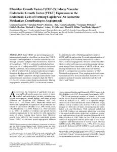
CRP Induces Release of Both Endothelial Microparticles and Circulating Endothelial Cells In Vitro and In Vivo: Further Evidence of Endothelial Dysfunction Sridevi Devaraj, Pappanaicken R. Kumaresan, and Ishwarlal Jialal*
BACKGROUND: Inflammation is pivotal in atherosclerosis. A key early event in atherosclerosis is endothelial dysfunction. C-reactive protein (CRP), the prototypic marker of inflammation in humans, is a risk marker for cardiovascular disease, and there is mounting evidence to support its role in atherothrombosis. CRP has been shown to promote endothelial dysfunction both in vitro and in vivo. Emerging biomarkers of endothelial dysfunction include circulating endothelial cells (CECs) and endothelial microparticles (EMPs). However, there is a paucity of data examining the effect of CRP on CEC and EMP production in vitro and in vivo. METHODS: In this report, we treated human aortic endothelial cells (HAECs) with increasing concentrations of CRP (0 –50 g/mL) or boiled CRP. We counted CECs and EMPs by flow cytometry. RESULTS:
Although CRP treatment resulted in a significant increase in release of both CECs and EMPs, boiled CRP failed to have an effect. Pretreatment of HAECs with sepiapterin or diethylenetriamine NONOate, both of which preserve nitric oxide (NO), resulted in attenuation of CRP’s effects on CECs and EMPs. CD32 and CD64 blocking antibodies but not CD16 antibody or lectin-like oxidized LDL receptor 1 small interfering RNA (LOX-1 siRNA) prevented CRP-induced production of CECs and EMPs. Furthermore, delivery of human CRP to Wistar rats compared with human serum albumin resulted in significantly increased CECs and EMPs, corroborating the in vitro findings.
CONCLUSIONS:
We provide novel data that CRP, via NO deficiency, promotes endothelial dysfunction by in-
Laboratory for Atherosclerosis and Metabolic Research, University of California– Davis Medical Center, Sacramento, CA, and VA Medical Center, Mather, CA. * Address correspondence to this author at: Laboratory for Atherosclerosis and Metabolic Research, UC Davis Medical Center, 4635, 2nd Ave., Room #3000, Research Building 1, Sacramento, CA 95817; Fax 916-734-6593; e-mail
[email protected]. Received June 1, 2011; accepted September 22, 2011.
ducing release of CECs and EMPs, which are biomarkers of endothelial dysfunction. © 2011 American Association for Clinical Chemistry
C-reactive protein (CRP),1 a member of the pentraxin family, is the prototypic marker of inflammation in humans. CRP is a valid marker of cardiovascular risk, and mounting data support a role for CRP in atherothrombosis (1– 4 ). Several studies have shown a significant relationship between CRP and endothelial dysfunction and that CRP impairs endothelial vasoreactivity in vivo (5, 6 ). CRP has been shown to inhibit endothelial nitric oxide synthase (eNOS) activity and bioactivity in vitro and in vivo, resulting in hypertension (7–10 ). In addition, CRP downregulates endothelial progenitor cell number and function in vitro (11 ). Endothelial microparticles (EMPs) are defined as small vesicles (100 nm to 1 mm in diameter) released from endothelial cells in response to activation or apoptosis by various stimuli (12 ), with a subset of important membrane proteins and phospholipids from their parent cells. Recent studies have demonstrated that EMPs can be used as a novel marker of endothelial injury (13 ). Accumulating evidence shows that circulating EMP levels are significantly increased in conditions associated with endothelial dysfunction, such as hypertension, dyslipidemia, diabetes mellitus, coronary artery disease, and heart failure (14 –18 ). Interestingly, endothelial NOS uncoupling contributes to EMP release (19 ), and CRP has been shown to promote eNOS uncoupling (7–10 ). Hence, in this study, we examined the effect of CRP on EMPs in vitro and in vivo. Another emerging biomarker of endothelial dysfunction is the quantification of ex-luminal circulating endothelial cells (CECs). Their presence indicates shedding of the damaged intimal endothelial layer into
Previously published online at DOI: 10.1373/clinchem.2011.169839 Nonstandard abbreviations: CRP, C-reactive protein; eNOS, endothelial nitric oxide synthase; EMP, endothelial microparticle; CEC, circulating endothelial cell; LPS, lipopolysaccharide; TLR4, Toll-like receptor 4; siRNA, small interfering RNA; HAEC, human aortic endothelial cell; BH4, tetrahydrobiopterin; DETA, diethylenetriamine; LOX-1, lectin-like oxidized LDL receptor 1; HuSA, human serum albumin; ACS, acute coronary syndrome.
1
1
Copyright (C) 2011 by The American Association for Clinical Chemistry
the blood, resulting in small numbers of these cells in health and increasing numbers in disease states that induce endothelial dysfunction such as diabetes and hypertension (20 –22 ). Increased numbers of CECs are present in other systemic inflammatory diseases, such as lupus and other vasculitides (23, 24 ). A high CEC level in the setting of acute coronary syndrome, for instance, has been shown to identify individuals at higher risk for subsequent cardiovascular events (25 ). However, there is a paucity of data examining the effects of CRP on CECs. Hence, in this study, we examined the effect of CRP on both EMPs and CECs.
E-selectin⫹, and CD42– or Annexin⫹, CD51⫹, CD61⫹, and CD42– per 5000 cells. FLOW CYTOMETRY FOR CECs
The flow cytometry protocol was based on a mononuclear cell analysis for size, nuclear complexity, and binding of specific antibodies conjugated to given fluorochromes. After excluding cell debris with a morphological gate, we stained cells with fluorescent-tagged antibodies to CD34, CD146, and CD105 and matched isotype controls. These cells in a subset were found to be negative for CD45 (to rule out lymphocytes). MECHANISTIC INSIGHTS
Methods CRP purified from human ascitic or pleural fluids was passed through a Detoxigel column to remove endotoxin [lipopolysaccharide (LPS)] and dialyzed extensively to remove azide as described (5, 7, 26 ). Our CRP preparations have been shown to have proinflammatory effects in Toll-like receptor 4 (TLR4) small interfering RNA (siRNA) knockdown cells, whereas LPS loses its effect in these cells (26 –28 ). Boiled CRP was also used as control (5, 7, 26 ). CELL CULTURE
We used human aortic endothelial cells (HAECs) between passages 3 and 6 as described (5, 7 ). HAECs were incubated overnight with different concentrations of CRP (0, 12.5, 25, and 50 g/mL) or boiled CRP (25 g/mL). For preparation of EMPs, culture supernatants were collected and cleared from cell fragments by centrifugation at 4300g for 5 min. The supernatant was then ultracentrifuged at 200 000g for 120 min at 10 °C. Pelleted EMPs were resuspended in 400 L PBS (pH 7.6, filtered) and used immediately for flow cytometry.
Because CRP has been shown to decrease eNOS activity by uncoupling (5, 7 ), we tested the effect of pretreatment with sepiapterin [a tetrahydrobiopterin (BH4) precursor, 100 mol/L] and diethylenetriamine (DETA) NONOate (nitric oxide donor, 100 mol/L) for 2 h on CRP-induced CECs and EMPs. Also, because CRP appears to mediate its effects in HAECs via CD32 and CD64, we pretreated HAECs with blocking antibodies to CD16, CD32, and CD64 before treatment with CRP. Finally, because data suggest that CRP binds to lectin-like oxidized LDL receptor 1 (LOX-1) (28 ), we examined the effect of LOX-1 siRNA on CRP’s effects on EMPs and CECs. Cells were transfected with scrambled or LOX siRNA before addition of CRP. IN VIVO EFFECTS OF CRP IN RAT CECs AND EMPs
Wistar rats were injected with either human CRP (20 mg/kg body weight) or vehicle control [human serum albumin (HuSA)] (n ⫽ 4/group) for 3 days as described (27, 28 ). We obtained blood for CEC and EMP quantification using rat-specific fluorochrome-labeled antibodies followed by flow cytometry, using isotype controls. STATISTICAL ANALYSIS
FLOW CYTOMETRY FOR EMPs
We incubated 10 L EMP suspension with 10 L antihuman/rat E-selectin, CD144, CD31, and CD42 or isotype control. We labeled another set of cells with antibodies to Annexin V, CD51, CD61, and CD42. After labeling, samples were analyzed by flow cytometry (29 ). For the EMP measurements, the flow cytometer was kept running for 30 min until the background events were ⬍1%. Microparticles present in PBS were analyzed regarding size and fluorescence. Briefly, on a log forward scatter/log side scatter dot plot, we defined the upper size limit of the microparticles using 1-m calibrant beads and drew a gate around the population. Only events included in this gate were further analyzed for their fluorescence. We collected 5000 events for each sample, expressed as CD144⫹, CD31⫹, 2
Clinical Chemistry 57:12 (2011)
We used Student paired t-test for normally distributed data and Wilcoxon signed rank test using Graph Pad Prizm software if data were nonnormal. ANOVA followed by appropriate multiple-comparisons posttest was carried out for experiments having ⬎2 experimental groups. Data are represented as mean (SD), and P ⬍ 0.05 was considered statistically significant. All experiments were performed at least 3 times in duplicate. Results CRP treatment dose-dependently increased the number of CECs, with significant increase at concentrations ⬎25 g/mL (ANOVA, P for trend ⫽ 0.012) (Fig. 1A). CRP treatment of HAECs resulted in increased EMPs, with a dose of 25 g/mL (P ⫽ 0.066) and a significant
CRP, EMPs, and CECs
Fig. 1. Effect of CRP on numbers of CECs (A) and EMPs (B) in vitro. Cells were treated with increasing concentrations of purified human CRP (0, 12.5, 25, 50 g/mL) or boiled CRP (50 g/mL) for 24 h, and numbers of CEC and EMPs were assessed by flow cytometry. *P ⬍ 0.05 compared to controls. n ⫽ 6 experiments. MFI, mean fluorescence intensity.
increase with a CRP concentration of 50 g/mL (P ⫽ 0.033) (Fig. 1B). Whereas we show increased positivity for CD144, CD31, and E-selectin (to phenotypically represent EMPs), we have also examined positivity of Annexin, CD51, and CD61 (markers expressed on EMPs) and obtained results similar to those seen in Fig. 1B. Addition of boiled CRP had no effect on either biomarker of endothelial function, showing that these are effects due to CRP, per se. We then examined if these effects of CRP were observed in our in vivo model. We have previously shown that human CRP injection into rats resulted in decreased eNOS due to uncoupling, resulting in impaired vasoreactivity (5 ). Here, we show that injection of human CRP, compared to HuSA, in vivo in rats significantly increased the number of circulating CECs (Fig. 2A) and EMPs (Fig. 2B). We have previously reported that with this system we achieve a plasma concentration of human CRP of around 20 g/mL (5, 26, 27 ). To obtain mechanistic insights, we tested the effect of sepiapterin and DETA NONOate on CRP-induced CECs and EMPs as well as blocking antibodies to Fc␥
Fig. 2. Effect of CRP on numbers of CECs (A) and EMPs (B) in vivo. Wistar rats were injected with human CRP or HuSA (20 mg/kg body weight) for 3 days and killed. Numbers of CECs and EMPs were assessed by flow cytometry in triplicate. *P ⬍ 0.001 compared to controls. n ⫽ 4 rats/group.
receptors (CD16, CD32, and CD64). As shown in Fig. 3A, 25 g/mL CRP significantly increased CEC production, and this was significantly attenuated with sepiapterin and DETA NONOate and antibodies to CD32 and CD64 but not CD16. Similar effects were seen for EMPs. CRP (50 g/mL) significantly increased EMP number, and this was significantly decreased by pretreatment with sepiapterin and DETA NONOate and antibodies to CD32 and CD64 but not CD16 (Fig. 3B). Transfection of cells with LOX1 siRNA failed to abrogate the effects of CRP (data not shown). Discussion It is becoming abundantly evident that CRP has a clear role in atherothrombosis, and most effects reported for CRP (azide-free and without endotoxin contamination) appear to bear a relationship to endothelial dysfunction and activation and the polarization of macroClinical Chemistry 57:12 (2011)
3
Fig. 3. Mechanisms by which CRP upregulates CECs (A) and EMPs (B). Cells were pretreated with sepiapterin (Sepi), DETA NONOate, or antibodies to CD16, CD32, or CD64 and then incubated with increasing concentrations of purified human CRP (0, 12.5, 25, 50 g/mL) or boiled CRP (50 g/mL) for 24 h. Numbers of CECs and EMPs were assessed by flow cytometry. *P ⬍ 0.05 compared to controls; #P ⬍ 0.05 compared to CRP 25 and/or 50 g/mL. n ⫽ 4 experiments. Ab, antibody.
phages to a proinflammatory phenotype (2, 27 ). We and others have shown that CRP impairs endothelial vasoreactivity, via decreased eNOS activity and bioactivity, mediated by eNOS uncoupling (4 –10 ). The initial reports in regard to CRP inhibition of eNOS activity and bioactivity raised the concern that CRP, by inducing endothelial dysfunction, could put patients at risk for hypertension and cardiovascular disease. Recently, this has been confirmed in vivo in rat models by at least 2 groups (10, 30, 31 ) Thus, additional biomarkers for assessing endothelial dysfunction have now emerged and include CECs and EMPs. In this report, we show for the first time, in vitro and in vivo, that CRP promotes significantly increased release of CECs and EMPs from aortic endothelial cells. One of the key early events in atherosclerosis is endothelial activation and dysfunction. During sustained endothelial cell activation, endothelial cells may physically detach from the vessel wall by a process involving the initiation of proapoptotic signaling cascades and loss of cell– cell contact and anchoring pro4
Clinical Chemistry 57:12 (2011)
teins. These detached endothelial cells, referred to as circulating endothelial cells, can be used as a marker of severe vessel wall damage. CECs have been shown to be increased in acute coronary syndrome (ACS) and in diabetes and heart failure, where CRP concentrations are also increased (20 –25 ). In this report, we show that CRP promotes CEC release from aortic endothelial cells in vitro and in rats in vivo, further supporting the data that endothelial dysfunction is induced by CRP. With regard to the effect of CRP on EMPs, Wang et al. (19 ) showed that in human umbilical vein endothelial cells, CRP treatment in vitro resulted in increased EMP generation, and this was associated with a downregulation of NO production secondary to decreased bioavailability of BH4 in endothelial cells. The purity of their CRP preparation was not reported, and the fact that they saw effects at lower concentrations than in the present report (20 vs 50 g/mL) raises the issue of contribution of contamination such as endotoxins. In this study, we have gone further and confirmed these findings in HAECs (the primary site of atherosclerosis) and also reported on the increase in CECs. In addition, we have confirmed these findings in vivo. Because we have observed increased E-selectin as well as increased Annexin staining, we believe that these EMPs are biomarkers of endothelial activation and apoptosis. We have previously shown that addition of sepiapterin, a BH4 precursor, reverses CRP inhibition of eNOS (7 ). In this study, a BH4 precursor and an NO donor, DETA NONOate, had similar effects, i.e., they were able to attenuate the effects of CRP on numbers of both CECs and EMPs. Thus, impaired eNOS activity appears to be proximal to these events, since rescuing eNOS from uncoupling and replenishing NO abrogates those effects. Also, CRP’s biological effects in endothelial cells appear to be mediated via the Fc␥ receptors, CD32 and CD64 (32 ). In this report, we also show that CRP’s effects in inducing EMPs and CECs is abrogated with antibodies to CD32 and CD64 but not CD16. These mechanistic pathways by which CRP exerts its effects on EMPs and CECs need to be confirmed in vivo in future studies. In conclusion, this study adds to the published literature demonstrating that CRP induces endothelial dysfunction by reporting on novel additional biomarkers, CECs and EMPs. These effects appear to be mediated by Fc␥ receptors, CD32 and CD64, and are due to NO deficiency induced by CRP. These events could result in pronounced endothelial dysfunction in vivo and promote clinical events such as hypertension and cardiovascular disease.
CRP, EMPs, and CECs
Author Contributions: All authors confirmed they have contributed to the intellectual content of this paper and have met the following 3 requirements: (a) significant contributions to the conception and design, acquisition of data, or analysis and interpretation of data; (b) drafting or revising the article for intellectual content; and (c) final approval of the published article.
Employment or Leadership: None declared. Consultant or Advisory Role: None declared. Stock Ownership: None declared. Honoraria: None declared. Research Funding: I. Jialal, NIH grant RO1 HL 074360 for an investigator-initiated proposal. Expert Testimony: None declared.
Authors’ Disclosures or Potential Conflicts of Interest: Upon manuscript submission, all authors completed the Disclosures of Potential Conflict of Interest form. Potential conflicts of interest:
Role of Sponsor: The funding organizations played no role in the design of study, choice of enrolled patients, review and interpretation of data, or preparation or approval of manuscript.
References 1. Bassuk SS, Rifai N, Ridker PM. High-sensitivity C-reactive protein: clinical importance. Curr Probl Cardiol 2004;29:439 –93. 2. Devaraj S, Singh U, Jialal I. The evolving role of C-reactive protein in atherothrombosis. Clin Chem 2009;55:229 –38. 3. Verma S, Devaraj S, Jialal I. Is C-reactive protein an innocent bystander or proatherogenic culprit? C-reactive protein promotes atherothrombosis. Circulation 2006;113:2135–50. 4. Cleland SJ, Sattar N, Petrie JR, Forouhi NG, Elliott HL, Connell JM. Endothelial dysfunction as a possible link between C-reactive protein levels and cardiovascular disease. Clin Sci (Lond) 2098: 531–5. 5. Hein TW, Singh U, Vasquez-Vivar J, Devaraj S, Kuo L, Jialal I. Human C-reactive protein induces endothelial dysfunction and uncoupling of eNOS in vivo. Atherosclerosis 2009;206:61– 8. 6. Venugopal SK, Devaraj S, Yuhanna I, Shaul P, Jialal I. Demonstration that C-reactive protein decreases eNOS expression and bioactivity in human aortic endothelial cells. Circulation 2002; 106:1439 – 41. 7. Singh U, Devaraj S, Vasquez-Vivar J, Jialal I. C-reactive protein decreases endothelial nitric oxide synthase activity via uncoupling. J Mol Cell Cardiol 2007;43:780 –91. 8. Teoh H, Quan A, Lovren F, Wang G, Tirgari S, Szmitko PE, et al. Impaired endothelial function in C-reactive protein overexpressing mice. Atherosclerosis 2008;201:318 –25. 9. Verma S, Wang CH, Li SH, Dumont AS, Fedak PW, Badiwala MV, et al. A self-fulfilling prophecy: C-reactive protein attenuates nitric oxide production and inhibits angiogenesis. Circulation 2002; 106:913–9. 10. Guan H, Wang P, Hui R, Edin ML, Zeldin DC, Wang DW. Adeno-associated virus mediated human C-reactive protein gene delivery causes endothelial dysfunction and hypertension in rats. Clin Chem 2009;55:274 – 84. 11. Verma S, Kuliszewski MA, Li SH, Szmitko PE, Zucco L, Wang CH, et al. C-reactive protein attenuates endothelial progenitor cell survival, differentiation, and function: further evidence of a mechanistic link between C-reactive protein and cardiovascular disease. Circulation 2004;109: 2058 – 67.
12. Jimenez JJ, Jy W, Mauro LM, Horstman LL, Bidot CJ, Ahn YS. Endothelial microparticles (EMP) as vascular disease markers. Adv Clin Chem 2005; 39:131–57. 13. Horstman LL, Jy W, Jimenez JJ, Ahn YS. Endothelial microparticles as markers of endothelial dysfunction. Front Biosci 2004;9:1118 –35. 14. Preston RA, Jy W, Jimenez JJ, Mauro LM, Horstman LL, Valle M, et al. Effects of severe hypertension on endothelial and platelet microparticles. Hypertension 2003;41:211–7. 15. Koga H, Sugiyama S, Kugiyama K, Watanabe K, Fukushima H, Tanaka T, et al. Elevated levels of VE-cadherin-positive endothelial microparticles in patients with type 2 diabetes mellitus and coronary artery disease. J Am Coll Cardiol 2005;45: 1622–30. 16. Bernal-Mizrachi L, Jy W, Jimenez JJ, Pastor J, Mauro LM, Horstman LL, et al. High levels of circulating endothelial microparticles in patients with acute coronary syndromes. Am Heart J 2003; 145:962–70. 17. Heloire F, Weill B, Weber S, Batteux F. Aggregates of endothelial microparticles and platelets circulate in peripheral blood: variations during stable coronary disease and acute myocardial infarction. Thromb Res 2003;110:173– 80. 18. Mallat Z, Benamer H, Hugel B, Benessiano J, Steg PG, Freyssinet JM, et al. Elevated levels of shed membrane microparticles with procoagulant potential in the peripheral circulating blood of patients with acute coronary syndromes. Circulation 2000;101– 8:841–3. 19. Wang JM, Wang Y, Huang JY, Yang Z, Chen L, Wang LC, et al. C-reactive protein-induced endothelial microparticle generation in HUVECs is related to BH4-dependent NO formation. J Vasc Res 2007;44:241– 8. 20. Lee KW, Lip GY, Tayebjee M, Foster W, Blann AD. Circulating endothelial cells, von Willebrand factor, interleukin-6 and prognosis in patients with acute coronary syndromes. Blood 2005; 105:526 –32. 21. Blann AD, Woywodt A, Bertolini F, Bull TM, Buyon JP, Clancy RM, et al. Circulating endothelial cells: biomarker of vascular disease. Thromb Haemost 2005;93:228 –35. 22. McClung JA, Naseer N, Saleem M, Rossi GP, Weiss MB, Abraham NG, et al. Circulating endo-
23.
24.
25.
26.
27.
28.
29.
30.
31.
32.
thelial cells are elevated in patients with type 2 diabetes mellitus independently of HbA(1)c. Diabetologia 2005;48:345–50. Clancy R, Marder G, Martin V, Belmont HM, Abramson SB, Buyon J. Circulating activated endothelial cells in systemic lupus erythematosus: further evidence for diffuse vasculopathy. Arthritis Rheum 2001;44:1203– 8. Woywodt A, Streiber F, de Groot K, Regelsberger H, Haller H, Haubitz M. Circulating endothelial cells as markers for ANCA-associated smallvessel vasculitis. Lancet 2003;361:206 –10. Boos CJ, Soor SK, Kang D, Lip GY. Relationship between circulating endothelial cells and the predicted risk of cardiovascular events in acute coronary syndromes. Eur Heart J 2007;28:1092– 1101. Dasu MR, Devaraj S, Du Clos TW, Jialal I. The biological effects of CRP are not attributable to endotoxin contamination: evidence from TLR4 knockdown human aortic endothelial cells. J Lipid Res 2007;48:509 –12. Devaraj S, Jialal I. C-reactive protein polarizes human macrophages to an m1 phenotype and inhibits transformation to the m2 phenotype. Arterioscler Thromb Vasc Biol 2011;31:1397– 402. Singh U, Dasu MR, Yancey PG, Afify A, Devaraj S, Jialal I. Human C-reactive protein promotes oxidized low density lipoprotein uptake and matrix metalloproteinase-9 release in Wistar rats. J Lipid Res 2008;49:1015–23. Fujita Y, Kakino A, Nishimichi N, Yamaguchi S, Sato Y, Machida S, et al. Oxidized LDL receptor LOX-1 binds to C-reactive protein and mediates its vascular effects. Clin Chem 2009;55:285–94. Jialal I, Devaraj S, Siegel D. CRP induces hypertension in animal models: homo sapiens says NO. Hypertens Res 201134:801–2. Pravenec M, Kajiya T, Zı´dek V, Landa V, Mlejnek P, Sima´kova´ M, et al. Effects of human C-reactive protein on pathogenesis of features of the metabolic syndrome. Hypertension 2011;57:731–7. Devaraj S, Du Clos TW, Jialal I. Binding and internalization of C-reactive, protein by Fcgamma receptors on human aortic endothelial cells mediates, biological effects. Arterioscler Thromb Vasc Biol 2005;25:1359 – 63.
Clinical Chemistry 57:12 (2011)
5