Scandinavian Journal of Surgery 97: 341–346, 2008
CURRENT CONCEPTS IN RECONSTRUCTION OF HAND FUNCTION IN TETRAPLEGIA J. Fridén, C. Reinholdt National Center of Reconstructive Hand Surgery in Tetraplegia, Department of Hand Surgery, Sahlgrenska University Hospital, Göteborg, Sweden
AbSTRACT
Several recent developments in the field of reconstructive hand surgery in tetraplegia have created a foundation for further refinements of both surgical techniques and postoperative training strategies to improve the outcome of restoration of upper extremity functions. A remarkable means of improving function is the immediate activation of transferred muscle after surgery. Early active training of new motors not only prevents the formation of adhesions but facilitates the voluntary recruitment of motors powering new functions before swelling and immobilization-induced stiffness restrain muscle contractions. A common observation internationally over the past years is that the number of incomplete tetraplegics increases. This shift towards more incomplete injuries with spasticity as a common feature in addition to the paralysis has expanded and changed the spectrum of surgeries in this group of patients and also emphasizes the need for a revisit and further development of the different strategies for reconstruction of hand function. Key words: Tetraplegia; hand function; surgical reconstruction; tendon transfer; spasticity; active training
INTRoDUCTIoN At the recent world congress in reconstructive hand surgery and rehabilitation in tetraplegia (Philadelphia, 2007), a resolution regarding assessment of individuals with tetraplegia was presented and accepted. Briefly, this resolution stated that every person who sustains a cervical spinal cord injury with tetraplegia should be examined, assessed and informed concerning the options of possible reconstruction of motor function of the hands and arms. It is of course a long way before this ambitious goal can be achieved but the resolution put forward by the leading experts in this field certainly stresses the necessity of increasing the awareness and improving the infraCorrespondence: Jan Fridén, M.D. Department of Hand Surgery Sahlgrenska University Hospital SE - 413 45 Göteborg, Sweden Email:
[email protected]
structure to meet patients’ demand of informed discussions of options for improvement of hand function. Aside from the brain, the tetraplegic individuals’ upper limbs are their most important functional resource and judged to be the most desirable ability to regain after cervical spinal cord injury (1). An increased awareness among patients and doctors of available surgical techniques to improve motor function has helped to increase the interest and scientific efforts in this field. During the last 30 years, a successful development has occurred in tetraplegia upper limb rehabilitation surgery (2–9) and an enhanced understanding of the complex interactions between muscles, tendons and joints in both normal and paralyzed individual has been a prerequisite for this development (10–11). Due to specialized spinal cord injury units and better coordination of the multiple levels of care required after injuries of cervical spinal cord, larger numbers of patients with normal life expectancy will be anticipated in the future. Individuals with tetraplegia
342
J. Fridén, C. Reinholdt TABlE 1
List of procedures that alone or combined are used to reconstruct various functions needed to improve the tetraplegic individual’s motor ability. Surgical procedures in reconstructive tetraplegia hand surgery • • • • • •
Muscle-tendon transfer Tenodeses Joint fusions (thumb only) Myo-tenotomies Tendon elongations Capsulodeses
are now better informed about the benefits with surgery probably because of easy access to comprehensive web pages on the Internet, a trend that will likely increase the demands on more surgical reconstructions in the future. The overall goal of treatment is to improve motor functions in order to achieve a higher degree of independence for the individual with tetraplegia. To take better advantage of remaining shoulder, arm and hand functions various philosophies have evolved. Reconstruction of upper limb motor functions in tetraplegia involve a multi-fold of surgeries (Table 1) but it is always necessary to keep in mind the goals of the surgeries i.e., to provide the individual with a better ability to perform activities in daily life. CURRENT TRENDS IN RECoNSTRUCTIvE TETRAPlEGIA HAND SURGERy The general trend is to direct the treatment towards the restoring of abilities more than just functions. In other words, the communication with the patient and her/his caregivers and relatives is critical in order to pinpoint the exact needs of improvement in daily life activities. It is not the functional improvement from the surgeon’s or the therapist’s perspective that is important but demands from the patient to be able to control as many as possible of the daily routines of life. Not until this attitude has been fully implemented into the preoperative assessment and goal discussion, we can truly provide the service requested.
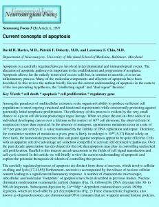
Based on the experience from more than approximately 700 reconstructions performed in the Swedish National Center for Reconstructive Hand Surgery in Tetraplegia, we prefer to primarily reconstruct grip function before reconstructions of the thumb and finger extensors. In our experience some individuals decide to only undergo one reconstruction and are satisfied with that. If they would choose to quit after one reconstruction, any further reconstructions after just having the extensor function restored would not benefit them functionally enough. on the other hand, if they settle after a grip reconstruction they have gained a lot of new abilities of daily life. It should also be kept in mind that we regularly reconstruct the intrinsics in the same operation as the reconstruction of grip, thus providing an acceptable opening of the hand. The most remarkable and effective strategy of improving function has been the consistent and immediate activation of transferred muscle after surgery. Early active training of new motors not only prevents the formation of adhesions but facilitates the voluntary recruitment of motors powering new functions before swelling and immobilization-induced stiffness restrain muscle contractions. Additionally, the patient will experience an early, spectacular and inspiring effect of the reconstruction, which will help motivate training during the demanding and sometimes painful initial postoperative period. Early activation of the transferred muscles requires reliable tendon-totendon attachments. We have cumulated experience of hundreds of side-to-side attachments using running sutures back and forth along both sides and with a minimum of 5 cm overlap (Fig. 1). This technique has proven extremely safe for allowing early active training and is now standard in our unit. Briefly, the day after surgery a removable splint replaces the cast and intermittent exercises commence. Training emphasizes on activating the donor muscles without any external resistance. During nights and between training sessions the patient wears a splint with a design that aims at unloading the attachment sites. During exercises “training splints” are used. The exact design of the splints depends on the actual combinations of surgeries performed. Four weeks postoperatively active use of the grip in activities is started, using only a splint during nights. The load is then gradually
Fig. 1. Close-up of attachment sites for donor and recipient tendons. Note the meticulous suturing technique with running sutures in both directions and on both sides.
343
Reconstruction of hand function in tetraplegia
increased up to three months postoperatively when the patient is allowed full active normal daily life including personal transfers. Another current trend is to power the new functions with active donors instead of active or passive tenodeses. A typical example is the current standard use of brachioradialis to power the thumb flexor (12) as opposed to the previously more often used tenodesis of FPl to radius (3). In addition, donor muscles of less power have been used lately in procedures aiming more at improvements of positioning than power-demanding joint motions. A striking example of restoration of positioning ability is the use of a relatively weak extensor digiti minimi (strength grade 2–4) to drive the abductor pollicis brevis. This transfer doesn’t require very much strength since the goal is to provide an ability to increase the opening of the hand as well as position the ulnar aspect of the thumb along the radial aspect of the index finger. Neither of these two functions entails much strength but a replacement for positional control, which, certainly makes a big difference for the ultimate manipulative ability of the tetraplegic’s hand. A common observation internationally over the past years is that the number of incomplete tetraplegics increases. Regardless of the reason for that development, it has expanded and changed the spectrum of surgeries in this group of patients and also emphasizes the need for a revisit and further development of the different strategies for reconstruction of hand function. The patients with incomplete injuries consistently demonstrate more or less debilitating spasticity or spasticity-induced tightness of the muscle-tendon units. The disability pattern in posttraumatic tetraplegia has shifted over the years. Incomplete tetraplegics with a somewhat new configuration and more complex functional loss demonstrate often various degrees of spasticity. The general approach therefore needs to be an even more careful assessment of remaining useful functions. We need to realize and utilize the possibilities of correcting stiffness, deformities and hypertonicity more actively to perform tendon lengthening and releases and to incorporate these lengthening procedures as natural parts of tendon transfers. Much of the focus of this article will be on surgical implications in the correction of spasticity-induced deformities.
cle must be of adequate strength, preferably not injured or re-innervated. In certain conditions, with limited available donor muscles, a weak muscle can be considered for transfer. When several donor muscles are available for transfer, additional factors are considered. These include the condition of soft tissue in which a transfer must cross, route and direction or the transfers, donor site morbidity, the architecture of the donor muscle and function desired, synergism of the tendon transfer, the experience and preference of the surgeon, and the consensus or preferences of an informed patient. optimally, the donor muscle must be healthy, synergistic, similar in architecture, and have an adequate soft tissue bed along the route of transfer. The basic procedures for reconstruction in tetraplegia include reconstruction of elbow extension, grip function and opening of the hand. Restoring active elbow extension for the spinal cord injury patient in terms of locomotion, transfers and accurately positioning the arm in space is critical and it improves the effect of more distal tendon transfers to restore hand function. While the details of reconstruction of elbow extensors and grip function are well covered in numerous publications (see e.g. 8 and 13) we believe that it is important to further emphasize strategies of improving the reach and grasp function. This knowledge is particularly imperative in the light of the increasing number of individuals presenting with close fist pattern of deformity after incomplete cervical spinal cord injuries (Fig. 2). In our unit altogether 40 patients with incomplete tetraplegia were treated surgically between 2002 and 2007. The number of surgical procedures performed on patients with incomplete tetraplegia increased from 20 in 2002 to 67 in 2007. Individual surgical procedures alone or in combination changed over the same time period. Tendon lengthenings of the deep and superficial flexor tendons almost tripled over the past 3 years. Releases of the interossei increased by a factor of 5 and the number of releases of adductor pollicis increased slightly during the same time period. There is also an increase in the number of teno-myotomies of the original motor releases in parallel with tendon transfers.
PlANNING oF RECoNSTRUCTIoN Before performing surgical rehabilitation of the upper limb, care must be taken to preserve the integrity and health of the upper limbs in the acute injury period (8). The principal therapy includes: preventing and treating hand edema, maintaining supple joints and controlling pain and spasticity. Preoperative evaluation of the upper limb includes muscle strength tests, joint range of motion tests, and sensation testing. Muscle testing is performed according to the British Research Council (see e.g. ref. 8) and the assessment of tetraplegia function groups that was modified at the 6th International Tetraplegia Meeting 1998, in Cleveland (Table 2). The donor mus-
TABlE 2 International classification of muscle function in tetraplegia. Group
lowest muscle grade > 4
00 01 02 03 04 05 06 07 08 09 10
No muscle below elbow Brachioradialis Extensor carpi radialis longus Extensor carpi radialis brevis Pronator Flexor carpi radialis Finger extensors Thumb extensors Partial finger flexors lacks only intrinsics Exceptions
344
J. Fridén, C. Reinholdt
Fig. 2. Typical close fist appearance in a patient with incomplete cervical spinal cord injury and spasticity.
Fig. 3. Method of examining intrinsic tightness of the interossei. PIP joint is passively flexed while MCP joint is extended.
RECoNSTRUCTIoN oF oPENING oF THE HAND
hands is often intricate. A particular challenge is diagnosing the intrinsic tightness i.e. stiff or spastic interossei muscles (Fig. 3). The powerful extrinsic digital flexors may obscure tight interossei by strongly flexing all finger joints. In other words, a spastic FDS muscle can usually overpower the extension power of interosseus muscles at the level the PIP joint. The final detection of intrinsic tightness may be done during surgery, i.e. after lengthening of the FDS tendons. Another important aspect of spasticity surgery in tetraplegia is to consider the original remaining spasticity when a function is powered by a new healthy motor. In those cases the original muscle must usually be detached from its distal tendon so that the
Reconstruction of opening of the hand is necessary to facilitate the ability to come around an object in order to grasp (Table 3). Many of the tetraplegic patients do not have this ability due to “tenodesis grip” with adhesions of the finger flexors and insufficient stretching of the fingers even with good passive wrist flexion. Improvement of the opening of the hand is particularly necessary in patients with finger flexor spasticity where gravity or remaining finger extension strength cannot overpower the finger flexion spasticity. It should also be kept in mind that examining and understanding the underlying mechanisms in tight
TABlE 3 Summary of selected surgical procedures to achieve patient’s ability goal of opening of the hand. ABIlITy GoAl
FUNCTIoNAl GoAl
PRoCEDURE
Reaching for objects e.g. a cup or glass positioning of thumb and fingers for improved grasp control
Opening of the hand
Reconstruction of thumb and finger extensors Passive opening CMC I arthrodesis EPl to extensor fascia attachment Active opening PT-EDC and EPl/APl
REHABIlITATIoN
8 weeks wrist and thumb in splint. Sessions with passive or active exercises of wrist from 1st postop day. 4 weeks wrist, fingers and thumb in splint. Sessions with active exercises of thumb and finger extension.
Reconstruction of intrinsics Passive opening Zancolli-lasso tenodesis Interossei tenodesis Active opening Active Interossei (powered by e.g. ECRl or FDS) EDM-APB
4 weeks of immobilization in intrinsic plus position. Sessions with passive training after 4 wks. Protection for 8 weeks needed. Active training from 1st postop day. Protection between training sessions for 8 weeks.
Reconstruction of hand function in tetraplegia
new motor can function without unwanted interaction and uncontrolled power from the original spastic muscle. An example can be replacement of a spastic FPl muscle by brachioradialis in cases with “thumb in palm” due to spasticity. To achieve full use of the normal brachioradialis activation pattern without disturbance of original FPl actions, the FPl will be myotomized i.e., disconnected from its distal tendon. SURGICAl TECHNIqUE
345
extension of the ring- and small finger. If the interossei function is restored using this technique in parallel with reconstruction of digital flexors, the interossei need to be protected during the postoperative training period. our postoperative training regime therefore allows only limited digital flexion motions. In practice, the patients actively flex the fingers not more than to a position with a 4 cm gap between fingertips and palm. Some of the patients belonging to Group 5 or 6 (Table 2) may have their intrinsic function of the thumb restored (volar abduction). To date we have successfully reconstructed this function in 22 cases by transferring extensor digiti minimi (quinti) (EDM) to the insertion of abductor pollicis brevis (APB). It is noteworthy that for this reconstruction full power of the EDM is not needed since the main goal of the reconstruction is to increase the opening of the hand and to position the thumb along the radial aspect of the index finger. None of these movements requires any substantial force but merely activation.
opening of the hand can be obtained by tenodesis surgery. An EPl tenodesis to extensor retinaculum or forearm fascia is performed and the tenodesis is then powered by actively or passively flexing the wrist. In our unit we now always attach the EPl tendon to the fascia because of its firmer and more reliable mechanical integrity compared to the extensor retinaculum. All thumb reconstructions include a split FPl-EPl tenodesis to prevent hyperflexion of the IP joint and obtain a favourable contact surface between thumb and index finger (14–15). An active hand opening can, in selected cases be performed by transferring PT to EPl, APl and EDC (Figs 4–5). It is also important to remember that reconstruction of intrinsics improves PIP joint extension and thereby opening of the hand (9). This is usually performed by using a tendon graft inserted into extensor hood at the radial aspect of the proximal phalanx of the index finger i.e., under the insertion tendon of the first dorsal interosseus muscle, brought proximally and volarly to the intermetacarpal ligament, dorsally to the neck of the metacarpal bone of long finger and deep to the extensor tendon, through the lumbrical canal and inserted into the radial aspect of the extensor hood of the long finger. The radial insertion will guarantee a pull of the fingers in radial direction when the tenodesis or, if actively powered by for example the ECRl, is tensioned during MCP flexion and simultaneous PIP extension. This route will ultimately facilitate the approach of the radial side of index finger to the thumb during key pinch. The same type of loop with an additional tendon graft is used for providing PIP joint
Tendon transfer procedures are optimally undertaken with a team approach, using the assistance of an occupational and a physical hand therapist as well as a surgical nurse. The essential hand therapist performs the “other half” of the surgical procedure, rehabilitation and retraining of the transferred tendons. The hand therapist promotes functional restoration, assists with edema control, contracture prevention, and muscle activation and strengthening (Fig. 6). The surgical nurse assists with preoperative planning and scheduling, with medications and wound management and contributes to general postoperative management. Many patients who undergo tendon transfer procedures have sustained devastating, life-changing injuries and they should be considered full members of the rehabilitation team. Their input is required in the preoperative planning so the patient understands operative options, alternatives, and appreciates the commitment required for successful rehabilitation.
Fig. 4. PT muscle is exposed and tendon graft (FDS tendon of ring finger) is attached. Distally the interposition graft will be sutured to EDC and EPl tendons.
Fig. 5. Effect of tenodesis on finger and thumb posture during passive flexion of the wrist is tested at the completion of the reconstruction of finger and thumb extensors demonstrated in Fig. 4.
TEAM APPRoACH
346
J. Fridén, C. Reinholdt
The patient also performs an active role in a home therapy program under the guidance of the hand therapist. The patient’s postoperative compliance, motivation, and efforts are vital for successful rehabilitation. REFERENCES 01. Anderson K: Targeting recovery: priorities of the spinal cordinjured population. J Neurotrauma 2004;21:1371–1383 02. Zancolli E: Surgery for the quadriplegic hand with active, strong wrist extension preserved. Clin orthop Rel Res 1975;112: 101–113 03. Moberg E: Surgical treatment for absent single-hand grip and elbow extension in quadriplegia. J Bone Joint Surg 1975;57A: 196–206 04. Moberg E: The upper limb in tetraplegia. A new approach to surgical rehabilitation. Thieme, Stuttgart, Germany, 1978 05. Moberg E: Surgical rehabilitation of the upper limb in tetraplegia. Paraplegia 1990;28:330–334 06. Moberg E, lamb D: Surgical rehabilitation of the upper limb in tetraplegia. Hand 1980;12:209–213 07. lamb DW, Chan KM: Surgical reconstruction of the upper limb in traumatic tetraplegia. A review of 41 patients. J Bone Joint Surg 1983; 65:291–298
08. Hentz vR, leclerq C: Surgical rehabilitation of the upper limb in tetraplegia. WB Saunders, london, 2002 09. House JH, Walsh T: Two-stage reconstruction of the tetraplegic hand. In: Master techniques in orthopaedic surgery. Ed. JW Strickland. lippincott-Raven Publishers, Philadelphia, USA (1998), pp 229–266 10. Fridén J, Ejeskär A, Dahlgren A, lieber Rl: Protection of the deltoid–to-triceps tendon transfer repair sites. J Hand Surg 2000;25A:144–149 11. Ward SR, Peace WJ, Fridén J, lieber Rl: Dorsal transfer of the brachioradialis to the flexor pollicis longus enables simultaneous powering of key pinch and forearm pronation. J Hand Surg 2006;31A:993–997 12. Freehafer A: Tendon transfers in patients with cervical spinal cord injury. J Hand Surg 1991;16A: 804–809 13. Fridén J: Tendon transfers in reconstructive hand surgery. Ed. J Fridén, Taylor & Francis, oxford, England, 2005 14. Mohammed KD, Rothwell AG, Sinclair SW, Willems SM, Bean AR: Upper-limb surgery for tetraplegia. J Bone Joint Surg 1992; 74B:873–879 15. Ejeskar A, Dahlgren A, Fridén J: Split distal flexor pollicis longus tenodesis: long-term results. Scand J Plast Reconstr Surg Hand Surg 2002;36:96–99
Received: November 24, 2008