Review For reprint orders, please contact
[email protected]
Current findings of fMRI in panic disorder: contributions for the fear neurocircuitry and CBT effects Expert Rev. Neurother. 10(2), 291–303 (2010)
Marcele Regine de Carvalho†, Gisele Pereira Dias, Fiammetta Cosci, Valfrido Leão de-Melo-Neto, Mário Cesar do Nascimento Bevilaqua, Patricia Franca Gardino and Antonio Egidio Nardi Author for correspondence Laboratory of Panic and Respiration, Institute of Psychiatry, Universidade Federal do Rio de Janeiro (UFRJ), INCT Translational Medicine (CNPq), Rio de Janeiro, Brazil Tel.: +55 212 436 8202 Fax: +55 212 523 6839
[email protected] †
www.expert-reviews.com
Thanks to brain imaging great advances have been made concerning the comprehension of neural substrates related to panic disorder (PD). This article aims to: review the recent functional MRI (fMRI) studies concerning PD; correlate the PD fMRI neurobiological findings with the fear neurocircuitry hypothesis; discuss the fear neurocircuitry hypothesis and link it to cognitive–behavior therapy findings; and comment on fMRI study limitations and suggest methodological changes for future research. As a whole, there is increasing evidence that brain structures such as the prefrontal cortex, the anterior cingulate cortex and limbic areas (hippocampus and amygdala) might play a major role in the panic response. Keywords : amygdala • anterior cingulate cortex • anxiety neurocircuitry • cognitive–behavior therapy • fMRI • hippocampus • panic disorder • prefrontal cortex
Current findings of functional MRI in panic disorder Relevance of functional MRI technique
New technologies, including functional MRI (fMRI), are helping to elucidate diverse issues related to neural circuits, especially concerning fear and anxiety. The primary efforts in applying brain techniques to the investigation of psychiatric disorders focused on schizophrenia [1] but important attention has been directed to anxiety disorders in the last few years [2] , which might provide better guidance for both clinical diagnosis and therapeutic interventions [3] . Functional MRI can show changes in brain functioning during noninvasive cognitive and behavioral tasks [4] and researchers can, thus, identify the brain regions that mediate the individual’s experiences at the moment they occur. In panic disorder (PD), scientists are especially interested in the brain regions related to fear response. Thus, as deeper subcortical and brainstem structures are importantly involved in PD circuitry, fMRI can be considered to be an appropriate kind of neuroimaging technology, as it has the anatomic resolution to distinguish between small and deep structures. Furthermore, it has better temporal resolution than PET and single photon emission computed tomography, 10.1586/ERN.09.161
making it possible to investigate PD circuitry during a real-time panic attack. Therefore, it can be useful to study PD neural networks. Information obtained from a fMRI scan makes it possible to compare two or more cognitive states. The technique is based on blood oxygenation; more activated brain regions require increased oxygen supply, which in turn increases the concentration of oxygenated hemoglobin (oxyhemoglobin). This is the reason why this technique is known as blood oxygen level dependent (BOLD). Functional studies allow brain functioning assessment under the influence of visual, auditory, tactile, gustatory or olfactory stimuli, as well as during the performance of both cognitive and affective tasks [5] . In order to determine the brain areas to be studied, either manual or automatic selection of regions of interest might be used. One of the advantages of using the region-of-interest method is that it offers substantial anatomic validity. On the other hand, some limitations should be pointed out, such as the fact that data analysis is a time-consuming procedure and that it is difficult to compare multiple brain regions or large samples. This method depends on the development of valid and reliable anatomic identifications, as well as evaluator training and experience and reliability among evaluators.
© 2010 Expert Reviews Ltd
ISSN 1473-7175
291
Review
de Carvalho, Dias, Cosci et al.
Despite the relevance of functional features, structural findings may also be considered crucial in the attempt to understand the biological basis of PD. Previous neuroanatomical features showed reduced volume of temporal lobes in PD patients [6–8] , although some other studies, such as Uchida et al. [9] , found increased left superior temporal gyrus, midbrain, pons and left insula gray matter volumes in 19 PD patients compared with 20 controls. Bilateral reduced amygdala volumes were detected in a study comparing 12 PD patients with 12 healthy controls [10] . Finally, other studies detected reduced gray matter volume in the left parahippocampal gyrus [11] and increased gray matter volume in the brain stem [12] . fMRI studies on PD
A bibliographical search was carried out in the Web of Science/ ISI and Medline/PubMed data banks to collect studies about panic disorder and functional investigation of its neurocircuitry with fMRI using the keywords ‘fMRI’ and ‘panic disorder’. Only articles written in English and published in the last 10 years were selected. Articles about regional lesions, epilepsy and panic attack challenges within healthy subjects were excluded. Nine articles were selected: eight originals and one case report. Two of them refer to fMRI genetic findings on PD. Marchand et al. used a motor activation paradigm to probe putamen function in 12 female subjects with PD and 18 healthy female controls using fMRI at 3 T [13] . The main objective was to determine whether functional abnormalities of the putamen occur in panic disorder. The task consisted of a 4-min run of pressing buttons with the first and third fingers simultaneously alternating with pressing button with the middle finger alone. There were six blocks of rest and six blocks of activity presented in a pseudorandom order. Each block was repeated once for each hand. It was controlled for possible bias from reduction of BOLD signal secondary to cerebral vasoconstriction resulting from possible hyperventilation among panic subjects. Panic and control subjects presented activation in the following regions: bilateral prefrontal, precentral, postcentral, cingulate, insular and cerebellar regions; right parietal, midbrain and brainstem and left temporal regions; bilateral putamen, caudate, globus pallidus internal and external segments and the thalamus. They found evidence for bilateral decreased putamen activation among subjects with PD compared with normal controls and concluded that it is possible that the subcortical-mediated fight or flight response might be abnormal in PD. Pfleiderer et al. elaborated an auditory habituation paradigm in which it was reported that a 26-year-old PD female patient experienced a spontaneous panic attack [14] . The experiment consisted of three stimulation cycles of emotionally neutral, digitally generated sine tones alternating with rest periods. The panic attack occurred in the third stimulation cycle of the fMRI at the 3 T paradigm. It was associated with a significant increase of activity in the right amygdala and of the right putamen compared with the resting state. The authors emphasize the role of the amygdala in the neurocircuitry associated with panic attack and the activation patterns of a spontaneous panic attack, which seems to be different from pharmacologically induced attacks. 292
Some studies concerning the visual stimulus paradigm were found. Pillay et al. studied eight patients with PD and eight healthy controls and investigated cingulate cortex and amygdala response to a fearful face [15] and a neutral face condition using fMRI at 1.5 T [16] . The affective stimuli consisted of six faces expressing the emotion of fear, including male and female posers. The scanning sequence consisted of five alternating 30-s stimulus/ rest periods. Three different face photographs were presented during each of two stimulation periods. PD patients activate the anterior cingulate cortex (ACC) and amygdala significantly less than the controls while viewing fearful faces. In the neutral face condition, compared with controls, PD subjects demonstrated greater activation of the cingulate cortex bilaterally. The results indicate a synchrony of function of the ACC and amygdala in response to fearful faces in PD. The authors concluded that reduced ACC and amygdala activation may be due to chronic hyperarousal, which may decrease attentional resources and emotional response in PD. They hypothesized that this response may represent a defense against hyper-responsiveness to anxiety-inducing stimuli. Pillay et al. aimed to describe possible differences in neural processing between PD patients and controls when faced with identifying a positive affect [17] . The authors investigated activation of the ACC and amygdala during the presentation of happy faces in eight PD patients in comparison with eight controls. The scan sequence began with a baseline condition and was followed by alternating task/resting-control conditions. The affective stimuli consisted of six faces displaying a similar emotional expression. Three different face photographs were presented during each of two stimulation periods. Each subject participated in both a happy and a neutral affect perception task. Using BOLD fMRI at 1.5T, increased ACC bilateral activation was found in PD patients when presented with happy faces. Curiously, there were no differences in amygdala activation between the groups, and no differences in activation of this structure were identified when patients were presented with neutral faces. In response to this set of neutral stimuli, greater bilateral ACC activation was also found, although not so intensively as that observed for the happy condition. The authors suggest that this data may be a result of enhanced attentional demands experienced by PD patients, which leads to the abnormal cognitive processing characteristic of this psychopathology. In an interesting experimental paradigm, Chechko et al. investigated if emotion perception was altered in 18 remitted PD patients as a trait feature in comparison with 18 healthy controls [18] . For this purpose, fMRI was used during the presentation of emotionally congruent and incongruent pairs of face/ words. Thus, for the congruent condition, subjects could be presented with a happy face with the German word for ‘happiness’ printed in bold capital red letters across the picture or to a fearful expression with the word for ‘fear’ across it. On the other hand, in the incongruent condition, for example, happy faces with the word for fear and fearful faces with the word for happiness were shown. In both situations, patients showed slower reactions, although not less accurate, and stronger behavioral interference in incongruent trials. Curiously, similar responses were found Expert Rev. Neurother. 10(2), (2010)
Current findings of fMRI in panic disorder
for controls and patients in relation to the effect of incongruence times congruence. However, stronger activation in the dorsal ACC (dACC) was detected in remitted PD patients in the context of preceding congruence. A dropout was observed of both the dACC and dorsomedial prefrontal cortex (PFC) preceding the incongruent condition, but activation of lower limbic structures, such as the amygdala and brainstem, was seen in the experimental group. According to these data, it may be proposed that the processing of emotional stimuli (specifically the order in which nonpanic-related facial expressions are presented) is impaired in PD patients despite clinical remission, a feature that might be associated with abnormal dACC/dorsomedial PFC activity, as well as limbic and brainstem activation. Van der Heuvel et al. used a cognitive and emotional Stroop task (consisting of congruent and incongruent color words), and obsessive–compulsive disorder (OCD)- and panic-related negative and neutral word paradigms to study three patient groups: PD (n = 15), OCD (n = 16) and hypochondriasis (n = 13), and a control group (n = 19), during fMRI at 1.5 T [19] . Stimuli were presented in 18 randomized blocks (three blocks of each condition), each containing 16 words. Each word was presented followed by a blank screen. Subjects were asked to respond by pressing the button corresponding to the color of the word ink, regardless of its meaning. Their objective was to investigate functional neural correlates and disease specificity of attentional bias across those three different anxiety disorders. PD patients showed increased frontal–striatal involvement during color naming OCD- and panic-related words. Color naming of panicrelated words was slowed in these patients and correlated with increased activation of the right amygdala and hippocampus. Within PD patients an increased distractibility for irrelevant information was observed compared with controls. Emotional interference effects involving ventral and dorsal brain regions were also identified, pointing to a hypothesis of increased cognitive elaboration and unconscious emotional stimulus processing in PD. Maddock et al. aimed to verify possible threat-related stimuli effects on cognition and memory in patients with PD, expecting that these effects could provide an insight into the cognitive components of vulnerability to this disorder [20] . The authors conducted a study of valence judgments of threat-related and neutral words within six patients with PD and eight healthy controls. The threat-related stimuli were ten words with meanings suggesting a threat to survival. The neutral words were matched for word length and frequency of usage. Each word was presented once in pseudorandom order in each of 16 blocks of ten words of the same type. Subjects were instructed to make a silent judgment of the valence of each word. fMRI at 1.5 T was used. Although PD patients showed significantly greater activation, both groups presented threat-related activation in the left posterior cingulate and left middle frontal cortices. According to the authors, this activation of the posterior cingulate cortex and dorsolateral prefrontal PFC is consistent with the hypothesis that PD patients engage in enhanced memory processing of threat-related stimuli and that an exaggerated influence of this kind of stimuli on cognition and www.expert-reviews.com
Review
memory may be a considerable component of the vulnerability to PD. For a summarized overview of the mentioned studies, please, see Table 1 and Figures 1 & 2 . fMRI genetic studies on PD
Increasing amounts of evidence has supported the interaction between genetic profile and life experience in both the etiology and development of anxiety disorders as a valuable paradigm for understanding the psychophysiological mechanisms underlining these conditions. The PD literature shows an estimated heritability of up to 48% [21] , seriously implicating a genetic contribution for this psychopathology. In this context, Domschke et al. studied the genetic influence of the val158met genotype [22] , a polymorphism that has already been associated with PD on emotional processing [23] . The val158met genotype is a polymorphism in COMT, characterized by an amino acid change from valine to methionine at position 158 in the COMT gene, with the valine allele (472G) associated with higher COMT activity in comparison with the methionine allele (472A). Thus, fearful, angry and happy faces were presented to PD patients, who were also genotyped for the COMT val158met polymorphism. In comparison to homozygotes for the 158val (472A), carriers of at least one risk allele (472G) presented higher activation in the right amygdala and left orbitofrontal cortex when presented with fearful faces and showed less deactivation in the left ventromedial PFC to angry faces. Carriers of the 472G risk allele also showed less deactivation in both ventromedial PFCs in response to happy faces. Considering that COMT is a methylation enzyme that takes part in metabolizing monoaminergic neurotransmitters, such as dopamine [24] , and that PD patients may benefit from pharmacological treatment with dopaminergic agents [25] , interesting considerations may be proposed. Brain regions found to be abnormally activated in PD patients were associated with the COMT valine allele and are relevant for emotional stimuli processing, which provides evidence for a role of the studied polymorphism on activation patterns for emotional cue processing in the fear circuit of PD patients. Future studies are required to unravel the specific mechanisms, including the influence on the dopaminergic system, by which this polymorphism contributes to altering neuronal processing of anxiety-related stimuli. Domschke et al. focused on the polymorphism encoding information for the serotonergic system, which has been widely reported as a candidate for taking part in the psychobiological basis of anxiety and depressive disorders [26] . Specifically, they studied the influence on PD of the -1019C/G serotonin (5-hydroxytrypyamine [5-HT]) 5-HT1A polymorphism, associated with reduced 5-HT1A receptor expression, and the 5-HTTLPR, a polymorphism linked to the serotonin transporter (5-HTT) expression, which can result in a short and a long variant allele, with the short one involved with abnormal levels of anxiety and neuroticism [27] . Facial stimuli with fearful, angry, happy and neutral expressions were presented to PD patients and genotyping for the mentioned polymorphisms was performed. Stimuli were presented in both masked and unmasked paradigms. Data showed no influence of 293
294
4/4 4/4
8P 8C
8P 8C
Pillay et al. (2007)
Pillay et al. (2006)
36 ± 8.3 31.6 ± 8.8
36 ± 8.3 25.8 ± 3.5
26
X
X
X
X
X
X
Visual
X
Auditory Rest
Stimulus
X Motor activation
Other
[16]
UHF: ACC activation No differences in amygdala activation UNF: ACC activation, although less remarkable than data found to UHF
UFF: ↑ ACC and amygdala Treatment: SSRI and BZD (6); significantly less than C SSRI, BZD and gabapentin (1); UNF: ↑ cingulate cortex bilaterally BZD (2)
[14]
[17]
The activation described refers to a panic attack that occurred in the third stimulation cycle of the fMRI paradigm, during digitally generated tones
Panic attack: ↑ right amygdala and right putamen compared with the resting state ↑ right parahippocampal gyrus compared with the first stimulation cycle
[13]
Ref.
Data should not be generalized to PD groups with comorbidities
Treatment: BZD (2-needed basis/wash out 24 h), SSRI (1), SSRI and BZD (3-needed basis) Comorbidities: GAD (1),GAD and DD (1), SP (1), SP and PTSD (1) The experimenters controlled for possible confounds from reduction of BOLD signal secondary to cerebral vasoconstriction
Comments
↓ right temporal/occipital region, bilateral putamen and surrounding regions
Findings
ACC: Anterior cingulate cortex; aPFC: Anterior prefrontal cortex; BOLD: Blood oxygen level dependent; BZD: Benzodiazepine; C: Control; dACC: Dorsal anterior cingulate cortex; dLPFC: Dorsolateral prefrontal cortex; dmPFC: Dorsomedial prefrontal cortex; DD: Dysthymic disorder; F: Female; GAD: Generalized anxiety disorder; H: Hypochondriac disorder; M: Male; OCD: Obsessive–compulsive disorder; OFC: Orbitofrontal cortex; P: Patients; PD: Panic disorder; PTSD: Post-traumatic stress disorder; SD: Standard deviation; SP: Social phobia; SSRI: Selective serotonin-reuptake inhibitor; UFF: Unmasked fearful faces; UHF: Unmasked happy faces; UNF: Unmasked neutral faces; vLPFC: Ventrolateral prefrontal cortex.
4/4 4/4
F
Pfleiderer 1 et al. (2007)
27.5 ± 4.9 26.45 ± 3.9
1.5 T 3 T
M/F Age Scanner (mean ± SD) type
F
Subjects (n)
Marchand 12 P et al. (2009) 18 C
Study
Table 1. Functional MRI findings on panic disorder.
Review de Carvalho, Dias, Cosci et al.
Expert Rev. Neurother. 10(2), (2010)
www.expert-reviews.com
8/10 34.4 ± 8 8/10 30 ± 6.4
Chechko 18 P et al. (2009) 18 C
X
X
Visual
X
Auditory Rest
Stimulus Other
Preceding congruence: ↑ dACC Preceding incongruence: ↓ dACC; ↓ dmPFC; ↑ lower limbic areas and brainstem
Experimental group composed of remitted PD patients Treatment: all patients were under SSRI medication No comorbidities
[18]
[20]
↑ left posterior cingulate and left middle frontal cortices ↑ right > left asymmetry of activation in the middle parahippocampal region
Ref.
[19]
Only one patient without agoraphobia
Comments
PD: ↑ frontal–striatal involvement during color naming for both OCD- and panic-related words ↑ right amygdala and hippocampus during color naming panic-related words ↑ bilaterally in the aPFC, ACC and inferior parietal cortex and right-sided in the dLPFC, vLPFC, OFC, thalamus, middle temporal cortex, amygdala and hippocampus during color naming panic-related words compared with neutral words, in contrast with C
Findings
ACC: Anterior cingulate cortex; aPFC: Anterior prefrontal cortex; BOLD: Blood oxygen level dependent; BZD: Benzodiazepine; C: Control; dACC: Dorsal anterior cingulate cortex; dLPFC: Dorsolateral prefrontal cortex; dmPFC: Dorsomedial prefrontal cortex; DD: Dysthymic disorder; F: Female; GAD: Generalized anxiety disorder; H: Hypochondriac disorder; M: Male; OCD: Obsessive–compulsive disorder; OFC: Orbitofrontal cortex; P: Patients; PD: Panic disorder; PTSD: Post-traumatic stress disorder; SD: Standard deviation; SP: Social phobia; SSRI: Selective serotonin-reuptake inhibitor; UFF: Unmasked fearful faces; UHF: Unmasked happy faces; UNF: Unmasked neutral faces; vLPFC: Ventrolateral prefrontal cortex.
X
X
34 ± ? 35 ± ?
2/4 3/5
Maddock 6P et al. (2003) 8 C
1.5 T 3 T
M/F Age Scanner (mean ± SD) type
X
Subjects (n)
van der 15 P (PD) 8/7 33.7 ± 2.5 Heuvel et al. 18 P (OCD) 6/12 33.4 ± 2.4 (2005) 14 P (H) 10/9 40.6 ± 3.2
Study
Table 1. Functional MRI findings on panic disorder (cont.).
Current findings of fMRI in panic disorder
Review
295
Review
de Carvalho, Dias, Cosci et al.
Brainstem
PFC in response to fearful masked faces was observed in high-risk homozygous patients. Interestingly, it can be noted that the influence of -1019C/G 5-HT1A polymorphism was found to be associated with specific right hemispheric prefrontal regions. Although neither masked nor unmasked fearful or angry faces were observed to be influenced by 5-HTTLPR polymorphism, this genetic variant, as well as -1019C/G 5-HT1A , might play a role in amygdala activation to positive emotional stimuli in PD patients: higher activation in this structure to positive stimuli may indicate unspecific neuronal amygdala response to emotional faces. Taken together, these data provided more evidence for the contribution of genetics for the features of PD: depending on the genetic variants of serotoninergic components, PD patients may be prone to impaired cerebral processing of anxiety-related stimuli, which in turn can lead to over-evaluation of emotional cues. For a summarized overview of the mentioned studies, please see Table 2.
OFC
Limitations & future studies
Putamen PFC Amygdala ACC Hippocampus Thalamus
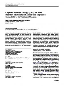
Several limitations have been recognized throughout the studies regarding PD and Visual stimulus fMRI. Among these, we can underline that most of the studies used a small number Expert Rev. Neurother. © Future Science Group (2010) of patients [26,17] . Additionally, some studies did not include healthy volunteers or Figure 1. Brain structures activated in panic-disorder patients by either visual or auditory stimuli. ACC activation can be observed when unmasked happy faces and control groups with other anxiety disorunmasked neutral faces stimuli are presented [16,17] ; unmasked fearful faces can activate ders [22,18] . Another concern is the extent the amygdale [16] . Color naming panic-related words activated the anterior PFC, ACC, to which medication effects may account PFC, OFC, thalamus, middle temporal cortex, amygdala and hippocampus [19] . Facial for interfering in activation patterns [17] . stimuli can also activate the ACC, in a preceding congruence task, and brainstem as well One important limitation with referas limbic areas, when preceding incongruence [18] . Concerning auditory stimuli, sine tones evoked right amygdala and right putamen activation compared with the resting ence to fMRI studies with PD subjects state [14] . Threat-related words activated the left posterior cingulate and left middle is that these patients report more anxiety frontal cortices [20] . during the scan than control subjects. The ACC: Anterior cingulate cortex; OFC: Orbitofrontal cortex; PFC: Prefrontal cortex. latter increase in anxiety may account for induced hypocapnia, which could result in the 5-HTTLPR polymorphism on brain activation in response to an overall decline in brain blood flow and a reduction in the size unmasked fearful or angry faces or to masked emotional stimuli. of BOLD responses of the patients. Consequently, there is a difOn the other hand, higher amygdala activation was observed in ficulty inferring whether the areas showing less activation reflect PD patients carrying at least one short allele compared with homo- real differences in neural responses [20] . The unpredictability zygotes for the long allele. In relation to the -1019C/G 5-HT1A and rapid onset and offset of panic attacks make it difficult to polymorphism, decreased activation in the right ventromedial PFC, directly investigate their various symptoms during spontaneous right orbitofrontal cortex and right ACC in response to unmasked attacks [28] . fMRI low temporal resolution is another concern [29] . fearful faces was found. Concerning the unmasked happy faces Some of the limitations found in the investigated studies have response, homozygous patients from the high-risk group showed been pointed out; therefore, some topics for improving future significantly higher activation in the left amygdala. During pro- investigations are indicated. Larger samples, studies with concessing of angry faces, a trend towards differential activation in the comitant cognitive measures, studies including both pre and post right ventromedial PFC region was identified for different genotype pharmacological and psychological treatment data are necessary to groups. Additionally, decreased activation in the right ventromedial enlarge preliminary findings [20] . Since agoraphobia is a common Temporal cortex
296
Audio stimulus
Expert Rev. Neurother. 10(2), (2010)
Current findings of fMRI in panic disorder
PD comorbidity, the lack of studies using these groups as experimental samples makes it necessary that future research considers this question. The same is true for the description of comorbidities presented by the patients. The experimental designs regarding PD patients must control for possible confounds from reduction of BOLD signal secondary to hypocapnia. Partial pressure of CO2 level measurement before and after the scan or calibration of BOLD responses with sensory or motor tasks are encouraged [20] . Fear neurocircuitry hypothesis & cognitive–behavioral therapy interventions
Review
changing dysfunctional thoughts regarding body sensations [36] . Behavior therapy uses exposure procedures to cause habituation and elimination of anxiety and agoraphobic responses [37] . CBT seems to control autonomic reactions involved in PD-response conscious processes (thought restructuring) and by eliciting of emotional responses until habituation occurs (exposure therapy). Psychotherapy can alter the brain and physiology of patients with psychiatric disorders [38–40] . Theoretical models of CBT action implicate top-down mechanisms that exert inhibitory effects on a variety of learned responses [29] . It is presumed that at least two different patterns of brain activity changes are plausible. One pattern considers that psychotherapy may lead to a normalization of previously abnormal activation. The other supposes that psychotherapy may lead to the recruitment of additional areas and, therefore, attenuate symptoms [41] . The amygdala plays a central role in the fear learning processes. Explicit emotional learning and memory are modulated by the amygdala [31] . The hippocampus is necessary to obtain explicit or declarative knowledge of the emotional properties of a stimulus [32] . Despite the controversial findings regarding hippocampal functions in the fear circuitry, some findings show that the hippocampus has a role in the mnemonic processes that underlie
According to Gorman et al., a deficiency in the coordination of the stimuli from the cortex and brainstem could lead to an abnormal activation of the amygdala, with a behavioral, autonomic and neuroendocrine stimulation [29] . One hypothesis about the neurocircuitry involved in PD is proposed by the author and is summarized here. Panic or anxiety may be precipitated by visuospatial/auditory stimuli or catastrophic cognitions [30] . The information may flow by two different pathways. In the ‘short loop’ pathway stimuli received by the sensory thalamus are transmitted to the lateral amygdala and are then transferred to its central nucleus [31] . In the ‘long loop’ pathway the sensory information projects to the thalamus. The thalamus sends this information to sensory cortex, insula and PFC for a finer analysis. The results of this analysis are sent to the amygdala [32,33] . From the amygdala, the information projects to the effector sites in the brainstem and hypothalamus, which produce the autonomic, behavioral and endocrine responses of acute fear [31] . The main effector sites are the parabrachial nucleus (increases respiratory rate), the locus ceruleus (increases norepinephrine release), the lateral and paraventricular nucleus of the hypothalamus (sympathetic discharge and increases in adrenocorticoid release, respectively) and the periaqueductal gray region (additional behavioral responses). The hipPFC Occipital region of pocampus communicates directly with the temporal cortex amygdala [29,34] . ACC The fMRI studies regarding PD to date Motor activation Putamen seem to show activation of some of the Visual stimulus regions that are included in this hypothesis, Temporal cortex especially the amygdala, as was shown in the previous section. The amygdala and the Expert Rev. Neurother. © Future Science Group (2010) PFC play a special role in this fear circuitry. Figure 2. Brain structures with decreased activation in panic disorder patients These regions, along with the hippocampus, under either visual or motor stimulation. Facial stimuli led to decreased activation of seem to be an important focus of attention the PFC, in a preceding incongruence task [18] . When asked to perform a motor task, for cognitive–behavior therapy (CBT) studpanic disorder patients showed decreased activation in the right temporal, occipital region, bilateral putamen and surrounding regions [13] . ies [35] . It is known that cognitive therapy ACC: Anterior cingulate cortex; PFC: Prefrontal cortex. achieves its goal in treating PD patients by www.expert-reviews.com
297
298
20
0
Domschke et al. (2008)
Domschke et al. (2006)
36.75 ± 9.39
36.75 ± 9.39
X
X
Age Scanner type (mean ± SD) 1.5 T 3 T
X Unmasked and masked emotional faces
X Unmasked emotional faces
Visual
Auditory Rest Other
Stimulus
For the 5-HT1A -1019C/G genotype group: UFF: ↓ activation in the right vMPFC; right OFC, and right ACC UAF: trend towards differential activation of right vMPFC UHF: ↑ left amygdala MP: ↓ right vMPFC in response to fearful faces For the 5-HTTLPR genotype group: UAF: ↑ amygdale activation in PD patients with at least one short allele
↑ right amygdala in carriers of at least one allele (158val) UFF: ↑ left OFC in carriers of at least one allele (158val) UAF: ↓ left vMPFC for patients carrying at least one of the risk alleles UHF: ↓ both vMPFC for carriers of the risk alleles
Findings
Treatment: SSRI (10) Comorbidities: SP (10); MDD ( 5) No influence of the 5-HTTLPR polymorphism on brain activation in response to unmasked fearful or angry face or to masked emotional stimuli
Neuronal activation following emotional stimulation Stimulation was used as an endophenotype and investigated for association with the COMT val158met polymorphism in PD Treatment: SSRI (10) Comorbidities: SP (10), MDD (5)
Comments
[26]
[22]
Ref.
ACC: Anterior cingulate cortex; C: Control; F: Female; M: Male; MDD: Major depression disorder; MP: Masked presentation of emotional faces; OFC: Orbitofrontal cortex; P: Patients; PD: Panic disorder; ROI: Region of interest; SD: Standard deviation; SP: Social phobia; SSRI: Selective serotonin-reuptake inhibitor; UAF: Unmasked angry faces; UFF: Unmasked fearful faces; UHF: Unmasked happy faces; VBM: Voxel-based morphometry; vMPFC: Ventromedial prefrontal cortex.
8/12
8/12
Subjects (n) M/F
Study
Table 2. Functional MRI genetic findings on panic disorder.
Review de Carvalho, Dias, Cosci et al.
Expert Rev. Neurother. 10(2), (2010)
Current findings of fMRI in panic disorder
acquisition, consolidation and retrieval of contextual fear [42] . Medial PFC functions relevant to fear include attention to the emotional states of the self and others and guidance of response selection by emotional states [43] . The medial PFC is also thought to regulate extinction of long-term memory [31] . Before CBT, the amygdala activation may be a consequence of misinterpretation of sensory information [29] .��������������� Reduced �������������� activity in the PFC might reflect a deficiency in top-down control of the fear response, allowing for the cascade of neural events that is experienced as a panic attack [44] .��������������������� �������������������� Hippocampal dysfunction might be associated with an overgeneralization of potential threat, that is, it might reveal a consequence of deficient������� ������ appreciation for the contextual specificity of potentially threatening stimuli [44] . It is accepted that with continued presentation of the conditioned stimuli, the medial PFC inhibits firing of amygdala neurons under the modulation of the hippocampus [31] . During exposure therapy the focus of attention is directed towards the threatening situation to facilitate PFC control over the amygdala. Inhibitory projections from the hippocampus are created by the meeting of new contextual emotional information associated with safety that is added in the emotional network [45] . As a consequence, cognitive, emotional and behavioral responses are modified. Through cognitive restructuring, CBT plays the role of strengthening the ability of the cortical projections, including medial PFC, to assert reason over automatic behavioral and physical responses [29] ; that is, the medial PFC strengthens its ability to inhibit the amygdala [46,47] , and changes in information processing and affective states are observed. To date, two neuroimaging studies have examined the modulation of the anxiety neurocircuitry with CBT in PD. fMRI studies with PD patients on CBT treatment were not found. Preliminary hypotheses indicate that CBT has the potential to modify the dysfunctional neural circuitry associated with PD, but there is still insufficient data regarding this issue. Prasko et al. investigated brain structures that could show changes in 18F-fluorodeoxyglucose PET during psychological and pharmacological treatment [48] . The researchers randomly assigned 12 patients with PD to either the CBT group or the antidepressant treatment group. Results of both treatment groups reported similar neural changes in the frontal and temporal regions, with prominent right–left difference. No change in subcortical regions was detected. Improvements of similar magnitude were observed in both groups, with a more rapid decrease in psychopathology with CBT. The CBT group showed decreases in regions of the right hemisphere in the inferior temporal gyrus, superior and inferior frontal gyrus. Increases in the left hemisphere (inferior frontal gyrus, middle temporal gyrus and insula) were also detected. Regarding the results, the authors suggested that successful CBT can positively modulate brain metabolism in PD patients. Sakai et al. investigated regional brain glucose metabolic changes associated with successful completion of CBT in PD patients using PET with 18F-fluorodeoxyglucose [49] . The regional glucose utilization was compared before and after CBT. Of the 12 patients www.expert-reviews.com
Review
11 improved after treatment. In these patients, increased glucose utilization was detected in the bilateral medial prefrontal cortices. Decreased glucose utilization was seen in the right hippocampus, left ACC, left cerebellum and pons. The results point to adaptive metabolic changes of the bilateral medial PFCs after CBT. Expert commentary
For an accurate diagnosis of PD, it must be considered that a panic attack (an episode of intense fear associated with several somatic symptoms) must also lead to apprehension regarding future attacks (anticipatory anxiety). These two conditions (panic attack and anticipatory anxiety) can be defined as ‘stressful’ events in a patient’s life. Stress can be defined as an actual or anticipated disrupting of homeostasis or anticipated threat to wellbeing [50] . In other words, it is the subjective state of sensing potentially adverse changes in the environment [51] . The physiological response to stress involves a set of interlocking systems with the objective of physiologic integrity maintenance [50] . The brain responds to psychogenic stressors based on prior experience or innate programs [50] . Psychological stressors engage stress mediators in brain regions that subserve emotion (the amygdala and PFC), learning and memory (the hippocampus) and decision-making (PFC) [51] . All of these regions are engaged in the pathophysiology of PD according to the neuroanatomical hypotheses, so it is predictable that these regions present functional abnormalities in PD patients. Based on animal studies of the effects of stress and emerging literature in the clinical neuroscience of anxiety disorders, Bremner and Charney have developed a working model for the neural circuitry of fear and anxiety [52] . Their model intends to explain how information related to a threatening stimulus is integrated into an image that activates memory traces of prior similar experiences and triggers a motor response. They emphasized that the cognitive response to threat, which involves placing the threatening object in space and time, is an aspect of the stress response as important as the incorporation of a person’s prior experience (memory) into the cognitive appraisal of stimuli. They also stated that several studies showed alterations in hippocampus and adjacent cortex (parahippocampus) function in PD, with no change in hippocampal volume. Patients with PD have had alterations of parahippocampal gyrus and other portions of extrahippocampal temporal lobe that may underlie declarative memory deficits. Increased cortisol release with stress in PD may result in cognitive dysfunction associated with this disorder [52] . Bremner and Charney explained that anxiety symptoms in PD patients may be related to fear responses to a traumatic cue, which suggests that the original fear-inducing stimulus is impossible to be determined [52] . They also noted that the amygdala plays an important role in conditioned fear and in the emotional response. The amygdala has been used as part of an animal model for stress-induced abnormalities of emotional memory. According to the neuroanatomical hypothesis of PD, conditioned fear would be the core subject of the neurobiological explanation of this disorder, and the amygdala stands as a key structure that coordinates autonomic and behavioral responses [29] . 299
Review
de Carvalho, Dias, Cosci et al.
The amygdala increases in volume under chronic stress. Given that the amygdala plays an important role in the detection of fear and threat, it is possible that throughout evolution increases in amygdala volume in response to stress might have improved the detection of threatening information and so increased survival probability [53] . According to the review article of Ulrich-Lai and Herman, the central nucleus of the amygdala is a key node for stress integration, but it is not activated by psychogenic stressors [50] . On the other hand, the medial and basolateral amygdala nuclei are preferentially activated by psychological stressors. The medial PFC has inhibitory inputs that decrease amygdala responsiveness and has been hypothesized to mediate extinction of fear responding. Brodmann area 25 also stimulates the peripheral cortisol and the sympathetic response to stress. Activation of this area has been shown to be a normal response to stress or increased emotionality. Bremner and Charney have hypothesized that dysfunction in this area may mediate increased emotionality and failure of extinction to fear-inducing cues in anxiety disorders, as evidenced by the failure of normal activation with yohimbineinduced provocation of anxiety in PD [52] . Potentiated release of norepinephrine with stressors in PD is expected to be associated with a relative decrease in function of neurons in this area [52] . According to the review article of Ulrich-Lai and Herman, the infralimbic PFC is involved in initiating autonomic and hypothalamus–pituitary–adrenal axis responses to psychogenic stimuli [50] . In the hippocampus and PFC, chronic restraint causes retraction of apical dendrites and reduces spine density in pyramidal cells. As can be seen, reduced activation of PFC and of the hippocampus would be predictable features of neuroimaging PD studies. Chronic stress can recruit pathways that are distinct from those involved in acute responses [50] . This would explain why panic attack neuroimaging studies find divergent activation patterns from those studies where there are no panic attacks during scanning. The PFC is the brain region that is most sensitive to the detrimental effects of stress exposure. Chronic stress disrupts the plastic relationship between the PFC and the hippocampus that is needed for flexible memory consolidation [54] . Enhanced memory for stressful or emotionally arousing events is a well-recognized, highly emotionally adaptive phenomenon that helps us to remember important information. Such memory enhancement is not limited to unpleasant experiences; pleasurable events also tend to be well remembered. The basolateral complex of the amygdala is an important locus of integration of modulatory influences on memory consolidation. Adrenal stress hormones modulate the consolidation of memory for emotionally arousing experiences, but do not affect memory consolidation of neutral information. Interactions between the basolateral complex of the amygdala and the hippocampus regulate emotional arousal effects on memory consolidation of spatial or contextual information. The basolateral complex of the amygdala also modulates cortical functioning involved in memory consolidation. This complex interacts with the entorhinal cortex and also with the insular cortex and ACC. Stress 300
exposure can induce amygdala activation in concert with excitatory and inhibitory effects on other brain regions, to create a brain state that on the one hand promotes the long-term storage of memories of these emotionally arousing events and, thus, preserves significant information, but on the other hand impairs memory retrieval and working memory [55] . Lupien et al. report that the impact of acute stressors depends on the level of glucocorticoid elevations, with small increases resulting in enhanced hippocampus-mediated learning and memory and larger, prolonged elevations impairing hippocampal function [53] . This impairment is related to spatial learning (or contextual fear in PD). The authors also correlate the hippo campus small volume findings with experiences of childhood trauma, and conclude that decreased hippocampal volume can be a marker of vulnerability to psychiatric disorders rather than a marker of the disorder itself. Furthermore, they explore two complementary hypotheses: the neurotoxicity and vulnerability hypotheses. The former implies that a reduced hippocampal size is the end product of years or decades of chronic stress. The second hypothesis suggests that reduced hippocampal volume in adulthood is not a consequence of chronic stress, but is a preexisting risk factor for stress-related disorders that is induced by genetics and/or early exposure to stress. The latter could be a possible explanation as to why hippocampal volumes and activation patterns are sometimes conflicting between different neuroimaging studies. It is necessary to explore the history of early stressful events in a more detailed fashion. The authors report that exposure to stress and/or adversity during the key vulnerable periods of life might slow down the development of those brain regions (especially the hippocampus and the PFC). During childhood and adolescence the brain undergoes a period of overproduction and pruning of synapses. The amygdala is one of the brain regions that show the slowest development over a person’s lifespan [53] . A previous study discussed structural putaminal abnormalities in PD patients. In that paper, the putaminal gray matter volume reduction was negatively correlated with the severity scores and duration of PD. It was discussed that the putamen was associated with the programming of learned sequential motor acts, such as avoidant responses, and in PD patients this could represent a diminished ability in implicit learning of avoidant behaviors via the dysfunction of motor learning processes. It was also discussed that there are several adrenergic receptors and dopamine receptors in the putamen, which exert an important role on sympathetic regulation and could account for the dysregulation of autonomic systems in PD [56] . This data could explain why in the Marchand et al. paradigm a bilateral decreased putamen function was observed [13] , whereas in the case report of Pfleiderer et al. increased activity in the right putamen was detected (Table 1) [14] . Five-year view
Replication studies on fMRI in PD patients should be conducted with the aim to control for possible limitations and produce clearer results. As stated previously, larger samples and subjects Expert Rev. Neurother. 10(2), (2010)
Current findings of fMRI in panic disorder
with agoraphobia and comorbid PD might be evaluated. More information on the characteristics of the sample should be collected to control for possible confounders (e.g., other psychiatric comorbid disorders, hypocapnia during fMRI scan, cognitive measures and a current smoking status) [57] . Moreover, pre- and post-fMRI scan pharmacological and/or psychological treatment have to be described. Besides replication studies, fMRI might be applied to PD subjects under different pharmacological treatments (e.g., serotoninergic antidepressants, atypical antipsychotic with serotoninergic activity and dopaminergic drugs), different psychological interventions (e.g., in vivo exposure, cognitive therapy and wellbeing therapy) or under sequential treatments (e.g., antidepressant and CBT) [37] . Furthermore, sequential treatment of mood and anxiety disorders to highlight if and how they might interfere with the activity of specific brain areas is recommended.
Review
Studies on the specific personality characteristics (e.g., anxiety sensitivity, neuroticism) of PD subjects should be implemented to clarify whether these traits might modulate the activation of brain circuitries. Finally, further contribution from genetic studies may derive from studies on GABA transporter 1 gene that has been considered a novel candidate for anxiety disorders with panic-like symptoms [58] . Financial & competing interests disclosure
The grant for this review was provided by the Brazilian Council for Scientific and Technological Development (CNPq) and INCT Translational Medicine (CNPq).���������������������������������������������������������������������� The authors have no other relevant affiliations or financial involve� ment with any organization or entity with a financial interest in or financial conflict with the subject matter or materials discussed in the manuscript apart from those disclosed. No writing assistance was utilized in the production of this manuscript.
Key issues • Functional MRI techniques are helping to elucidate a number of issues related to neural circuits, especially concerning fear and anxiety. • Functional MRI data shows that brain structures, such as the prefrontal cortex, the anterior cingulate cortex, limbic areas (hippocampus and amygdala) and brainstem structures, might play a major role in the panic circuitry. • The understanding of the neurobiological basis of panic disorder has benefited from genetic studies, which show serotoninergic- and dopaminergic-related polymorphisms associated to panic responses. • Future studies are required to address some limitations. These studies should include: larger samples; concomitant cognitive measures; both pre- and post-pharmacological and psychological treatment data; and agoraphobic patients as experimental samples. • The experimental designs regarding panic disorder patients must control for possible confounds from reduction of blood oxygen level dependent (BOLD) signal secondary to hypocapnia. • Increasing evidence point that cognitive–behavior therapy has the potential to modify the dysfunctional neural circuitry associated with anxiety disorders. • Cognitive restructuring and exposure therapy are cognitive–behavior therapy techniques that facilitate prefrontal cortex control over the amygdala. Additionally, inhibitory projections from the hippocampus are created by the meeting of new contextual emotional information associated with safety. As a consequence, cognitive, emotional and behavioral responses are modified.
References
6
Ontiveros A, Fontaine R, Breton G, Elie R, Fontaine S, Déry R. Correlation of severity of panic disorder and neuroanatomical changes on magnetic resonance imaging. J. Neuropsychiatry Clin. Neurosci. 1(4), 404–408 (1989).
7
Fontaine R, Breton G, Déry R, Fontaine S, Elie R. Temporal lobe abnormalities in panic disorder: an MRI study. Biol. Psychiatry 27(3), 304–310 (1990).
8
Vythilingam M, Anderson ER, Goddard A et al. Temporal lobe volume in panic disorder – a quantitative magnetic resonance imaging study. Psychiatry Res. 99(2), 75–82 (2000).
9
Uchida RR, Del-Ben CM, Busatto GF et al. Regional gray matter abnormalities in panic disorder: a voxel-based morphometry study. Psychiatry Res. 63(1), 21–29 (2008).
10
Massana J, Mercader JM, Gómez B, Tobeña A, Salamero M. Amygdalar atrophy in panic disorder patients detected by volumetric magnetic resonance imaging. Neuroimage 19(1), 80–90 (2003).
Papers of special note have been highlighted as: •• of considerable interest 1
Andreasen NC. Evaluation of brain imaging techniques in mental illness. Ann. Rev. Med. 39, 335–345 (1988).
2
Bremner JD. Brain imaging in anxiety disorders. Expert Rev. Neurother. 4(2), 275–284 (2004).
3
Brambilla P, Barale F, Caverzasi E, Soares JC. Anatomical MRI findings in mood and anxiety disorders. Epidemiol. Psichiatr. Soc. 11(2), 88–100 (2002).
4
Rocha ET, Alves TCTF, Garridoc GEJ, Buchpiguel R, Nitrinid R, Filho GB. Novas técnicas de neuroimagem em psiquiatria: qual o potencial de aplicações na prática clínica? Rev. Bras. Psiquiatr. 23(Suppl. I), 58–60 (2001).
5
Amaro Jr. E, Yamashita H. Aspectos básicos de tomografia computadorizada e ressonância magnética. Rev. Bras. Psiquiatr. 23, 2–3 (2001).
www.expert-reviews.com
11
Massana G, Serra-Grabulosa JM, Salgado-Pineda P et al. Parahippocampal gray matter density in panic disorder: a voxel-based morphometric study. Am. J. Psychiatry 160(3), 566–568 (2003).
12
Protopopescu X, Pan H, Tuescher O et al. Increased brainstem volume in panic disorder: a voxel-based morphometric study. Neuroreport 17(4), 361–363 (2006).
13
Marchand WR, Lee JN, Healy L et al. An fMRI motor activation paradigm demonstrates abnormalities of putamen activation in females with panic disorder. J. Affect. Disord. 116(1–2), 121–125 (2009).
14
Pfleiderer B, Zinkirciran S, Arolt V, Heindel W, Deckert J, Domschke K. fMRI amygdala activation during a spontaneous panic attack in a patient with panic disorder. World J. Biol. Psychiatry 8(4), 269–272 (2007).
•• Represents an important contribution by reviewing the recent advances on functional brain imaging studies in psychiatry.
301
Review
de Carvalho, Dias, Cosci et al.
15
Ekrnan P, Friesen W. Pictures of Facial Affect. Consulting Psychologists Press, Palo Alto, CA, USA (1975).
16
Pillay SS, Gruber SA, Rogowska J, Simpson N, Yurgelun-Todd DA. fMRI of fearful facial affect recognition in panic disorder: the cingulate gyrus-amygdala connection. J. Affect. Disord. 94(1–3), 173–181 (2006).
27
Lesch KP, Bengel D, Heils A et al. Association of anxiety-related traits with a polymorphism in the serotonin transporter gene regulatory region. Science 274, 1527–1531 (1996).
41
Linden DE. Brain imaging and psychotherapy: methodological considerations and practical implications. Eur. Arch. Psychiatry Clin. Neurosci. 258 (5), 71–75 (2008).
17
Pillay SS, Rogowska J, Gruber SA, Simpson N, Yurgelun-Todd DA. Recognition of happy facial affect in panic disorder: an fMRI study. J. Anxiety Disord. 21(3), 381–393 (2007).
28
Schunck T, Erb G, Mathis A et al. Functional magnetic resonance imaging characterization of CCK-4-induced panic attack and subsequent anticipatory anxiety. Neuroimage 31(3),1197–1208 (2006).
42
Sanders MJ, Wiltgen BJ, Fanselow MS. The place of the hippocampus in fear conditioning. Eur. J. Pharmacol. 463(1–3), 217–223 (2003).
43
18
Chechko N, Wehrle R, Erhardt A, Holsboer F, Czisch M, Sämann, PG. Unstable prefrontal response to emotional conflict and activation of lower limbic structures in remitted panic disorder. PLOS One 4(5), 1–11 (2009).
29
Gorman JM, Kent JM, Sullivan GM, Coplan JD. Neuroanatomical hypothesis of panic disorder, revised. Am. J. Psychiatry 157(4), 493–505 (2000).
Miller LA, Taber KH, Gabbard GO, Hurley RA. Neural underpinnings of fear and its modulation: implications for anxiety disorders. J. Neuropsychiatry Clin. Neurosci. 17(1), 1–6 (2005).
44
Cannistraro PA, Rauch SL. Neural Circuitry of anxiety: evidence from structural and functional neuroimaging studies. Psychopharmacol. Bull. 37(4), 8–25 (2003).
45
De Raedt R. Does neuroscience hold promise for the further development of behavior therapy? The case of emotional change after exposure in anxiety and depression. Scand. J. Psychol. 47, 225–236 (2006).
19
20
21
22
23
24
25
26
van den Heuvel OA, Veltman DJ, Groenewegen HJ et al. Disorder-specific neuroanatomical correlates of attentional bias in obsessive-compulsive disorder, panic disorder, and hypochondriasis. Arch. Gen. Psychiatry 62(8), 922–933 (2005). Maddock RJ, Buonocore MH, Kile SJ, Garrett AS. Brain regions showing increased activation by threat-related words in panic disorder. Neuroreport 14(3), 325–328 (2003). Hettema JM, Neale MC, Kendler KS. A review and meta-analysis of the genetic epidemiology of anxiety disorders. Am. J. Psychiatry 158, 1568–1578 (2001). Domschke K, Ohrman P, Braun M et al. Influence of cathecol-O-methyltransferase val158met genotype on amygdala and prefrontal cortex emotional processing in panic disorder. Psychiatry Res. 163, 13–20 (2008). Schulman R, Griffiths J, Diewold P. Cathecol-O-methyltransferase activity in patients with depressive illness and anxiety states. Br. J. Psychiatry 132, 133–138 (1978). Weinshilboum RM, Otterness DM, Szumlanski CL. Methylation pharmacogenetics: cathecol-Omethyltransferase, thiopurine methyltransferase, and histamine N-methyltransferase. Annu. Rev. Pharmacol. Toxicol. 39, 19–52 (1999). Simon NM, Emmanuel N, Ballenger J, Worthington JJ, Kinrys G, Korbly B, Farach, FJ, Pollack MH. Bupropion sustained release for panic disorder. Psychopharmacol. Bull. 37, 66–72 (2003). Domschke K, Braun M, Ohrman P et al. Association of the functional -1019C/G 5-HT1A polymorphism with prefrontal
302
cortex and amygdala activation measured with 3 T fMRI in panic disorder. Int. J. Neuropsychopharmacol. 9, 349–355 (2006).
•• Represents an important contribution to the etiological hypothesis of panic disorder. 30
Coplan JD, Lydiard RB. Brain circuits in panic disorder. Biol. Psychiatry 44(12), 1264–1276 (1998).
31
Garakani A, Mathew SJ, Charney DS. Neurobiology of anxiety disorders and implications for treatment. Mt Sinai J. Med. 73(7), 941–949 (2006).
32
Gazzaniga MS, Ivry RB, Mangun GR. Cognitive Neuroscience: the Biology of the Mind. WW Norton & Company, NY, USA (1998).
33
LeDoux, J. Fear and the brain: where have we been, and where are we going? Biol. Psychiatry 44(12), 1229–1238 (1998).
disorders: Empirical and methodological review. Clin. Psychol. Rev. 28, 228–246 (2008).
•• Represents an important contribution to the understanding of the neurobiological hypothesis of psychotherapy mechanisms of change. 46
Roffman JL, Marci CD, Glick DM, Dougherty DD, Rauch SL. Neuroimaging and the functional neuroanatomy of psychotherapy. Psychol. Med. 35, 1385–1398 (2005).
47
McNally RJ. Mechanisms of exposure therapy: How neuroscience can improve psychological treatments for anxiety disorders. Clin. Psychol. Rev. 27, 750–759 (2007).
48
Prasko J, Horácek J, Záleský R et al. The change of regional brain metabolism (18FDG PET) in panic disorder during the treatment with cognitive behavioral therapy or antidepressants. Neuro Endocrinol. Lett. 25(5), 340–348 (2004).
49
Sakai Y, Kumano H, Nishikawa M et al. Changes in cerebral glucose utilization in patients with panic disorder treated with cognitive–behavioral therapy. Neuroimage 33(1), 218–226 (2006).
34
Stein DJ. The neurobiology of panic disorder: toward an integrated model. CNS Spectr. 10(9), 5–13 (2005).
35
De Carvalho MR, Rosenthal M, Nardi AE. The fear circuitry in panic disorder and its modulation by cognitive-behaviour therapy interventions. World J. Biol. Psychiatry (2009) (In press).
36
Clark DM. A cognitive approach to panic. Behav. Res. Ther. 24, 461–470 (1986).
37
Fava GA, Ruini C, Rafanelli C. Sequential treatment of mood and anxiety disorders. J. Clin. Psychiatry 66(11), 1392–1400 (2005).
38
Kumari V. Do psychotherapies produce neurobiological effects? Acta Neuropsychiatrica 18, 61–70 (2006).
39
Beauregard M. Mind does really matter: Evidence from neuroimaging studies of emotional self-regulation, psychotherapy, and placebo effect. Prog. Neurobiol. 81, 218–236 (2007).
50
Frewen PA, Dozois DJA, Lanius RA. Neuroimaging studies of psychological interventions for mood and anxiety
Ulrich-Lai YM, Herman JP. Neural regulation of encodrine and autonomic stress response. Nat. Rev. 10, 397–409 (2009).
51
Joëls M, Baram TZ. The neuro-symphony of stress. Nat. Rev. 10, 459–466 (2009).
40
Expert Rev. Neurother. 10(2), (2010)
Current findings of fMRI in panic disorder
52
Bremner D, Charney DS. Neural circuits in fear and anxiety. In: Textbook of Anxiety Disorders (Second Edition). Stein DJ, Hollander E, Rothbaum BO (Eds). American Psychiatric Publishing Inc., Washington, DC, USA (2010).
53
Lupien SJ, McEwen BS, Gunnar MR, Heim C. Effects of stress throughout the lifespan on the brain, behaviour and cognition. Nat. Rev. 10, 434–445 (2009).
54
Arnsten AFT. Stress signaling pathways that impair prefrontal cortex structure and function. Nat. Rev. 10, 410–422 (2009).
55
Roozendaal B, McEwen BS, Chattarji S. Stress, memory and the amygdala. Nat. Rev. 10, 423–433 (2009).
56
Yoo HK, Kim MJ, Kim SJ et al. Putaminal gray matter volume decrease in panic disorder: an optimized voxel-based morphometry study. Eur. J. Neurorsci. 22, 2089–2094 (2005).
57
Yalachkov Y, Kaiser J, Naumer MJ. Brain regions related to tool use and action knowledge reflect nicotine dependence. J. Neurosci. 29(15), 4922–4929 (2009).
58
Thoeringer CK, Ripke S, Unschuld PG et al. The GABA transporter 1 (SLC6A1): a novel candidate gene for anxiety disorders. J. Neural Transm. 116(6), 649–657 (2009).
www.expert-reviews.com
Affiliations •
•
•
Marcele Regine de Carvalho Laboratory of Panic and Respiration, Institute of Psychiatry, Universidade Federal do Rio de Janeiro (UFRJ), Rio de Janeiro, Brazil and INCT Translational Medicine (CNPq), Brazil Av. das Américas, 1155/ 2005, 22631-000, Rio de Janeiro, Brazil Tel.: +55 212 436 8202 Fax: +55 212 523 6839
[email protected] Gisele Pereira Dias Laboratory of Panic and Respiration, Institute of Psychiatry, Universidade Federal do Rio de Janeiro (UFRJ), Rio de Janeiro , Brazil and INCT Translational Medicine (CNPq), Brazil Av. das Américas, 1155/ 2005, 22631-000, Rio de Janeiro, Brazil and Laboratory of Neurobiology of the Retina, Universidade Federal do Rio de Janeiro (UFRJ), Rua Mal Ramon Castilla, Rio de Janeiro 199/806, 22290-175, Brazil Tel.: +55 212 229 4445 Fax: +55 212 523 6839
[email protected] Fiammetta Cosci Department of Neurology and Psychiatry, University of Florence, Via di San Salvi, 12, Florence 50134, Italy and Department of Psychology, University of Florence, Florence 50137, Italy Tel.: +39 055 623 7811 Fax: +39 055 623 6047
[email protected]
Review
•
Valfrido Leão de-Melo-Neto Laboratory of Panic and Respiration, Institute of Psychiatry, Universidade Federal do Rio de Janeiro (UFRJ), Rio de Janeiro, Brazil and INCT Translational Medicine (CNPq), Rua Eng. Mario de Gusmão, 674/ 404. Zip Code: 57035-000, Maceió, AL, Brazil Tel.: +55 823 313 3043 Fax: +55 212 523 6839
[email protected]
•
Mário Cesar do Nascimento Bevilaqua Laboratory of Neurobiology of the Retina, Universidade Federal do Rio de Janeiro (UFRJ), Rua Cerqueira Daltro, 934, Rio de Janeiro 21380-320, Brazil Tel.: +55 212 229 3904 Fax: +55 212 562 6594
[email protected]
•
Patricia Franca Gardino Laboratory of Neurobiology of the Retina, Universidade Federal do Rio de Janeiro (UFRJ), Rua Corrêa Dutra 44 apto 203, Rio de Janeiro 22210–050, Brazil Tel.: +55 212 557 5387 Fax: +55 212 562 6594
[email protected]
•
Antonio Egidio Nardi Laboratory of Panic and Respiration, Institute of Psychiatry, Universidade Federal do Rio de Janeiro (UFRJ), Rua. Visconde de Pirajá, 407/702, Rio de Janeiro 22410-003, Brazil Tel.: +55 212 521 6147 Fax: +55 212 523 6839
[email protected]
303