Acta Pharmaceutica Sinica B 2013;3(2):86–96 Institute of Materia Medica, Chinese Academy of Medical Sciences Chinese Pharmaceutical Association
Acta Pharmaceutica Sinica B www.elsevier.com/locate/apsb www.sciencedirect.com
REVIEW
Current strategies for drug delivery to the inner ear Hongzhuo Liua,b,n, Jinsong Haoa, Kevin S. Lia a
Division of Pharmaceutical Sciences, College of Pharmacy, University of Cincinnati, Cincinnati 45267, Ohio, USA School of Pharmacy, Shenyang Pharmaceutical University, Shenyang 110016, China
b
Received 15 December 2012; revised 17 January 2013; accepted 3 February 2013
KEY WORDS Inner ear; Drug delivery; Intratympanic; Intracochlear; Gene; Stem cell
Abstract For many years, drug delivery to the inner ear has been a challenge to physicians in the treatment of inner ear disorders. In the past decade, the field of inner ear drug delivery has emerged with the development of new biomaterials and drug delivery technologies to improve the effectiveness of inner ear drug therapy. This paper reviews a number of inner ear drug delivery strategies including systemic, intratympanic, and intracochlear delivery. A focus of this review is the recent advances in intratympanic delivery of medications; approaches utilizing novel biomaterials as well as other recent developments are also discussed. Biotechnology-based approaches, such as gene and stem cell therapy methods are also reviewed. Among the various strategies, local drug delivery approaches including intratympanic and intracochlear drug delivery methods that limit systemic exposure are particularly promising. These inner ear drug delivery systems provide a new opportunity to improve the treatment of inner ear disorders. & 2013 Institute of Materia Medica, Chinese Academy of Medical Sciences and Chinese Pharmaceutical Association. Production and hosting by Elsevier B.V. All rights reserved.
Abbreviations: BLB, blood labyrinth barrier; AAV, adeno-associated virus; ABR, auditory brainstem response; AIED, autoimmune inner ear disease; BDNF, brain-derived neurotrophic factor; BP, betamethasone phosphate; BBB, blood brain barrier; GDNF, glial-derived neurotrophic factor; GFP, green fluorescent protein; HBPL, hyperbranched poly L-lysine; LNCs, lipid core nanocapsules; MRP-1, multidrug resistance-related protein-1; PEG, poly (ethylene glycol); P-gp, P-glycoprotein; PEG-b-PCL, poly (ethylene glycol)-b-poly (e-caprolactone); PLGA, poly lactic/ glycolic acid; RMW, round window membrane; ST, scala tympani; SGNs, spiral ganglion neurons; SSNHL, sudden sensorineural hearing loss; SPIONs, superparamagnetic iron oxide nanoparticles n Corresponding author at: School of Pharmacy, Shenyang Pharmaceutical University, Shenyang 110016, China. Tel.: þ86 24 23986258. E-mail address:
[email protected] (Hongzhuo Liu). Peer review under responsibility of Institute of Materia Medica, Chinese Academy of Medical Sciences and Chinese Pharmaceutical Association.
2211-3835 & 2013 Institute of Materia Medica, Chinese Academy of Medical Sciences and Chinese Pharmaceutical Association. Production and hosting by Elsevier B.V. All rights reserved. http://dx.doi.org/10.1016/j.apsb.2013.02.003
Current strategies for drug delivery to the inner ear 1.
87
Introduction
Inner ear drug delivery has been a challenge to physicians in the treatment of inner ear disorders. In the past decade, new biomaterials and drug delivery technologies have been developed for inner ear delivery. Swan et al.1 published a comprehensive review on inner ear drug delivery in 2008. In the same year, Borkholder2 reviewed the status of inner ear delivery via the intratympanic and intracochlear routes. Since then, the field of inner ear drug delivery has gained a lot of interest and advanced rapidly. Several other review articles on inner ear drug delivery have been published in the past five years. Table 1 provides a list of these papers. These reviews focus either on a particular drug delivery strategy, a specific type of material or disease, or a combination of these topics in the field. The goal of the present review is to provide an updated general overview of inner ear drug delivery. We first review the administrative routes for inner ear drug delivery and then compare their advantages and disadvantages, discussing their potential as a result of recent advances in biomaterials, delivery technologies, and biotechnology methods. Recent developments in inner drug delivery strategies such as intratympanic and biotechnology-based approaches are highlighted. The strategies of inner ear drug delivery reviewed in this paper are listed in Table 2.
2.
Anatomy relevant to inner ear drug delivery
To understand the challenges of drug delivery to the inner ear, the anatomy relevant to inner ear drug delivery is first discussed. The inner ear in humans consists of the bony labyrinth, a system of passages comprising two main parts: (a) the cochlea dedicated to hearing and (b) vestibular system Table 1
dedicated to balance. Blood labyrinth barrier (BLB) and round window membrane (RWM) are described below because these parts constitute the barriers of drug delivery to the inner ear. 2.1.
Blood labyrinth barrier
BLB is a major barrier separating the inner ear from systemic circulation with tight junctions, made up of capillary endothelial cells that line blood vessels located in the stria vascularis13–15. It plays an important role in maintaining the microhomeostasis of the inner ear fluid and protecting the inner ear integrity similar to the function of the blood brain barrier (BBB) to the brain16. BLB functions not only as a physical barrier but also as a biochemical barrier with efflux pump systems, including P-gp (P-glycoprotein) and MRP-1 (multidrug resistance-related protein-1). These efflux systems further protect the inner ear17. Thus, the BLB is often considered as the rate-limiting barrier in the permeation of therapeutic agents from systemic circulation to the inner ear. The current knowledge on the processes of drug transport through BLB in the literature is limited. The BLB consisting of tight junctions generally permits only the permeation of small lipid-soluble molecules1. Although the actual mechanism of tight junction opening of BLB is not clear, a number of factors were found to lead to tight junction opening of the BLB, including the presence of ototoxic drugs18, noises19 and inflammation20. It has been reported that aminoglycosides given systemically in combination with diuretics such as furosemide or ethacrynic acid result in hearing loss and inner ear damage faster than that of aminoglycosides alone21,22. This suggests that the diuretics can enhance drug penetration across the BLB. Particularly, osmotic agents were suggested to
Review articles related to inner ear drug delivery.
Review subject of focus
Year
Author
Inner ear drug delivery applications and methods Transtympanic and intracochlear drug delivery Pharmacokinetics of inner ear Clinical opinion on inner ear drug delivery and current status of inner ear disease treatment Cochlear implant Nanoparticles Intracochlear drug delivery Biodegradable materials Protective agents against sensorineural hearing loss Nanoparticles Sensorineural hearing loss, animal model for evaluation New technologies on drug delivery device
2008 2008 2009 2010
Swan et al.1 Borkholder2 Salt and Plontke3 McCall et al.4
2010 2010 2011 2011 2011 2011 2012 2012
Staecker et al.5 Chen et al.6 Borenstein7 Nakagawa and Ito8 Mukherjea et al.9 Pyykko et al.10 Rivera et al.11 Pararas et al.12
Table 2
Comparison of various strategies in inner ear drug delivery.
Strategy
Efficiency
Safety
Systemic strategies Intratympanic strategies Intracochlear strategies
Low Moderate, produce variable outcome High
Low safety, side effects caused by high systemic doses over time High safety, minimally invasive procedure that can be performed in a physician’s office Limited safety, precise surgery is needed and potential of serious complications
88
Hongzhuo Liu et al.
induce endothelial cell ‘‘shrinkage’’ and thereby lead to the opening of BLB tight junction23. Glycerol, an osmotic agent, was shown to increase the concentrations of drugs such as vasodilators and steroids in the lymph of the inner ear after systemic administration. In addition to chemicals, hearing disorders such as autoimmune inner ear disease (AIED), Meniere’s disease, meningitis-associated labyrinthitis, and genetic diseases can also affect the BLB. 2.2.
Round window membrane
The round window membrane (RMW) is a soft tissue barrier separating the middle ear from the inner ear. It is the main passage for drug delivery from the middle ear cavity to the inner ear. The RWM is made up of an outer epithelial layer facing the middle ear cavity, middle connection layer, and inner cellular layer facing the scala tympani (ST) perilymph24–26. The variable RWM thickness and conditions of the membrane across the patient population is believed to be a factor leading to patient-to-patient variability in intratympanic inner ear drug delivery. Additionally, the pseudomembrane of the RWM (plugs of connective or adipose tissue) also accounts for this variability27. The RWM acts like a semipermeable membrane. It is permeable primarily to low molecular weight molecules such as aminoglycoside antibiotics and corticosteroids. Large molecules, such as horseradish peroxidase (MW 45,000), can also diffuse through RWM under normal physiological conditions28. While larger molecules like albumin (MW 70,000) cannot diffuse across the RWM easily under normal conditions, such large molecules have been shown to penetrate the RWM into the inner ear during the early phase of inflammation29. Besides the molecular weights of the drugs and disease states of the ear, other factors including the integrity of the RWM and drug lipophilicities and charges also affect the rates at which the molecules diffuse across the RWM14. In addition, the permeability of RWM is affected by local treatments with anesthetics30, endotoxins and exotoxins31, histamine32, osmotic disturbances, and benzyl alcohol (a commonly used preservative)33. 3. 3.1.
Administration routes for inner ear drug delivery
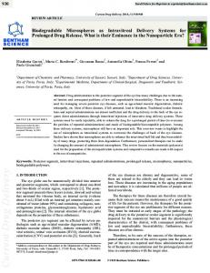
term systemic steroid therapy37. Several studies have demonstrated that systemic administration of lidocaine can relieve tinnitus, but the treatment of tinnus by lidocaine systemically involves the risks of arrhythmia and central nervous system excitation or depression38. Another treatment of inner ear disorders via the systemic route is the use of streptomycin and gentamicin, which are ototoxic, in severe bilateral Meniere’s diseases39,40. The lack of selective pharmacological effects of systemic streptomycin and gentamicin on hearing and balance has led to hearing loss in clinical interventions of Meniere’s diseases41. Other drugs that have been delivered systemically to the inner ear in conjunction with gentamicin are glutathione42, salicylate43, alpha-tocopherol44,45, trimetazine46 and flavonoids47 for their protective effects on hearing loss or histological damages in gentamicin treatment. Encouraged by the successful application of nanoparticles targeted to the brain, researchers have investigated the potentials of nanoparticles in inner ear drug delivery by systemic administration. Tamura et al.48 found that the systemic application of poly lactic/glycolic acid (PLGA) nanoparticles of rhodamine provided targeted delivery of rhodamine to the liver but not the cochlea. Horie et al.49 discussed the limited capability of nanoparticles for sustained and/or targeted delivery of drugs to cochlea after systemic application, which might be related to rapid clearance of the nanoparticles from circulation by the mononuclear phagocyte system (MPS) in the liver and spleen. They subsequently examined the efficacy of stealth nanoparticles encapsulating betamethasone phosphate (BP) for the treatment of noiseinduced sensorineural hearing loss in mice. The results in their study demonstrated that stealth-nano-BP could deliver higher level of BP to the cochlea than free BP without the nanoparticles (Fig. 1). In addition to the pharmacokinetic data, immunohistochemistry for the glucocorticoid receptor showed remarkable enhancement of glucocorticoid receptor nuclear translocation in outer hair cells of cochlea treated with the stealth-nano-BP. This treatment provided functional and histological protection of the cochlea from the trauma of noise as compared to those with free BP. However, this method was still accompanied by a high drug concentration in the blood plasma as well as in organs such as the liver, which might lead to adverse systemic effects. With the problems in systemic drug delivery, there is a need to design a safer and more effective drug delivery system for the treatment of inner ear disorders.
Systemic route 3.2.
Generally, drugs are delivered to the inner ear via the systemic route, but only a few drugs can reach the target site of action at therapeutic concentrations in the inner ear because of the presence of BLB. In order to achieve therapeutic levels of drugs in the inner ear, high systemic doses are required, which are often associated with undesirable side effects1,4,34. Such systemic toxicities and side effects can range from minor nuisances to potentially lifethreatening situations4. Despite these adverse effects, systemic delivery through oral, intravenous, and intramuscular routes is still considered as the most convenient method of drug administration to the inner ear and is currently accepted as the first line approach in the treatment of inner ear disorders. Systemic corticosteroids, for example, are used in the management of sudden sensorineural hearing loss (SSNHL) and AIED35,36 in spite of potential side effects such as hypertension, irritability, cushingoid appearance and organ damage associated with long-
Intratympanic route
In the last two decades, the topic of treating inner-ear disorders by local drug delivery has attracted considerable interest. Intratympanic delivery to the inner ear was performed via the injection or perfusion of the drug to the middle ear with the aim of drug diffusion through the RWM into the inner ear. This route of drug delivery was introduced more than half a century ago for the treatment of Meniere’s disease with local anesthetics50 and antibiotics51,52 and has been widely used in clinics since 1990s. This approach possesses several advantages over systemic drug delivery as this local drug delivery method can bypass the BLB, and therefore result in higher drug concentrations in the inner ear fluids and avoid undesired systemic exposure1,4,34. A number of clinical studies have been published on intratympanic injections of corticosteroids for Meniere’s diseases53–55 and SSNHL56,57. Obstacles of intratympanic drug
Current strategies for drug delivery to the inner ear
Figure 1 Betamethasone phosphate (BP) concentrations in the cochlea after systemic applications of stealth-nano-BP or free BP. The concentrations of BP in the cochlea after stealth-nano-BP application (diamonds) were significantly higher than those after free BP application (squares). The level of BP in the cochlea after stealth-nano-BP application was also maintained for a longer period of time. Data were obtained from Horie et al.49
delivery include the anatomic barriers to drug absorption from the middle ear to the inner ear such as the RWM, loss of drug in the middle ear through the Eustachian tube, and highly variable or unknown pharmacokinetic profiles of medications delivered via this route27,34,58. The percentage of drugs entering the inner ear following intratympanic injections can be relatively low. For example, one study showed that gentamicin reaching the basal turn was in the order of 2.5% of the applied drug after intratympanic injection59. Drug clearance in the cochlea is also a major factor that leads to a base–apex concentration gradient. Due to this concentration gradient, it may be difficult to treat hearing disorders in the middle and lower frequency ranges (disorders in the apex area) by the intratympanic drug delivery approaches. In addition, it is difficult to predict the amounts of drugs delivered to the cells in different turns of the cochlea even though computational simulations have been used to study the base–apex concentration gradient60–62. Effective drug delivery to the inner ear via the intratympanic route also relies on the contact time of the drug solution (or drug delivery system) with the RWM. Unfortunately, large portions of the administered drugs are usually eliminated through the Eustachian tube following intratympanic drug delivery. There have been efforts to overcome this limitation through the development of devices and sustained-release drug delivery systems. This is the focus of the present review paper and is discussed in Section 4. 3.3.
89 systems. Direct injection is the injection of drug solution directly into the cochlea through the RWM with a microsyringe and a narrow-gauge needle. This provides accurate drug delivery for acute drug application to the base of the cochlea. Cochlear implant is a device inserted into the ST in the treatment of inner ear disorders. The basic mechanism behind this treatment is to directly simulate SGNs with electrical pulses through an electrode, overcoming the loss of hair cells in the cochlea5. However, it was recognized that the process of electrode insertion deep into the cochlea could destroy the remaining acoustic hearing due to various complications associated with implantation5. Thus, this method is most used for the treatment of severe to profound SSNHL. A recent development with cochlear implant was the concurrent use of drugs with implant to reduce trauma to the inner ear or to prevent further degeneration of hearing after implantation63,64. Osmotic mini-pumps are used to directly deliver drugs into ST via cannula, and reciprocating perfusion systems are similar to osmotic mini-pumps with the main difference that there is no accompanying net volume change of fluid in the reciprocating perfusion systems65. Recently, Sewell et al.66 developed a implantable reciprocating inner ear drug delivery system which could provide time-sequence release of multiple agents for therapeutic applications. This approach is suggested to be best suited for controlled automatically complex dosing of numerous compounds. Although intracochlear strategies are more efficient than intratympanic delivery, they carry significant risks7. Based on observations in surgical procedures that involve perforation of the inner ear such as stapedectomy or the cochlear implantation on patients, it is well known that perforation and surgical manipulation of the ear leads to a significant risk of deafness67,68. Currently, a safe and robust technique for intracochlear delivery of drugs is not available.3 These technologies have been summarized in a recent review paper7 and will not be discussed here in detail.
4. 4.1.
Intratympanic approaches to inner ear drug delivery Cannula-based delivery systems
Several cannula-based delivery systems are available commercially for sustained delivery of drugs to the middle ear. These devices include Silverstein Microwick and Round Window Microcatheter. Potential problems and adverse effects of these devices include the persistent perforation of the tympanic membrane, risk of infection in the middle ear or external ear, and tissue growth in the middle ear either in the form of fibrosis or epithelial ingrowth leading to cholesteatoma69,70.
Intracochlear route
Like intratympanic delivery, the intracochlear delivery approach provides an alternative to systemic drug delivery to the inner ear. Direct intracochlear drug delivery can bypass the middle ear and allow drugs to get to their intended sites directly. Intracochlear delivery can substantially increase drug bioavailability in the inner ear and has the highest efficiency among the inner ear delivery methods discussed in this review. Numerous intracochlear delivery technologies are being developed to improve the efficiency of drug delivery to the inner ear. They include direct injections, cochlear implants, osmotic mini-pumps, as well as reciprocating perfusion
4.1.1. Silverstein Microwick The Microwick (Micromedics, Inc., Eagan, MN) is a singlewick device placed in the round window niche through a tympanostomy opening71. The distal end of wick is in the external auditory canal, where the patients can instill medications several times a day for several weeks. Recent studies have demonstrated the reliability and consistency of gentamicin and methylprednisolone delivery by this device72. Longterm followed-up of 69 Meniere’s disease patients treated with gentamicin three times per day using this system has demonstrated vertigo control in 53 patients (76.8%)73.
90
Hongzhuo Liu et al.
4.1.2. Round Window Microcatheter The Round Window Microcatheter is a double lumensustained microcatheter inserted into the round window niche designed to deliver drugs to the inner ear.74 The catheter can be inserted into the middle ear and connected to a pump. This system has the advantage of delivering drugs to the middle ear continuously for several days to a few weeks. For example, by using a minipump attached to a microcatheter, gentamicin could be delivered for 10 days74. In another report, Panomat pump provided continuous delivery of glucocorticoid for a period of up to 4 weeks75. The microcatheter system has also been used for intratympanic infusion of steroids in patients with sensorineural hearing loss who have failed in the treatment with intravenous steroids and vasodilators76. When the microcatheter was used to deliver gentamicin to the RWM to treat the symptoms in Meniere’s diseases, 60–83% patients got relief of their tinnitus and 89–100% patients recovered from vertigo symptoms, which is a significant improvement over the cure rates reported with conventional intratympanic administration of gentamicin77. 4.2.
Sustained-release systems
Sustained-release drug delivery systems can increase the residence time of a drug in the middle ear and provide controlled drug delivery to the inner ear. A number of systems including hydrogels and nanoparticles have been studied for this application (Table 3). These systems generally sustain drug delivery by the mechanisms of slow degradation of the material, slow drug diffusion, or a combination of both. 4.2.1. Hydrogels Gelfoams is a biodegradable gelatin polymer that was first introduced in middle ear surgery and recently used as a drug delivery system for the inner ear. A recent study has demonstrated improved outcome of Meniere’s disease treatment by the placement of Gelfoams soaked in gentamicin on the RWM, which eliminated vertigo and tinnitus in 75% and 48% of the patients, respectively78. In another study, Havenith et al.79 applied Gelfoams infiltrated with brain-derived neurotrophic factor (BDNF) onto the RWM of deafened guinea pigs and evaluated the effect of this treatment by structural and functional measures. In the 2 weeks of Gelfoams BDNF treatment, survival of spiral ganglion neurons (SGNs) in the inner ear was observed in the low turn of the cochlea, but no significant improvement was observed in the apical turn of the cochlea, probably due to the small amount of drug reaching the apical turn. This suggests that local delivery of BDNF using Gelfoams can protect the SGNs in parts of the cochlea. However, Richardson et al.24 did not achieve SGN survival after 4 weeks of treatment using Gelfoams loaded with neurotrophin-3 (NT-3) on the RWM of deafened guinea pigs. Several hydrogel systems for inner ear drug delivery employ hyaluronic-based chemistry. Hyaluronic acid (HA) is an anionic, nonsulfated glycosaminoglycan polysaccharide that is present in human body. It has been used as material in otology due to its well-known safety profile80. A commercial hydrogel SeprapackTM (hyaluronic acid–carboxymethyl cellulose polymer, Genzyme Corporation) has been examined for the delivery of dexamethasone to the inner ear. The use of dexamethasone with
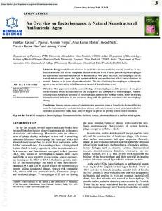
SeprapackTM via the intratympanic route on the RWM was found to reduce both low and high frequency hearing loss associated with trauma of cochlear implantation81. Similarly, nacetyl cysteine with SeprapackTM was shown to be effective to protect the residual hearing after implant surgery in the high frequency hearing region near the point of implantation82. James et al.83 investigated the concentration of dexamethasone in the cochlea after intratympanic delivery with SeprapackTM and found significantly higher and sustained concentration of the drug in the cochlea than that by the drug alone without SeprapackTM. These results were similar to those reported by Borden et al.84 who utilized the thiol-modified HA gelatin hydrogel as a drug delivery system. Saber et al.80 suggested that hyaluronic gel had no toxic effect on the hair cells but found that the gel temporary increased the thickness of RWM. The membrane returned to normal after 4 weeks, indicating that this material caused no permanent structural damage to the RWM. Several other hydrogels were also studied for inner ear drug delivery through the intratympanic route. For example, a hydrogel of glutaraldehyde cross-linked with porcine type-I collagen was examined for the sustained delivery of BDNF to the inner ear. This treatment was shown to provide a protective effect manifested by stable auditory brainstem response (ABR) thresholds and preservation of spiral ganglion neuron densities in animals85. In another study, gelatin hydrogel loaded with hepatocyte growth factor was found to provide sustained drug delivery to the inner ear in guinea pigs86. Saber et al.87 compared the feasibility of three structurally different chitosans as bioadhesives to deliver drugs to the inner ear through the RWM. All these bioadhesive gel formulations provided effective neomycin delivery to the cochlea over 7 days. The chitosans were free from any detectable toxicity on the cochlear tissue. Among the chitosans studied, glycosylated chitosan was considered as a promising biomaterial for inner ear therapy due to its safety and efficiency. Another type of hydrogels for sustained drug delivery to the inner ear is temperature sensitive polymers (e.g., sol–gel polymers). These polymers provide the advantage of solution to gel transition near body temperature. Particularly, the sol–gel polymer with a drug can be delivered as a solution through intratympanic injection using a narrow-gauge needle at room temperature, and the polymer solution then becomes a gel when it reaches the RWM in the middle ear at body temperature. Among these sol– gel polymers, poloxamer solution is liquid at room temperature and forms a gel after intratympanic injection, increasing the residence time of the drug in the middle ear. Salt et al.88 evaluated the potential of a poloxamer hydrogel formulation containing dexamethasone in intratympanic delivery to the RWM of guinea pigs and analyzed the effects of the duration of drug application on drug concentration in the cochlea of guinea pigs and humans. Besides enhancing drug delivery to the inner ear, the increase in residence time of the drug on the RWM could also lead to more uniform drug distribution in the cochlea. Particularly, the drug concentration gradient along the length of the cochlea was shown to be significantly affected by the duration of drug application (i.e., residence time on RWM): higher drug concentration and smaller drug concentration gradient in the cochlea were observed with prolonged drug delivery on the RWM (Fig. 2). In addition to the study by Salt et al.88, Wang et al.89 also utilized poloxamer combined with micronized dexamethasone in inner ear delivery to prolong drug residence in the ear. Paulson et al.90 developed a
Current strategies for drug delivery to the inner ear
Table 3
91
Brief review of recent developments in intratympanic drug delivery.
Drug delivery system
Drug
Significant finding
Ref.
Gelfoams
Gentamicin
78
Gelfoams
Gelfoams
Brain-derived neurotrophic factor (BDNF) Neurotrophins-3
Provided more consistent outcomes with transtympanic delivery of gentamicin, controlled vertigo in 75% of cases and improved tinnitus in 48% of the cases Local BDNF treatment enhanced the survival of cells in basal turn of the cochlea
Hyaluronic gel
Neomycin
SeprapackTM
Dexamethasone
SeprapackTM
N-acetyl cysteine
SeprapackTM
Dexamethasone
Thiol-modified Hyaluronic acid Glutaradehyde cross linking of porcine type-collagen
Dexamethasone Brain-derived neurotrophic factor
Gelatin
Growth factor
Chitosan glycosylated derivative
Neomycin
Poloxamer 407
Dexamethasone
Poloxamer 407
Dexamethasone
Chitosan glycerophosphate hydrogel
Dexamethasone
Nanoparticles Lipid core nanocapsules poly L-lysine (HBPL) nanoparticles Superparamagnetic iron oxide nanoparticles Superparamagnetic iron oxide nanoparticles encapsulated Pluronic F127 copolymer Polymersome Superparamagnetic iron oxide nanoparticles encapsulated PLGA nanoparticles PLGA nanoparticles Silica nanoparticles Lipid nanocapsules
Dextran
Rhodamine
No protective effect on the cells in the cochlea of guinea pigs Had no toxic effect on the hair cells but temporary increased the thickness of RWM Provided protection of hearing across the entire frequency domain (2–32 kHz) Increased the level of residual hearing at 24–32 kHz 4 weeks post-surgery compared to the controls Provided higher and sustained drug concentrations in the cochlear fluid Provided higher and sustained perilymph drug concentration Spiral ganglion neuron (SGN) densities were greater than controls in the basal turn of the cochlea 3 months after implantation Reduced the noise exposure-induced ABR threshold shifts and the loss of outer hair cells in the basal portion of cochlea A safe and effective carrier for inner ear therapy although causes the round window membrane (RWM) to swell Provided more uniform distribution of drug in the inner ear Sustained drug levels in the inner ear for a period of at least 1–2 weeks Provided measurable dexamethasone in perilymph for 5 days, and auditory testing revealed a temporary hearing loss, which resolved by the 10th postoperative day
79
24 80 81 82 83 84 85
86
87
88 89 90
Distributed throughout the human inner ear cell populations through the RWM of human temporal bone Distributed through a 3-cell layer RWM model under an external magnetic field Nanoparticles were seen throughout the inner ear cells
91
Provided specific targeting and binding affinity to SGNs, Schwann cells, and nerve fibers Provided nanoparticle delivery in RWM models (in vitro cell culture, in vivo rat and guinea pig, and in vitro human temporal bone) under a magnetic field Higher distribution of rhodamine in the cochlea Cy3-labeled nanoparticles were found in the sensory hair cells and the SGNs Provided delivery to SGNs, organ of Corti and lateral wall with no hearing impairment, cell death, or morphological changes in the inner ear
94
Gelfoams: gelatin sponge; SeprapackTM: hyaluronic acid–carboxymethlcellulose polymer.
92 93
95
48 96 97,98
92 drug delivery system based on a chitosan glycerophosphate hydrogel, a biodegradable matrix that is temperature-sensitive to achieve sustained delivery of dexamethasone from the RWM over 5 days. The chitosan glycerophosphate hydrogel system was found to be safe, with no identified toxicities or complications from the procedure in a murine model. Ten days after the treatment, hearing thresholds returned to pretreatment baseline levels after an initial transient elevation of the threshold. This temporary hearing loss was suggests to be a result of conductive hearing loss related to the presence of the hydrogel in the middle ear and/or fluid associated with immediate post-operative changes.
4.2.2. Nanoparticles In addition to hydrogel systems, nanoparticles have been studied for drug delivery through the RWM to the inner ear. Because nanoparticles can offer targeted drug delivery to specific cells in the cochlea, they provide certain advantages over conventional drug delivery methods. Roy et al.91 compared the ability of three types of nanoparticles to permeate the RWM on fresh frozen human temporal bone: polymersome nanoparticles of amphiphilic poly (ethylene glycol)-b-poly (e-caprolactone) (PEG-bPCL) block copolymers, lipid core nanocapsules (LNCs) of lecithin and stearate of PEG, and nanoparticles of hyperbranched poly L-lysine (HBPL). It was found that these nanoparticles can pass through the RWM in vitro. Mondalek et al.92 tested a delivery system of superparamagnetic iron oxide nanoparticles (SPIONs) through a 3-cell layer RWM model in vitro. The results showed that SPIONs distributed throughout the model membrane under an external magnetic field. In another study, Thaler et al.93 investigated the capability of ferrogel consisting of SPIONs and Pluronic F127 with an imaging tag for the delivery of therapeutic agents across the RWM of cadaver human temporal bones as well as in organotypic explant cultures of mouse inner ears. It was found that the SPIONs were in the cytoplasm in organotypic explant culture, suggesting that the nanoparticle system can be a suitable cell specific drug delivery vehicle that prevents drug degradation in the cell endolysosomal compartment during drug delivery. Roy et al.94 examined the cell targeting ability and toxicity of nerve growth factor-derived ligand functionalized polymersome nanoparticles for specific cell targeting to SGNs in mouse cochlear
Hongzhuo Liu et al. organotypic culture and observed specific targeting to SGNs, Schwann cells and nerve fibers in the cochlear culture. However, due to the potential difference between RWM permeability in vitro and in vivo, the presence of cochlear clearance in vivo, and the differences between in vitro cell culture models and human cells in vivo, it is unclear if the in vitro permeation and cell culture results can be extrapolated to humans in vivo. With the differences between in vitro and in vivo experiments, in vivo studies of inner ear drug delivery are preferred. Among the nanoparticle studies in vivo, superparamagnetic iron oxide encapsulated in PLGA nanoparticles were investigated and found to distribute throughout all turns of the cochlea of chinchilla95. Tamura et al.48 showed higher distribution of rhodamine to the cochlea after application of rhodamine encapsulated PLGA nanoparticles on RWM as compared to systemic application, illustrating that PLGA nanoparticles can be a useful drug carrier for inner ear delivery. Besides PLGA-based nanoparticles, Praetorius and co-workers reported that silica nanoparticles labeled with fluorescent cyanine dye could be delivered to inner hair cells, vestibular hair cells, and the spiral ganglia by the application of the nanoparticle solution to the RWM96. These nanoparticles were observed not to alter hearing threshold, and no cytotoxicity was found to be associated with the treatment. Zou et al.97 evaluated the ability of LNCs for inner ear drug delivery and found that these nanocapsules rapidly reached the spiral ganglion cells, nerve fibers, hair cells and spiral ligament fibrocytes after placement on the RWM. The biocompatibility of these nanocapsules in inner ear drug delivery was also evaluated. It was shown that the administration of nanocapsules did not cause hearing loss, cell death or morphological changes in the inner ear for up to 28 days after the application98. In summary, nanoparticles are a promising approach for inner ear drug delivery via intratympanic administration, especially for gene delivery because of their cellular uptake properties.
5.
Biotechnology-based approaches of inner ear drug delivery
Biotechnology-based drug delivery has been used extensively in various fields of drug delivery. For inner ear delivery, biological therapies of inner ear disorders have been investigated to provide long-term drug delivery with the advantages
Figure 2 Effect of the duration of dexamethasone (Dex) application to the round window of guinea pig (A) and human (B) upon the concentration gradient of Dex along the length of the cochlea. The data suggest an increase in the contact time of the drug delivery system with the RWM, e.g., via sustained drug delivery systems, can provide a more uniform drug distribution in the cochlea. Symbols: 30 min application, squares; 6 h application, triangles; 24 h application, diamonds. Data were obtained from Salt et al.88.
Current strategies for drug delivery to the inner ear of cell specificity, cell regeneration and/or cell replacement4,58. Biotechnology-based approaches to inner ear treatments include gene and stem cell therapies. 5.1.
damaged sensory epithelia in humans that might be an alternative to gene therapy to reverse profound deafness. In spite of these successes, an effective delivery method for stem cells to the inner ear is needed.
Gene therapy
Studies on the cellular and molecular mechanisms governing hair cell differentiation and regeneration in animal model systems have identified genes that may be targets of genetic manipulation in humans that can lead to protection, replacement, and/or regeneration of functional hair cells, supporting cells, spiral neurons and strial cells99. Until recently, systems for gene transfer to the inner ear have mainly focused on the utility of replication defective viral vectors, including adenovirus, adeno-associated virus (AAV), and herpes virus100. For example, adenoviruses were shown to transfer functional marker genes such as beta-galactosidase (b gal) and green fluorescent protein (GFP) as well as genes that alter the biology of the inner ear, such as glial-derived neurotrophic factor (GDNF), to the auditory system101,102. Besides viral vectors, liposomes have been investigated as a non-viral delivery system but were found to be less efficient in gene transfer than viral vectors100. Recently, Praetorius and coworkers96 suggested that silica nanoparticles might be a potential non-viral vector for the sensory hair cells and spiral ganglion cells in the cochlea and the vestibular organ when the nanoparticles were administered to the round window niche. Other recent development includes the findings by Tan et al. of polycationic-mediated cochlear gene transfer with linear polyethylenimine via cochleostomy and osmotic pump infusion method103 but the polyethylenimine has relatively low transfection efficiency as compared with viral vectors. The identification of gene targeting for inner ear disorder and the demonstration of effective gene transfer for gene delivery have shown promise in gene therapy for inner ear disorders. However, successful cochlear gene delivery relies not only on the gene delivery systems but also on the delivery routes58. Different routes of administration to the cochlea have been investigated with various purposes, such as maximizing transduced efficacy, reducing cochlear toxicity, and/or preserving hearing function. Viral transgenes injected directly into the ST can provide reporter gene expression in SGNs, hair cells, and supporting cells in the organ of Corti104–106. Injection of gene transfer vehicles directly into SM can result in efficient transduction of hair cells and supporting cells107,108. On the other hand, the intratympanic route that requires gene delivery across the RWM by diffusion can preserve the cochlear integrity, but offer less efficient transduction in the inner ear cells62. 5.2.
93
Stem cell therapy
Stem cell therapy to treat hearing loss has recently received attention due to its potential to replace and/or protect hair cells and SGNs after deafness. The feasibility of stem cell therapy in the treatment of inner ear disorders to replace damaged hair cells has been previously reported109. It was suggested that the implantation of embryonic stem cells, fetal dorsal root ganglion and otocyst cells in the inner ear could be used to restore damaged hair cells110,111. These findings for cell transplantation have shown an opportunity to repopulate
6.
Concluding observations and future trends
The prospect of efficacious inner ear drug delivery in the future lies within either the non-invasively administered sustained release systems via the ntratympanic approach or systems in which drugs are administered via targeted and highly efficacious intracochlear routes. This is because systemic drug delivery can lead to systemic adverse effects. Although the use of systemic nanoparticles (via systemic delivery) to achieve greater drug distribution to the cochlea has shown promise, adverse effects from the drugs or nanoparticles in systemic circulation and/or other organs remain a main concern. While the intracochlear approach can deliver drugs directly to the inner ear, it involves high risks such as inner ear trauma and hearing loss. Intratympanic drug delivery can bypass the BLB, resulting in higher drug levels in the inner ear with less systemic drug exposure. This approach is relatively safe compared to intracochlear drug delivery. However, inner ear drug delivery via the intratympanic route relies on drug permeation through the RWM, the barrier between the middle ear and inner ear. In addition, intratympanic drug delivery to the inner ear can be significantly affect by the residence time of drugs in contact with RWM, which in turn can impact the clinical outcome. The main problem encountered in intratympanic drug delivery is the variability in drug delivery and unpredictable bioavailability via this route, partly due to the variable residence time of drugs in the middle ear. To overcome this problem, various sustained drug release systems have been employed to increase the residence time of drugs in the middle ear and prolong drug contact with the RWM. This includes the use of polymers, hydrogels, and nanoparticles as the sustained release systems. Another problem in intratympanic drug delivery is related to the rapid clearance of drugs in the cochlea, resulting in low drug concentration in the apical section of the cochlea. Hence, it is more difficult to treat hearing loss of low frequencies via this route. Reports on inner ear drug delivery systems have suggested that nanoparticles loaded with drugs can be distributed broadly in the inner ear and enhance the delivery of drugs to the hair cells as well as spiral neutron ganglions. The results in these studies are encouraging and suggest that this approach can be the future of inner ear disorder treatment. Gene delivery and stem cell transplantation are also promising approaches that can be used to treat inner ear diseases. While recent studies using virus vectors for gene delivery have shown no significant toxicity, this risk remains a potential problem in clinical application because the inner ear is delicate and is located near the brain. Thus, until a complete understanding of the safety profiles of virus vectors, non-virus vectors might be better suited in gene transfer in the ear for clinical use. In addition, there is a lack of effective methods to deliver these gene delivery systems and stem cells to the inner ear, which needs to be addressed. In order to develop an effective intratympanic drug delivery system, an adequate evaluation method for inner ear drug
94
Hongzhuo Liu et al.
delivery is required. To date, researchers have employed both in vitro and in vivo methods to investigate drug delivery systems. For in vitro studies, generally, their purpose is to characterize the physical properties of the drug delivery systems before the in vivo animal studies. Consequently, the ability of in vitro studies to predict in vivo results through, e.g., in vitro/in vivo correlation, is important. However, there are few reports comparing the in vitro and in vivo methods. Particularly, potential differences between the RWM permeability in vitro and in vivo and the lack of cochlear clearance in vitro can be significant factors in the prediction of perilymph pharmacokinetics. Future research and development of an effective evaluation method for inner ear drug delivery are needed. For in vivo studies, guinea pigs are commonly used as an animal model since the morphology of guinea pig inner ear is similar to that of human. However, the interpretation of the pharmacokinetic data in guinea pigs requires the consideration of the routes by which drugs entered the cochlea and the distance from the basal turn to the apical turn in guinea pigs compared to humans, which could influence drug distribution in the cochlea. Accurate measurements of drug levels in the inner ear fluids can also be complicated. Considering the small volume of cochlear fluid in guinea pigs (i.e., less than 10 mL), the determination of drug concentration at different locations in the cochlea (e.g., different turns of the cochlea) can present a technical problem and be a major source of errors in advancing our understanding of perilymph pharmacokinetics. Thus, caution must be exercised in the analysis of the pharmacokinetic results in the literature and the generalization of the results from in vitro evaluation methods and in vivo animal models to humans. Besides the lack of an established method in inner ear drug pharmacokinetics research, the toxicity and safety of new inner ear drug delivery systems are generally not well established. The inner ear is a complicated and subtle organ. Adverse effects and complications in inner ear drug delivery can lead to severe side effects such as hearing loss. Potential ototoxicity of novel drug delivery systems should be carefully examined. To be truly clinically useful, the drug delivery systems should be both effective and safe, short of any major risks, during inner ear disease treatment. These are the current major hurdles of effective drug delivery to the inner ear.
References 1. Swan EE, Mescher MJ, Sewell WF, Tao SL, Borenstein JT. Inner ear drug delivery for auditory applications. Adv Drug Deliv Rev 2008;60:1583–99. 2. Borkholder DA. State-of-the-art mechanisms of intracochlear drug delivery. Curr Opin Otolaryngol Head Neck Surg 2008;16:472–7. 3. Salt AN, Plontke SK. Principles of local drug delivery to the inner ear. Audiol Neurootol 2009;14:350–60. 4. McCall AA, Swan EE, Borenstein JT, Sewell WF, Kujawa SG, McKenna MJ. Drug delivery for treatment of inner ear disease: current state of knowledge. Ear Hear 2010;31:156–65. 5. Staecker H, Jolly C, Garnham C. Cochlear implantation: an opportunity for drug development. Drug Discov Today 2010;15:314–21. 6. Chen G, Zhang X, Yang F, Mu L. Disposition of nanoparticlebased delivery system via inner ear administration. Curr Drug Metab 2010;11:886–97.
7. Borenstein JT. Intracochlear drug delivery systems. Expert Opin Drug Deliv 2011;8:1161–74. 8. Nakagawa T, Ito J. Local drug delivery to the inner ear using biodegradable materials. Ther Deliv 2011;2:807–14. 9. Mukherjea D, Rybak LP, Sheehan KE, Kaur T, Ramkumar V, Jajoo S, et al. The design and screening of drugs to prevent acquired sensorineural hearing loss. Expert Opin Drug Discov 2011;6:491–505. 10. Pyykko I, Zou J, Zhang W, Zhang Y. Nanoparticle-based delivery for the treatment of inner ear disorders. Curr Opin Otolaryngol Head Neck Surg 2011;19:388–96. 11. Rivera T, Sanz L, Camarero G, Varela-Nieto I. Drug delivery to the inner ear: strategies and their therapeutic implications for sensorineural hearing loss. Curr Drug Deliv 2012;9:231–42. 12. Pararas EE, Borkholder DA, Borenstein JT. Microsystems technologies for drug delivery to the inner ear. Adv Drug Deliv Rev 2012;64:1650–60. 13. Jahnke K. Permeability barriers of the inner ear. Fine structure and function. Fortschr Med 1980;98:330–6. 14. Jahnke K. The blood–perilymph barrier. Arch Otorhinolaryngol 1980;228:29–34. 15. Juhn SK. Barrier systems in the inner ear. Acta Otolaryngol Suppl 1988;458:79–83. 16. Juhn SK, Rybak LP. Labyrinthine barriers and cochlear homeostasis. Acta Otolaryngol 1981;91:529–34. 17. Saito T, Zhang ZJ, Tokuriki M, Ohtsubo T, Noda I, Shibamori Y, et al. Expression of p-glycoprotein is associated with that of multidrug resistance protein 1 (MRP1) in the vestibular labyrinth and endolymphatic sac of the guinea pig. Neurosci Lett 2001;303:189–92. 18. Yamasoba T, Suzuki M, Kaga K. Influence of chronic kanamycin administration on basement membrane anionic sites in the labyrinth. Hear Res 1996;102:116–24. 19. Suzuki M, Yamasoba T, Ishibashi T, Miller JM, Kaga K. Effect of noise exposure on blood-labyrinth barrier in guinea pigs. Hear Res 2002;164:12–8. 20. Kastenbauer S, Klein M, Koedel U, Pfister HW. Reactive nitrogen species contribute to blood–labyrinth barrier disruption in suppurative labyrinthitis complicating experimental pneumococcal meningitis in the rat. Brain Res 2001;904:208–17. 21. McFadden SL, Ding D, Jiang H, Woo JM, Salvi RJ. Chinchilla models of selective cochlear hair cell loss. Hear Res 2002;174:230–8. 22. Tran Ba Huy P, Manuel C, Meulemans A, Sterkers O, Wassef M, Amiel C. Ethacrynic acid facilitates gentamicin entry into endolymph of the rat. Hear Res 1983;11:191–202. 23. Juhn SK, Prado S, Pearce J. Osmolality changes in perilymph after systemic administration of glycerin. Arch Otolaryngol 1976;102:683–5. 24. Richardson RT, Noushi F, O’Leary S. Inner ear therapy for neural preservation. Audiol Neurootol 2006;11:343–56. 25. Banerjee A, Parnes LS. The biology of intratympanic drug administration and pharmacodynamics of round window drug absorption. Otolaryngol Clin North Am 2004;37:1035–51. 26. Goycoolea MV, Lundman L. Round window membrane. Structure function and permeability: a review. Microsc Res Tech 1997;36:201–11. 27. Salt AN, Plontke SK. Local inner-ear drug delivery and pharmacokinetics. Drug Discov Today 2005;10:1299–306. 28. Kim CS, Cho TK, Jinn TH. Permeability of the round window membrane to horseradish peroxidase in experimental otitis media. Otolaryngol Head Neck Surg 1990;103:918–25. 29. Hamaguchi Y, Morizono T, Juhn SK. Round window membrane permeability to human serum albumin in antigen-induced otitis media. Am J Otolaryngol 1988;9:34–40. 30. Hoft J. The permeability of the round window membrane and its changes by pantocaine (tetracaine). Arch Klin Exp Ohren Nasen Kehlkopfheilkd 1969;193:128–37.
Current strategies for drug delivery to the inner ear 31. Ikeda K, Morizono T. Changes of the permeability of round window membrane in otitis media. Arch Otolaryngol Head Neck Surg 1988;114:895–7. 32. Chandrasekhar SS, Rubinstein RY, Kwartler JA, Gatz M, Connelly PE, Huang E, et al. Dexamethasone pharmacokinetics in the inner ear: comparison of route of administration and use of facilitating agents. Otolaryngol Head Neck Surg 2000;122:521–8. 33. Mikulec AA, Hartsock JJ, Salt AN. Permeability of the round window membrane is influenced by the composition of applied drug solutions and by common surgical procedures. Otol Neurotol 2008;29:1020–6. 34. Bowe SN, Jacob A. Round window perfusion dynamics: implications for intracochlear therapy. Curr Opin Otolaryngol Head Neck Surg 2010;18:377–85. 35. Lasak JM, Sataloff RT, Hawkshaw M, Carey TE, Lyons KM, Spiegel JR. Autoimmune inner ear disease: steroid and cytotoxic drug therapy. Ear Nose Throat J 2001;80:808–11. 815–6,818 passim. 36. Wei BP, Mubiru S, O’Leary S. Steroids for idiopathic sudden sensorineural hearing loss. Cochrane Database Syst Rev 2006;25:CD003998. 37. Sakamoto T, Nakagawa T, Horie RT, Hiraumi H, Yamamoto N, Kikkawa YS, et al. Inner ear drug delivery system from the clinical point of view. Acta Otolaryngol Suppl 2010;563:101–4. 38. Murai K, Tyler RS, Harker LA, Stouffer JL. Review of pharmacologic treatment of tinnitus. Am J Otol 1992;13:454–64. 39. Berryhill WE, Graham MD. Chemical and physical labyrinthectomy for Meniere’s disease. Otolaryngol Clin North Am 2002;35: 675–82. 40. Sataloff RT, McCarter A, Spiegel JR. Very high-dose streptomycin labyrinthectomy. Ear Nose Throat J 1996;75:239–43. 41. Sajjadi H, Paparella MM. Meniere’s disease. Lancet 2008;372:406–14. 42. Lautermann J, McLaren J, Schacht J. Glutathione protection against gentamicin ototoxicity depends on nutritional status. Hear Res 1995;86:15–24. 43. Sha SH, Schacht J. Salicylate attenuates gentamicin-induced ototoxicity. Lab Invest 1999;79:807–13. 44. Fetoni AR, Sergi B, Ferraresi A, Paludetti G, Troiani D. alphaTocopherol protective effects on gentamicin ototoxicity: an experimental study. Int J Audiol 2004;43:166–71. 45. Fetoni AR, Sergi B, Scarano E, Paludetti G, Ferraresi A, Troiani D. Protective effects of alpha-tocopherol against gentamicin-induced Oto-vestibulo toxicity: an experimental study. Acta Otolaryngol 2003;123:192–7. 46. Unal OF, Ghoreishi SM, Atas A, Akyurek N, Akyol G, Gursel B. Prevention of gentamicin induced ototoxicity by trimetazidine in animal model. Int J Pediatr Otorhinolaryngol 2005;69:193–9. 47. Long M, Smouha EE, Qiu D, Li F, Johnson F, Luft B. Flavanoid of Drynaria fortunei protects against gentamicin ototoxicity. Phytother Res 2004;18:609–14. 48. Tamura T, Kita T, Nakagawa T, Endo T, Kim TS, Ishihara T, et al. Drug delivery to the cochlea using PLGA nanoparticles. Laryngoscope 2005;115:2000–5. 49. Horie RT, Sakamoto T, Nakagawa T, Ishihara T, Higaki M, Ito J. Stealth-nanoparticle strategy for enhancing the efficacy of steroids in mice with noise-induced hearing loss. Nanomedicine (Lond) 2010;5:1331–40. 50. Ersner MS, Spiegel EA, Alexander MH. Transtympanic injection of anesthetics for the treatment of Meniere’s syndrome. AMA Arch Otolaryngol 1951;54:43–52. 51. Lange G. Gentamicin and other ototoxic antibiotics for the transtympanic treatment of Meniere’s disease. Arch Otorhinolaryngol 1989;246:269–70. 52. Blakley BW. Clinical forum: are view of in tratympanic therapy. Am J Otol 1997;18:520-6; discussion 527–31.
95 53. Barrs DM. Intratympanic corticosteroids for Meniere’s disease and vertigo. Otolaryngol Clin North Am 2004;37:955–72. [v]. 54. Dodson KM, Woodson E, Sismanis A. Intratympanic steroid perfusion for the treatment of Meniere’s disease: a retrospective study. Ear Nose Throat J 2004;83:394–8. 55. Barrs DM, Keyser JS, Stallworth C, McElveen Jr. JT. Intratympanic steroid injections for intractable Meniere’s disease. Laryngoscope 2001;111:2100–4. 56. Banerjee A, Parnes LS. Intratympanic corticosteroids for sudden idiopathic sensorineural hearing loss. Otol Neurotol 2005;26:878–81. 57. Rauch SD. Intratympanic steroids for sensorineural hearing loss. Otolaryngol Clin N Am 2004;37:1061–74. 58. Richardson RT, Wise AK, Andrew JK, O’Leary SJ. Novel drug delivery systems for inner ear protection and regeneration after hearing loss. Expert Opin Drug Deliv 2008;5:1059–76. 59. Plontke SK, Mynatt R, Gill RM, Borgmann S, Salt AN. Concentration gradient along the scala tympani after local application of gentamicin to the round window membrane. Laryngoscope 2007;117:1191–8. 60. Plontke SK, Wood AW, Salt AN. Analysis of gentamicin kinetics in fluids of the inner ear with round window administration. Otol Neurotol 2002;23:967–74. 61. Salt AN, Ma Y. Quantification of solute entry into cochlear perilymph through the round window membrane. Hear Res 2001;154:88–97. 62. Stover T, Yagi M, Raphael Y. Cochlear gene transfer: round window versus cochleostomy inoculation. Hear Res 1999;136:124–30. 63. Rejali D, Lee VA, Abrashkin KA, Humayun N, Swiderski DL, Raphael Y. Cochlear implants and ex vivo BDNF gene therapy protect spiral ganglion neurons. Hear Res 2007;228:180–7. 64. Richardson RT, Thompson B, Moulton S, Newbold C, Lum MG, Cameron A, et al. The effect of polypyrrole with incorporated neurotrophin-3 on the promotion of neurite outgrowth from auditory neurons. Biomaterials 2007;28:513–23. 65. Pararas EE, Chen Z, Fiering J, Mescher MJ, Kim ES, McKenna MJ, et al. Kinetics of reciprocating drug delivery to the inner ear. J Control Release 2011;152:270–7. 66. Sewell WF, Borenstein JT, Chen Z, Fiering J, Handzel O, Holmboe M, et al. Development of a microfluidics-based intracochlear drug delivery device. Audiol Neurootol 2009;14:411–22. 67. Adunka O, Unkelbach MH, Mack M, Hambek M, Gstoettner W, Kiefer J. Cochlear implantation via the round window membrane minimizes trauma to cochlear structures: a histologically controlled insertion study. Acta Otolaryngol 2004;124:807–12. 68. Nadol Jr. JB, Shiao JY, Burgess BJ, Ketten DR, Eddington DK, Gantz BJ, et al. Histopathology of cochlear implants in humans. Ann Otol Rhinol Laryngol 2001;110:883–91. 69. Hochman J, Blakley B, Abdoh A, Aleid H. Post-tympanostomy tube otorrhea: a meta-analysis. Otolaryngol Head Neck Surg 2006;135:8–11. 70. Licameli G, Johnston P, Luz J, Daley J, Kenna M. Phosphorylcholine-coated antibiotic tympanostomy tubes: are post-tube placement complications reduced?. Int J Pediatr Otorhinolaryngol 2008;72:1323–8. 71. Light JP, Silverstein H. Transtympanic perfusion: indicati ons and limitations. Curr Opin Otolaryngol Head Neck Surg 2004;12:378–83. 72. Silverstein H. Use of a new device, the MicroWick, to deliver medication to the inner ear. Ear Nose Throat J 1999;78:595–8. 600. 73. Hill 3rd SL, Digges EN, Silverstein H. Long-term follow-up after gentamicin application via the Silverstein MicroWick in the treatment of Meniere’s disease. Ear Nose Throat J 2006;85:494. 96, 98. 74. DeCicco MJ, Hoffer ME, Kopke RD, Wester D, Allen KA, Gottshall K, et al. Round-window microcatheter-administered
96
75.
76.
77.
78.
79.
80.
81.
82.
83.
84.
85.
86.
87.
88.
89.
90.
91.
Hongzhuo Liu et al. microdose gentamicin: results from treatment of tinnitus associated with Meniere’s disease. Int Tinnitus J 1998;4:141–3. Plontke S, Lowenheim H, Preyer S, Leins P, Dietz K, Koitschev A, et al. Outcomes research analysis of continuous intratympanic glucocorticoid delivery in patients with acute severe to profound hearing loss: basis for planning randomized controlled trials. Acta Otolaryngol 2005;125:830–9. Lefebvre PP, Staecker H. Steroid perfusion of the inner ear for sudden sensorineural hearing loss after failure of conventional therapy: a pilot study. Acta Otolaryngol 2002;122:698–702. Marks S, Arenberg IK, Hoffer ME. Round window microcatheter administered microdose of gentamycin: an alternative in the treatment of tinnitus in patients with Meniere’s disease. Laryngorhinootologie 2000;79:327–31. Silverstein H, Arruda J, Rosenberg SI, Deems D, Hester TO. Direct round window membrane application of gentamicin in the treatment of Meniere’s disease. Otolaryngol Head Neck Surg 1999;120:649–55. Havenith S, Versnel H, Agterberg MJ, de Groot JC, Sedee RJ, Grolman W, et al. Spiral ganglion cell survival after round window membrane application of brain-derived neurotrophic factor using gelfoam as carrier. Hear Res 2011;272:168–77. Saber A, Laurell G, Bramer T, Edsman K, Engmer C, Ulfendahl M. Middle ear application of a sodium hyaluronate gel loaded with neomycin in a Guinea pig model. Ear Hear 2009;30:81–9. Eastwood H, Chang A, Kel G, Sly D, Richardson R, O’Leary SJ. Round window delivery of dexamethasone ameliorates local and remote hearing loss produced by cochlear implantation into the second turn of the guinea pig cochlea. Hear Res 2010;265:25–9. Eastwood H, Pinder D, James D, Chang A, Galloway S, Richardson R, et al. Permanent and transient effects of locally delivered n-acetyl cysteine in a guinea pig model of cochlear implantation. Hear Res 2010;259:24–30. James DP, Eastwood H, Richardson RT, O’Leary SJ. Effects of round window dexamethasone on residual hearing in a Guinea pig model of cochlear implantation. Audiol Neurootol 2008;13:86–96. Borden RC, Saunders JE, Berryhill WE, Krempl GA, Thompson DM, Queimado L. Hyaluronic acid hydrogel sustains the delivery of dexamethasone across the round window membrane. Audiol Neurootol 2011;16:1–11. Endo T, Nakagawa T, Kita T, Iguchi F, Kim TS, Tamura T, et al. Novel strategy for treatment of inner ears using a biodegradable gel. Laryngoscope 2005;115:2016–20. Inaoka T, Nakagawa T, Kikkawa YS, Tabata Y, Ono K, Yoshida M, et al. Local application of hepatocyte growth factor using gelatin hydrogels attenuates noise-induced hearing loss in guinea pigs. Acta Otolaryngol 2009;129:453–7. Saber A, Strand SP, Ulfendahl M. Use of the biodegradable polymer chitosan as a vehicle for applying drugs to the inner ear. Eur J Pharm Sci 2010;39:110–5. Salt AN, Hartsock J, Plontke S, LeBel C, Piu F. Distribution of dexamethasone and preservation of inner ear function following intratympanic delivery of a gel-based formulation. Audiol Neurootol 2011;16:323–35. Wang X, Dellamary L, Fernandez R, Harrop A, Keithley EM, Harris JP, et al. Dose-dependent sustained release of dexamethasone in inner ear cochlear fluids using a novel local delivery approach. Audiol Neurootol 2009;14:393–401. Paulson DP, Abuzeid W, Jiang H, Oe T, O’Malley BW, Li D. A novel controlled local drug delivery system for inner ear disease. Laryngoscope 2008;118:706–11. Roy S, Glueckert R, Johnston AH, Perrier T, Bitsche M, Newman TA, et al. Strategies for drug delivery to the human inner ear by multifunctional nanoparticles. Nanomedicine (Lond) 2012;7:55–63.
92. Mondalek FG, Zhang YY, Kropp B, Kopke RD, Ge X, Jackson RL, et al. The permeability of SPION over an artificial threelayer membrane is enhanced by external magnetic field. J Nanobiotechnol 2006;4:4. 93. Thaler M, Roy S, Fornara A, Bitsche M, Qin J, Muhammed M, et al. Visualization and analysis of superparamagnetic iron oxide nanoparticles in the inner ear by light microscopy and energy filtered TEM. Nanomedicine 2011;7:360–9. 94. Roy S, Johnston AH, Newman TA, Glueckert R, Dudas J, Bitsche M, et al. Cell-specific targeting in the mouse inner ear using nanoparticles conjugated with a neurotrophin-derived peptide ligand: potential tool for drug delivery. Int J Pharm 2010;390:214–24. 95. Ge X, Jackson RL, Liu J, Harper EA, Hoffer ME, Wassel RA, et al. Distribution of PLGA nanoparticles in chinchilla cochleae. Otolaryngol Head Neck Surg 2007;137:619–23. 96. Praetorius M, Brunner C, Lehnert B, Klingmann C, Schmidt H, Staecker H, et al. Transsynaptic delivery of nanoparticles to the central auditory nervous system. Acta Otolaryngol 2007;127:486–90. 97. Zou J, Saulnier P, Perrier T, Zhang Y, Manninen T, Toppila E, et al. Distribution of lipid nanocapsules in different cochlear cell populations after round window membrane permeation. J Biomed Mater Res B Appl Biomater 2008;87:10–8. 98. Zhang Y, Zhang W, Lobler M, Schmitz KP, Saulnier P, Perrier T, et al. Inner ear biocompatibility of lipid nanocapsules after round window membrane application. Int J Pharm 2011;404:211–9. 99. Tang LS, Montemayor C, Pereira FA. Sensorineural hearing loss: potential therapies and gene targets for drug development. IUBMB Life 2006;58:525–30. 100. Staecker H, Brough DE, Praetorius M, Baker K. Drug delivery to the inner ear using gene therapy. Otolaryngol Clin North Am 2004;37:1091–108. 101. Raphael Y, Frisancho JC, Roessler BJ. Adenoviral-mediated gene transfer into guinea pig cochlear cells in vivo. Neurosci Lett 1996;207:137–41. 102. Yagi M, Magal E, Sheng Z, Ang KA, Raphael Y. Hair cell protection from aminoglycoside ototoxicity by adenovirusmediated overexpression of glial cell line-derived neurotrophic factor. Hum Gene Ther 1999;10:813–23. 103. Tan BT, Foong KH, Lee MM, Ruan R. Polyethyleniminemediated cochlear gene transfer in guinea pigs. Arch Otolaryngol Head Neck Surg 2008;134:884–91. 104. Li Duan M, Bordet T, Mezzina M, Kahn A, Ulfendahl M. Adenoviral and adeno-associated viral vector mediated gene transfer in the guinea pig cochlea. Neuroreport 2002;13:1295–9. 105. Praetorius M, Knipper M, Schick B, Tan J, Limberger A, Carnicero E, et al. A novel vestibular approach for gene transfer into the inner ear. Audiol Neurootol 2002;7:324–34. 106. Luebke AE, Steiger JD, Hodges BL, Amalfitano A. A modified adenovirus can transfect cochlear hair cells in vivo without compromising cochlear function. Gene Ther 2001;8:789–94. 107. Ishimoto S, Kawamoto K, Kanzaki S, Raphael Y. Gene transfer into supporting cells of the organ of Corti. Hear Res 2002;173:187–97. 108. Wenzel GI, Xia A, Funk E, Evans MB, Palmer DJ, Ng P, et al. Helper-dependent adenovirus-mediated gene transfer into the adult mouse cochlea. Otol Neurotol 2007;28:1100–8. 109. Kojima K, Murata M, Nishio T, Kawaguchi S, Ito J. Survival of fetal rat otocyst cells grafted into the damaged inner ear. Acta Otolaryngol Suppl 2004;551:53–5. 110. Tateya I, Nakagawa T, Iguchi F, Kim TS, Endo T, Yamada S, et al. Fate of neural stem cells grafted into injured inner ears of mice. Neuroreport 2003;14:1677–81. 111. Hu Z, Wei D, Johansson CB, Holmstrom N, Duan M, Frisen J, et al. Survival and neural differentiation of adult neural stem cells transplanted into the mature inner ear. Exp Cell Res 2005;302:40–7.