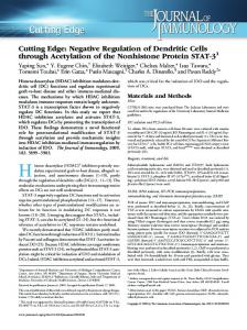
●
Cutting Edge: Myeloid Complement C3 Enhances the Humoral Response To Peripheral Viral Infection1 Admar Verschoor,*§ Mark A. Brockman,‡ David M. Knipe,‡ and Michael C. Carroll2†§
HSV-1 is the causative agent of cutaneous lesions, commonly referred to as cold sores. Primary exposure to the virus ordinarily occurs through the periphery, in particular through abraded skin or mucosal membranes. Under certain circumstances (e.g., in neonatals or AIDS patients), the infection becomes disseminated, often with severe consequences. Spread of HSV-1 is limited by virus-specific Ab. The development of an efficient humoral response to the virus is dependent on innate immunity component complement C3. The liver is the major source of C3, but there are also extrahepatic origins of C3 such as lymphoid macrophages. In the present study, the significance of C3 synthesis by bone marrow-derived cells was assessed by the transfer of wild-type bone marrow into irradiated C3-deficient mice. Using these chimeric mice, extrahepatic C3 was determined sufficient to initiate specific Ab and memory responses to a peripheral HSV-1 infection. The Journal of Immunology, 2001, 167: 2446 –2451.
H
erpes simplex virus 1 infects its host through mucosal surfaces or abraded skin. Following primary infection, HSV-1 can establish a life-long latent infection within the sensory neurons that innervate the peripheral site. The virus can reactivate from latency, resulting in a recurrent disease at or near the initial site of infection. The presence of oral-facial epithelial lesions, commonly referred to as cold sores or fever blisters, is a widely recognized symptom of recurrence (1). HSV infection can in some cases spread beyond the sensory neurons to produce a disseminated infection. This is particularly the case in neonates or immunocompromised individuals, such as AIDS patients or organ transplant recipients, where multiorgan involvement or infection of the CNS can lead to high mortality (2–9). In various studies, the presence of anti-HSV-1 serum Ab has been shown to protect against complications of HSV infection, presumably through limiting systemic viral spread (10 –12). Specific Ab elim-
Departments of *Pathology, †Pediatrics, and ‡Microbiology and Molecular Genetics, Harvard Medical School; and §The Center for Blood Research, Boston, MA 02115 Received for publication June 6, 2001. Accepted for publication July 6, 2001. The costs of publication of this article were defrayed in part by the payment of page charges. This article must therefore be hereby marked advertisement in accordance with 18 U.S.C. Section 1734 solely to indicate this fact. 1
inates virus by Ab-dependent cellular cytotoxicity and viral neutralization, which can be complement dependent (13) or independent (14 –16). However, specific Ab titers are not associated with clearance of HSV-1 infection, and latent virus can persist in its host. Humoral responses to HSV-1 are thymus-dependent (TD)3; Th cells are required to provide a costimulatory signal to activate B cells. Cytokine profiles and IgG isotype patterns indicate that T lymphocyte help in HSV-1 infection is skewed toward Th-1 (17, 18). The humoral response to HSV-1 is dependent on the complement system. Deficiencies in complement component C3, C4 (required to activate C3), or its receptor (CD21/CD35) result in impaired humoral responses to infectious virus (19). The importance of the innate immune system, including the complement system, in the adaptive immune response has only recently become fully appreciated (20 –24). The development of knockout mice for complement protein C3 (C3!/!) or complement receptors CD21 and CD35 (Cr2!/!) allowed for detailed studies of the impaired responses. C3!/! mice were found to have an impairment at the B lymphocyte level, as T lymphocyte responses appeared normal (25). Deficiency in CD21/CD35 resulted in impaired B cell activation and secondary humoral response (26, 27). Activation and covalent attachment of C3 to Ag enhances the B cell response by at least two mechanisms. First, coligation of the CD21/CD19/CD81 coreceptor by C3-Ag adducts lowers the threshold for B cell activation both in vivo and ex vivo (28, 29) and enhances the survival of activated B cells within follicles and germinal centers (GC) (30). Second, complement receptor-dependent retention of Ag on follicular dendritic cells (FDC) provides a source of Ag for clonal selection within GC (24, 31, 32). Direct coupling of C3d to hen egg lysozyme protein lowered the amount of protein required for an optimal response by as much as 10,000fold (33). Similarly, coupling C3d to influenza hemagglutinin in a DNA vaccine enhanced the humoral response (34). These combined experiments clearly underlined the role of complement in the humoral response to Ag. Complement C3 is produced at a variety of sites in the body. The liver is the primary source of C3 with serum concentrations of "1 mg/ml (35). However, a number of extrahepatic sources of C3 synthesis have been described. These include myeloid lineage cells such as monocytic cells (36) and polymorphonuclear leukocytes (37) as well as non-bone marrow (non-BM)-derived sources such as epidermal keratinocytes (38),
This work was supported by National Institutes of Health Grant AI-42257.
2
Address correspondence and reprint requests to Dr. Michael C. Carroll, Center for Blood Research, 200 Longwood Avenue, Boston, MA 02115. E-mail address:
[email protected] Copyright © 2001 by The American Association of Immunologists
●
3 Abbreviations used in this paper: TD, thymus-dependent; GC, germinal center; BM, bone marrow; WT, wild type; FDC, follicular dendritic cells; i.d., intradermal(ly).
0022-1767/01/$02.00
The Journal of Immunology kidney tubular epithelial cells (39, 40), and umbilical vein endothelial cells (41). The potential of macrophage-derived C3 in the humoral response to protein Ag was determined using C3!/! BM chimeras that received wild-type (WT) BM (WT BM3 C3!/!). Significantly, the humoral response to protein Ags introduced i.v. was fully restored in the absence of circulating C3. Combined immunohistochemistry and in situ hybridization strongly indicated MOMA2# macrophages, located in the splenic white pulp, as major producers of C3 mRNA (31). In the present study, C3!/! mice reconstituted with WT BM were used to assess the importance of myeloid-derived C3 in the more physiologically relevant model of intradermal (i.d.) HSV-1 infection. Our results identify nonsystemic BM-derived C3 as sufficient to restore full humoral responses to peripheral infection with HSV-1 in the absence of systemic C3. In complement C3 chimeras (WT BM3 C3!/!), C3 synthesis and deposition were detected in lymphoid organs. These findings indicate a critical role for myeloid-produced C3 in enhancement of B cell activation and production of viral specific Ab.
Materials and Methods Mice Mice deficient in complement component C3 (C3!/!) were constructed by gene targeting using homologous recombination in embryonic stem cells as previously reported (42), and were maintained on C57BL/6 or mixed C57BL/6 $ 129/sv backgrounds. C57BL/6 or mixed C57BL/6 $ 129/sv mice were used as WT controls. Although C57B/6 mice are considered to be resistant to HSV infection, the host immune response by resistant and susceptible mouse strains (i.e., BALB/c) has been shown to be identical (43). All mice were housed in a specific pathogen-free facility. Studies were performed according to institutional guidelines for animal use and care. Animals received food and water ad libitum.
Construction of chimeras BM cells of littermates were isolated by flushing their femurs and tibias with cold HBSS (Life Technologies, Grand Island, NY) using a 26-gauge needle. Via retro orbital sinus injection, 107 BM cells were introduced into anesthetized (300 !l Avertin, i.p.; mixture of 2,2,2-tribromoethanol and 2-methyl-2-butanol (Sigma, St. Louis, MO)) 6- to 8-wk-old mice that were previously lethally irradiated (2 $ 650 rad). Reconstituted mice were allowed to rest for 5 wk before they were used in experiments.
Virus and inoculations KOS 1.1, which has been used as a WT HSV-1 strain in immunization studies (19, 44) because it is attenuated but very immunogenic, was propagated and titrated on VERO cells. Virus stocks were concentrated by pelleting and resuspending extracellular virions in DMEM (Life Technologies) with 1% FCS and 15% glycerol (Sigma, St. Louis, MO) and then stored at !80°C. Mice were inoculated i.d. with 50 !l containing 2 $ 106 PFU in the rear flank near the base of the tail. Mice received identical second and third inoculations at wk 3 and 6 postprimary infection. Some groups of mice were inoculated with an equivalent volume and quantity of UV-irradiated virus. UV treatment reduced the viral titer by at least 2000-fold.
ELISA Plates were coated with HSV-1 Ag (Advanced Biotechnologies, Columbia, MD) overnight at 4°C at 50 ng/well in 0.05 M carbonate-bicarbonate buffer, pH 9.6 (Sigma) on 96-well plates. Plates were blocked with PBS containing 5% milk at 37°C for 1 h, then washed three times with PBS containing 5% milk and 0.05% Tween 20. Plates were incubated with serial 2-fold dilutions of mouse sera for 2 h at 37°C, washed, then incubated for 1 h with 1/1000 goat anti-mouse IgG Ab conjugated to alkaline phosphatase (Sigma), washed, and developed using SigmaFast pNitrophenyl Phosphate (Sigma), and the OD405 was read. Ab titers represent the final 2-fold dilution yielding an OD reading greater than 0.2 U above background and are expressed as the geometric mean % SEM. Similarly, C3 serum titers were determined by ELISA. Mice were bled, and blood was kept at 4°C. Serum was obtained by centrifuging for 15 min at 4°C at 15,000 $ g. Supernatant was applied for 2 h at 37°C to 96-well plates that had been coated overnight at 4°C with rat anti-mouse C3 mAb (provided by Dr. E.
2447 Kremmer, GSF-National Research Center for Environment and Health, Institute of Immunology, Munich, Germany) and blocked with 1% BSA in PBS for 1 h at 37°C. For detection, biotinylated polyclonal goat anti-mouse C3 (CAPPEL ICN, Costa Mesa, CA) was applied for 1 h at 37°C. After applying streptavidin-alkalin phosphatase (Sigma), enzymatic detection was performed with SigmaFast and OD405 values were compared with a standard of pooled WT mouse serum.
C3 production C3 production was detected using in situ hybridization specific for C3 mRNA. In situ hybridization was used to confirm the presence of C3 producing BM-derived cells in the lymphoid compartment and was performed as previously described (31).
C3 localization Localization of C3 was analyzed by immunohistochemistry on OCTembedded (Tissue-Tek) cryosections (Sakura Finetek, Torrance, CA). Lymph nodes were harvested and snap-frozen in an OCT-filled mold on a liquid nitrogen-cooled metal surface. Samples were stored at !80°C until the cryosections were cut. Cross-reactive FITC-conjugated rabbit antihuman complement C3d polyclonal Ab (DAKO, Carpinteria, CA) was used to detect mouse C3d on sections. Specific signal enhancement for this Ab was obtained by use of the Alexa Fluor 488 Signal-amplification Kit for Fluorescein-conjugated probes (Molecular Probes, Eugene, OR). GC were detected with biotinylated peanut agglutinin (EY Laboratories, San Mateo, CA) with secondary staining by Streptavidin- Cy-Chrome (BD PharMingen, San Diego, CA). T lymphocytes were stained using PE-labeled purified anti-mouse CD3" (CD3 "-chain) (BD PharMingen).
Results Reconstitution of C3-deficient mice with WT BM restores humoral responses to peripheral infection We infected mice with 2 $ 106 PFU of HSV-1 i.d., and re-exposed them to equal doses at wk 3 and 6 postprimary infection. At weekly intervals throughout the experimental period, sera from the mice were analyzed for their specific anti HSV-1 IgG titers. In WT mice, virus-specific IgG levels followed the well-characterized trend for a humoral response to a TD Ag or virus (Fig. 1A). By contrast, C3!/! animals responded with highly impaired primary and secondary IgG responses to HSV-1. This phenomenon in C3!/! mice has been reported both for inert (25) as well as infectious TD (19) and TI (45) agents (Fig. 1A). To examine whether the humoral immune response to a peripheral infection depends on C3 produced by BM-derived cells, lethally irradiated C3!/! mice were reconstituted with WT BM (WT BM3 C3!/!) and exposed to dermal HSV-1 infection. Both primary and memory responses were restored in these C3!/! chimeras. Antiviral IgG levels were nearly equal to those of chimeric WT mice reconstituted with WT BM (WT BM3 WT) mice (Fig. 1B). Measured differences in antiHSV-1 titers between both chimeras were not statistically significant at any measured time point, as determined by Student’s t test ( p && 0.05). At their peak responses, a week after the second boost, WT BM3 WT and WT BM3 C3!/! produced virus-specific IgG titers that were, respectively, 16- and 13-fold higher than that of C3!/! mice. Humoral responses are directed to infectious HSV-1 To confirm that immune responses were directed against viral Ag expressed from infected cells and not merely against input viral Ag, we compared Ab responses to equal doses of either infectious or UV-inactivated HSV-1. Infectious viral particles elicited a "20fold higher secondary antiviral response in WT mice when compared with the UV-inactivated virus (Fig. 2). Therefore, infectious virus is responsible for the bulk of the humoral response, confirming the validity of the inoculation procedure and measured responses as a model for dermal HSV-1 infection. As predicted, the response to both infectious and noninfectious viral Ags was impaired in C3!/! mice (Fig. 2) (19, 25).
2448
CUTTING EDGE: COMPLEMENT IN VIRAL IMMUNITY
FIGURE 2. Infectious HSV-1 induces significantly greater Ab response than noninfectious virus. WT and C3!/! mice were injected with either 2 $ 106 PFU live virus (black bars) or with equal input of UV-treated virus (PFU had dropped "2000-fold) (white bars). Primary and secondary virusspecific IgG titers in WT mice injected with UV-treated virus are lower than those of C3!/! animals exposed to live virus (light gray bars). C3!/! mice exposed to UV-inactivated HSV-1 show the lowest humoral responses (dark gray bars). All titers are expressed as reciprocal dilutions, with titer equal to the last serum dilution resulting in an ELISA reading above background % SEM.
FIGURE 1. Impaired humoral response to HSV-1 in C3!/! mice is corrected by engraftment with C3#/# BM. A, WT (f), but not C3!/! ("), mice mount normal humoral response to i.d. HSV-1. B, Reconstitution of C3!/! mice with WT BM restores normal IgG response. Mice were inoculated i.d. with 2 $ 106 PFU of HSV-1 (strain KOS1.1) at wk 0, 3, and 6, as indicated by the arrows. Serum was collected weekly throughout a 7-wk period starting with the first viral exposure. Virus-specific IgG titers were determined by ELISA. Titers are expressed as reciprocal dilutions, with titer equal to the last serum dilution resulting in an ELISA reading above background % SEM.
Synthesis of C3 within lymph nodes, but negligible levels in blood To verify C3 synthesis within the lymphoid compartment of WT and WT BM3 C3!/! animals, we harvested lymph nodes and prepared cryosections for analysis by in situ hybridization. Specific C3 mRNA staining in WT (Fig. 3a), but not C3!/! animals (Fig. 3b), was detected within the cortex of lymph nodes. Previous studies with chimeric mice indicated MOMA2# macrophages as the major source of C3 in lymphoid tissue (31). Fig. 3c shows that WT BM engraftment restored local lymphoid C3 production in C3!/! mice. Analysis of lymph node cryosections by fluorescent immunohistochemistry was used to assess whether restored local C3 production resulted in deposition of C3 within the follicles of draining lymph nodes isolated from experimental animals. For purpose of orientation, organ architecture was visualized by costaining the cryosections for T cell zones (red) and GC (blue). In WT (Fig. 3d) and WT BM3 C3!/! animals (Fig. 3f), but not in C3!/! mice (Fig. 3e), C3 protein (green) was detected at various sites in the lymphoid compartment. Beside C3 deposition in GC, abundant C3 is observed in the subcapsular sinus and various vessel structures in both B and T cell zones of WT animals. This most likely represents liver-derived C3 from the circulation. As expected, serum C3 is absent at these sites in the WT BM3 C3!/! animals. Here,
by contrast, C3 protein is found primarily within the peanut agglutinin-positive (blue) GC region of the follicle. Previous studies have identified Cr2-dependent uptake of C3-Ag complexes on the FDC surface (30, 32). Serum levels of C3 were examined for each group of mice. Samples obtained at weekly intervals throughout the experiment showed no significant serum C3 fluctuations occurring within the different groups of mice (data not shown). Typical systemic C3 mean serum levels of "0.8 mg/ml serum were identified in WT and WT BM3 WT chimeras (Fig. 4). By contrast, background levels of C3 protein were detected in the serum of C3!/! and WT BM3 C3!/! chimeras, despite restored local C3 synthesis within the lymphoid compartment in the latter group of mice (Figs. 3c and 4). This observation suggested that the C3 contribution to the serum by BM-derived cells was negligible.
Discussion We find that local production of C3 by BM-derived cells, presumably macrophages, is sufficient to restore the humoral response to infectious HSV-1 in complement C3-deficient mice. Radiation chimeras were prepared by reconstituting either lethally irradiated C3!/! or WT mice with WT BM, and chimeric mice were infected i.d. with HSV-1, to closely mimic a natural infection with the virus. Memory responses to the infection were evaluated by ELISA. Importantly, both groups of chimeric mice developed a normal humoral response to infectious virus (Fig. 1, A and B). Histological examination of draining lymph nodes harvested from infected chimeric mice identified synthesis of C3 mRNA within the cortex region (Fig. 3, a– c). Similarly, Fischer et al. (31) identified C3 synthesis within the splenic follicular region of C3!/! chimeric mice immunized with haptenated protein. In their study, the cellular source was identified as MOMA2# macrophages, which localized within both splenic and lymph node tissues. The finding of C3 protein deposition within lymph node GC of infected C3-deficient mice reconstituted with WT BM (WT BM3 C3!/!) suggests that sufficient C3 is being synthesized to insure activation and coupling to viral Ags (Fig. 3, d and f).
The Journal of Immunology
2449
FIGURE 3. WT BM engraftment into C3!/! mice restores C3 synthesis and C3 protein deposition in lymph nodes. Representative examples are shown. a– c, In situ hybridization detection of C3 mRNA within cryosections of draining inguinal lymph nodes, following i.d. HSV-1 infection in the flank. WT BM engraftment of C3!/! animals shows partially restored C3 production in the draining lymph node (c), as determined by the intensity and number of localized staining when compared with WT (a). C3!/! control mice show no specific C3 mRNA detection in the lymphoid compartment (b). Hybridized DIG-labeled C3 RNA probes were detected with anti-DIG Ab conjugated with alkalin-phosphatase (anti-DIG-AP). Sections were developed with the appropriate alkalin-phosphatase substrate. d–f, Detection of C3 protein within lymph node follicles by immunohistochemistry. In addition to C3 (green) deposition in the GC (blue), WT animals show abundant C3 presence at various sites in the lymphoid compartment that presumably represents C3 from circulation (e.g., in the subcapsular sinus and various vessel structures in both B (no stain) and T (red) cell zones (d). C3!/! mice (e) are negative for C3 staining. Restored production and retention of C3 in the lymph nodes of WT BM3 C3!/! mice correlates with restored humoral responses to i.d. infection with HSV-1 (Fig. 1b).
Because C3 serum levels are negligible (Fig. 4), it is most likely that C3 is activated and coupled to viral Ags within the local lymph node that drains the site of infection. Although the regulation of C3 synthesis by macrophages in vivo is not known, studies with human and murine cell lines indicate that C3 secretion can be induced by multiple cytokines such as IFN-# (46, 47), IL-6 (48), TNF (38), IL-1 (49), and IL-2 (40). Macrophages secrete other components of complement in addition to C3 and in particular C4, C2, C1r, C1s, and C1q (36, 50, 51). Like C3, C1q is synthesized by splenic macrophages in vivo; it is likely
FIGURE 4. Serum C3 titers are negligible in C3!/! mice reconstituted with WT BM. Serum C3 concentrations were determined by ELISA. The values shown for chimeras were determined at wk 7 postprimary infection. Whereas WT (first bar) and WT BM3 WT control chimeras (second bar) showed normal serum C3 levels throughout the experiments, WT BM reconstitution of C3!/! mice (WT BM3 C3!/!) (fourth bar) did not significantly elevate the blood C3 concentrations of these mice above the lower ELISA detection limit as determined for C3!/! mice (third bar). C3 concentrations are in milligrams per milliliter % SEM.
that this cell type is the major source of the necessary early components for classical pathway activation of C3 and its deposition on viral Ags (50). One advantage of a regulated source of complement within the lymph nodes is that relatively high local concentrations could be produced in response to pathogens. Activation of C3 and covalent attachment to viral Ags within the lymph nodes may enhance the humoral response by several mechanisms. Studies with mice chimeric for CD21/CD35 expression in which their B cells are Cr2# but the FDC are Cr2!/! support an important role for the B cell coreceptor CD21/CD19/CD81 in the short-term memory response (27). Ligation of the coreceptor is important for at least two stages in B cell activation: lowering the threshold for initial activation and enhancement of survival within GC (31, 52). A second important role for complement enhancement is deposition of Ag on FDC. Clonal selection of GC B cells is dependent on Ag localization, and complement-dependent retention increases the availability of Ag (32). It will be important to examine mice chimeric for expression of CD21/CD35 on FDC vs B cells in the HSV-1-infectious model. Whether C3 synthesis within the lymph node alone is responsible for an optimal response to virus infection via the skin remains to be determined. Earlier studies in C3- or C4-deficient guinea pigs demonstrated that reconstitution of the circulation with the relevant complement protein before immunization was sufficient to restore the Ab response to protein Ags administered i.v. or i.p. (22). Ags administered in this manner are rapidly taken up in the spleen where exposure to serum complement is not limiting. By contrast, viral infections via the periphery have limited exposure to serum complement. Although some exposure could occur in the lymph system, it is likely that the concentration of both complement and recognition proteins, such as
2450 natural Ab, that trigger complement activation, are reduced relative to serum. Therefore, synthesis of early complement proteins within the lymph node in the presence of specific IgM may be essential for providing sufficient concentrations to insure efficient covalent attachment of activated C3 and, consequently, enhancement of the B cell response. Members of the herpes virus family have evolved a multitude of mechanisms to evade the host response. For example, HSV-1 diverts presentation of viral peptides in the context of MHC by expression of a viral protein, ICP47, that blocks peptide transport into the endoplasmic reticulum via the TAP transporter (53, 54). To evade innate immunity, the virus expresses a membrane-bound protein, glycoprotein C, which binds C3b and accelerates its decay. It thus prevents amplification of the complement system that ultimately would result in viral neutralization (55, 56). Given the importance of complement C3 in enhancement of humoral immunity, an alternative role for glycoprotein C may be to evade the B cell response.
Acknowledgments We thank Michelle Bray, for assistance in editing, and members of the laboratory, including Mihaela Gadjeva, Robert Barrington, and Tom Schneider, for a critical reading of the manuscript. We also thank Emily Warren and Tina Barkley for breeding maintenance of the mouse colony, and Michelle Lowe for confocal microscopy.
References 1. Roizman, B. 1993. The Human Herpes Viruses: Biology, Pathogenesis, and Treatment. Lippincott-Raven Publishers, New York. 2. Tan, S. V., R. J. Guiloff, F. Scaravilli, P. E. Klapper, G. M. Cleator, and B. G. Gazzard. 1993. Herpes simplex type 1 encephalitis in acquired immunodeficiency syndrome. Ann. Neurol. 34:619. 3. Young, T. L., J. B. Robin, G. N. Holland, R. L. Hendricks, J. F. Paschal, R. E. Engstrom, and J. Sugar. 1989. Herpes simplex keratitis in patients with acquired immune deficiency syndrome. Ophthalmology 96:1476. 4. Chretien, F., L. Belec, L. Wingerstmann, P. de Truchis, M. Baudrimont, C. Perronne, and F. Gray. 1997. Central nervous system infection due to Herpes simplex virus in AIDS. Arch. Anat. Cytol. Pathol. 45:153. 5. Cinque, P., L. Vago, R. Marenzi, B. Giudici, T. Weber, R. Corradini, D. Ceresa, A. Lazzarin, and A. Linde. 1998. Herpes simplex virus infections of the central nervous system in human immunodeficiency virus-infected patients: clinical management by polymerase chain reaction assay of cerebrospinal fluid. Clin. Infect. Dis. 27:303. 6. Moulignier, A., M. Baudrimont, M. L. Martin-Negrier, J. Mikol, C. Lapresle, and B. DuPont. 1996. Fatal brain stem encephalitis due to herpes simplex virus type 1 in AIDS. J. Neurol. 243:491. 7. Pannuti, C. S., M. Cristina, D. S. Finck, R. S. Grimbaun, L. M. Sumita, A. L. Almeida, N. F. Rezende, M. P. Gomes, J. R. Pinho, and L. V. Kirchhoff. 1997. Asymptomatic perianal shedding of herpes simplex virus in patients with acquired immunodeficiency syndrome. Arch. Dermatol. 133:180. 8. Safrin, S., R. Ashley, C. Houlihan, P. S. Cusick, and J. Mills. 1991. Clinical and serologic features of herpes simplex virus infection in patients with AIDS. Aids 5:1107. 9. Schiff, D., and M. K. Rosenblum. 1998. Herpes simplex encephalitis (HSE) and the immunocompromised: a clinical and autopsy study of HSE in the settings of cancer and human immunodeficiency virus-type 1 infection. Hum. Pathol. 29: 215. 10. Simmons, A., and A. A. Nash. 1985. Role of antibody in primary and recurrent herpes simplex virus infection. J. Virol. 53:944. 11. Shimeld, C., T. J. Hill, W. A. Blyth, and D. L. Easty. 1990. Passive immunization protects the mouse eye from damage after herpes simplex virus infection by limiting spread of virus in the nervous system. J. Gen. Virol. 71:681. 12. Deshpande, S. P., U. Kumaraguru, and B. T. Rouse. 2000. Dual role of B cells in mediating innate and acquired immunity to herpes simplex virus infections. Cell. Immunol. 202:79. 13. Sullivan, V., and G. L. Smith. 1988. The herpes simplex virus type 1 US7 gene product is a 66K glycoprotein and is a target for complement-dependent virus neutralization. J. Gen. Virol. 69:859. 14. Peng, T., M. Ponce-de-Leon, H. Jiang, G. Dubin, J. M. Lubinski, R. J. Eisenberg, and G. H. Cohen. 1998. The gH-gL complex of herpes simplex virus (HSV) stimulates neutralizing antibody and protects mice against HSV type 1 challenge. J. Virol. 72:65. 15. Navarro, D., P. Paz, and L. Pereira. 1992. Domains of herpes simplex virus I glycoprotein B that function in virus penetration, cell-to-cell spread, and cell fusion. Virology 186:99. 16. Muggeridge, M. I., T. T. Wu, D. C. Johnson, J. C. Glorioso, R. J. Eisenberg, and G. H. Cohen. 1990. Antigenic and functional analysis of a neutralization site of HSV-1 glycoprotein D. Virology 174:375.
CUTTING EDGE: COMPLEMENT IN VIRAL IMMUNITY 17. Carmack, M. A., L. L. Yasukawa, S. Y. Chang, C. Tran, F. Saldana, A. M. Arvin, and C. G. Prober. 1996. T cell recognition and cytokine production elicited by common and type-specific glycoproteins of herpes simplex virus type 1 and type 2. J. Infect. Dis. 174:899. 18. Brubaker, J. O., C. M. Thompson, L. A. Morrison, D. M. Knipe, G. R. Siber, and R. W. Finberg. 1996. Th1-associated immune responses to $-galactosidase expressed by a replication-defective herpes simplex virus. J. Immunol. 157:1598. 19. Da Costa, X. J., M. A. Brockman, E. Alicot, M. Ma, M. B. Fischer, X. Zhou, D. M. Knipe, and M. C. Carroll. 1999. Humoral response to herpes simplex virus is complement-dependent. Proc. Natl. Acad. Sci. USA 96:12708. 20. Pepys, M. B. 1972. Role of complement in induction of the allergic response. Nat. New Biol. 237:157. 21. Ochs, H. D., R. J. Wedgwood, S. R. Heller, and P. G. Beatty. 1986. Complement, membrane glycoproteins, and complement receptors: their role in regulation of the immune response. Clin. Immunol. Immunopathol. 40:94. 22. Bitter-Suermann, D., and R. Burger. 1990. C3 deficiencies. Curr. Top. Microbiol. Immunol. 153:223. 23. Nussenzweig, V., C. Binco, P. Dukor, and A. Eden. 1971. Receptors for C3 on B lymphocytes: possible role in the immune response. In Progress in Immunology. B. Amos, ed. Academic Press, New York, pp. 73– 81. 24. Carroll, M. 2000. The role of complement in B cell activation and tolerance. Adv. Immunol. 74:6187. 25. Fischer, M. B., M. Ma, S. Goerg, X. Zhou, J. Xia, O. Finco, S. Han, G. Kelsoe, R. G. Howard, T. L. Rothstein, et al. 1996. Regulation of the B cell response to T-dependent antigens by classical pathway complement. J. Immunol. 157:549. 26. Molina, H., V. Holers, B. Li, Y. Fung, S. Marianthasan, J. Goellner, J. Strauss-Schoenberger, R. Karr, and D. Chaplin. 1996. Markedly impaired humoral immune response in mice deficient in complement receptors 1 and 2. Proc. Natl. Acad. Sci. USA 93:3357. 27. Ahearn, J., M. Fischer, D. Croix, S. Goerg, M. Ma, J. Xia, X. Zhou, R. Howard, T. Rothstein, and M. Carroll. 1996. Disruption of the Cr2 locus results in a reduction in B-1a cells and in an impaired B cell response to T-dependent antigen. Immunity 4:251. 28. Fearon, D., and R. Carter. 1995. The CD19/CR2/TAPA-1 complex of B lymphocytes: linking natural to acquired immunity. Annu. Rev. Immunol. 13:127. 29. Fearon, D., and M. Carroll. 2000. Regulation of B lymphocyte responses to foreign and self-antigens by the CD19/CD21 complex. Annu. Rev. Immunol. 18: 393. 30. Fischer, M. B., S. Goerg, L. Shen, A. P. Prodeus, C. C. Goodnow, G. Kelsoe, and M. C. Carroll. 1998. Dependence of germinal center B cells on expression of CD21/CD35 for survival. Science 280:582. 31. Fischer, M., M. Ma, N. Hsu, and M. C. Carroll. 1998. Local synthesis of C3 within the splenic lymphoid compartment can reconstitute the impaired immune response in C3-deficient mice. J. Immunol. 160:2619. 32. Fang, Y., C. Xu, Y. Fu, V. Holers, and H. Molina. 1998. Expression of complement receptors 1 and 2 on follicular dendritic cells is necessary for the generation of a strong antigen-specific IgG response. J. Immunol. 160:5273. 33. Dempsey, P. W., M. E. Allison, S. Akkaraju, C. C. Goodnow, and D. T. Fearon. 1996. C3d of complement as a molecular adjuvant: bridging innate and acquired immunity. Science 271:348. 34. Ross, T., Y. Xu, R. Bright, and H. Robinson. 2000. C3d enhancement of antibodies to hemagglutinin accelerates protection against influenza virus challenge. Nat. Immunol. 1:127. 35. Alper, C., A. Johnson, A. Birtch, and F. Moore. 1969. Human C’3: evidence for the liver as the primary site of synthesis. Science 163:286. 36. Whaley, K. 1980. Biosynthesis of the complement components and the regulatory proteins of the alternative complement pathway by human peripheral blood monocytes. J. Exp. Med. 151:501. 37. Botto, M., D. Lissandrini, C. Sorio, and M. J. Walport. 1992. Biosynthesis and secretion of complement component (C3) by activated human polymorphonuclear leukocytes. J. Immunol. 149:1348. 38. Terui, T., K. Ishii, M. Ozawa, N. Tabata, T. Kato, and H. Tagami. 1997. C3 production of cultured human epidermal keratinocytes is enhanced by IFN-# and TNF-% through different pathways. J. Invest. Dermatol. 108:62. 39. Gerritsma, J. S., A. F. Gerritsen, C. Van Kooten, L. A. Van Es, and M. R. Daha. 1996. Interleukin-1% enhances the biosynthesis of complement C3 and factor B by human kidney proximal tubular epithelial cells in vitro. Mol. Immunol. 33:847. 40. Brooimans, R. A., A. P. Stegmann, W. T. van Dorp, A. A. van der Ark, F. J. van der Woude, L. A. van Es, and M. R. Daha. 1991. Interleukin 2 mediates stimulation of complement C3 biosynthesis in human proximal tubular epithelial cells. J. Clin. Invest. 88:379. 41. Brooimans, R. A., A. A. van der Ark, W. A. Buurman, L. A. van Es, and M. R. Daha. 1990. Differential regulation of complement factor H and C3 production in human umbilical vein endothelial cells by IFN-# and IL-1. J. Immunol. 144:3835. 42. Wessels, M. R., P. Butko, M. Ma, H. B. Warren, A. L. Lage, and M. C. Carroll. 1995. Studies of group B streptococcal infection in mice deficient in complement component C3 or C4 demonstrate an essential role for complement in both innate and acquired immunity. Proc. Natl. Acad. Sci. USA 92:11490. 43. Brenner, G. J., N. Cohen, and J. A. Moynihan. 1994. Similar immune response to nonlethal infection with herpes simplex virus-1 in sensitive (BALB/c) and resistant (C57BL/6) strains of mice. Cell. Immunol. 157:510. 44. Morrison, L. A., and D. M. Knipe. 1997. Contributions of antibody and T cell subsets to protection elicited by immunization with a replication-defective mutant of herpes simplex virus type 1. Virology 239:315.
The Journal of Immunology 45. Ochsenbein, A. F., D. D. Pinschewer, B. Odermatt, M. C. Carroll, H. Hengartner, and R. M. Zinkernagel. 1999. Protective T cell-independent antiviral antibody responses are dependent on complement. J. Exp. Med. 190:1165. 46. Celada, A., M. Klemsz, and R. Maki. 1989. Interferon-# activates multiple pathways to regulate the expression of the genes for major histocompatibility class II I-A$, tumor necrosis factor and complement component C3 in mouse macrophages. Eur. J. Immunol. 19:1103. 47. Mitchell, T., M. Naughton, P. Norsworthy, K. Davies, M. Walport, and B. Morley. 1996. IFN-# up-regulates expression of complement components C3 and C4 by stabilization of mRNA. J. Immunol. 156:4429. 48. Katz, Y., M. Revel, and R. C. Strunk. 1989. Interleukin 6 stimulates synthesis of complement proteins factor B and C3 in human skin fibroblasts. Eur. J. Immunol. 19:983. 49. Barnum, S. R., J. L. Jones, and E. N. Benveniste. 1993. Interleukin-1 and tumor necrosis factor-mediated regulation of C3 gene expression in human astroglioma cells. Glia 7:225. 50. Schwaeble, W., M. K.-H. Scha¨ fer, F. Petry, T. Fink, D. Knebel, E. Weihe, and M. Loos. 1995. Follicular dendritic cells, interdigitating cells, and cells of the monocyte-macrophage lineage are the C1q-producing sources in the spleen: iden-
2451
51. 52. 53. 54. 55. 56.
tification of specific cell types by in situ hybridization and immunohistochemical analysis. J. Immunol. 155:4971. Cole, F. S., W. J. Matthews, Jr., T. H. Rossing, D. J. Gash, N. A. Lichtenberg, and J. E. Pennington. 1983. Complement biosynthesis by human bronchoalveolar macrophages. Clin. Immunol. Immunopathol. 27:153. Carter, R., and D. Fearon. 1992. CD19: lowering the threshold for antigen receptor stimulation of B lymphocytes. Science 256:105. Fruh, K., K. Ahn, H. Djaballah, P. Sempe, P. M. van Endert, R. Tampe, P. A. Peterson, and Y. Yang. 1995. A viral inhibitor of peptide transporters for antigen presentation. Nature 375:415. Hill, A., P. Jugovic, I. York, G. Russ, J. Bennink, J. Yewdell, H. Ploegh, and D. Johnson. 1995. Herpes simplex virus turns off the TAP to evade host immunity. Nature 375:411. Lubinski, J., L. Wang, D. Mastellos, A. Sahu, J. D. Lambris, and H. M. Friedman. 1999. In vivo role of complement-interacting domains of herpes simplex virus type 1 glycoprotein gC. J. Exp. Med. 190:1637. Lubinski, J. M., L. Wang, A. M. Soulika, R. Burger, R. A. Wetsel, H. Colten, G. H. Cohen, R. J. Eisenberg, J. D. Lambris, and H. M. Friedman. 1998. Herpes simplex virus type 1 glycoprotein gC mediates immune evasion in vivo. J. Virol. 72:8257.