ANTICANCER RESEARCH 31: 3151-3158 (2011)
Cyclin B1 Expression and p53 Status in Squamous Cell Carcinomas of the Head and Neck THOMAS K. HOFFMANN1*, SOKRATIS TRELLAKIS1*, KORNELIA OKULICZ1,2, PATRICK SCHULER1, JENS GREVE1, JUDITH ARNOLDS1, CHRISTOPH BERGMANN1, MURAT BAS3, STEPHAN LANG1, GÖTZ LEHNERDT1, SVEN BRANDAU1, STEFAN MATTHEIS1, KATHRIN SCHECKENBACH2, OLIVIERA J. FINN4, THERESA L. WHITESIDE5 and ENIKÖ SONKOLY6 1Department
of Otorhinolaryngology University of Duisburg-Essen, Essen, Germany; of Otorhinolaryngology, Heinrich-Heine-University, Duesseldorf, Germany; 3Department of Otorhinolaryngology, Technical University of Munich, Munich, Germany; 4Department of Immunology, University of Pittsburgh School of Medicine, Pittsburgh, PA, U.S.A.; 5Department of Pathology, Hillman Cancer Center, University of Pittsburgh, Pittsburgh, PA, U.S.A.; 6Dermatology and Venerology Unit, Department of Medicine, Karolinska Institute, Karolinska University Hospital Stockholm, Sweden 2Department
Abstract. Background: The cyclin B1/CDC2 complex governs entry into mitosis by regulating the G2/M checkpoint, and it can be repressed by the tumor suppressor p53. We aimed to determine cyclin B1 expression in squamous cell carcinomas of the head and neck (SCCHN) and correlate it with p53 status and clinicopathological parameters. Patients and Methods: Cyclin B1 and p53 protein expression was analyzed by immunohistochemistry, and p53 mutation analyses were performed. Results: Cytoplasmic expression of cyclin B1 was found in all 26 SCCHN studied. In contrast, nuclear staining was seen in the basal layers of normal mucosa. A total of 46% of tumors showed high cyclin B1 expression. p53 was overexpressed in 53.8% of cases, and of these 79% carried a p53 gene mutation. High cyclin B1 expression significantly correlated with the high tumor grade, but not with gender, tumor size, nodal status, local tumor recurrence or p53 expression. Conclusion: Cyclin B1 is frequently overexpressed in SCCHN, and its high expression is significantly associated with a high tumor grade. These data suggest that cyclin B1 may serve as a potential prognostic biomarker in SCCHN.
This article is freely accessible online. *These Authors contributed equally to this work. Correspondence to: Thomas K. Hoffmann, MD, Department of Otorhinolaryngology, University of Duisburg-Essen, Hufelandstrasse 55, 45147 Essen, Germany. Tel: +49 2017232481, Fax: +49 2017235903, e-mail:
[email protected] Key Words: Cyclin B1, cell cycle, p53, squamous cell carcinoma, head and neck cancer.
0250-7005/2011 $2.00+.40
Uncontrolled proliferation is a hallmark of malignant cells. In normal cells, progression through the cell cycle is controlled by cyclins. They bind to corresponding cyclindependent kinases and initiate a complex signaling cascade that regulates the timing of cell cycle phase transitions. Cyclin levels are tightly regulated to coordinate peak expression with kinase activation. Cyclins A, D and E regulate the passage from G1 phase to S phase, whereas cyclins A and B regulate the transition from G2 phase to M phase. Specifically, the onset of mitosis is regulated by the activation of a complex of cyclin B1 and CDC2 (1, 2). Since CDC2 is generally present abundantly throughout the cell cycle, synthesis and deactivation of cyclin B1 is the main mechanism for controlling the activity of the cyclin B1/CDC2 complex. Hence, inappropriate expression of cyclin B1 may result in premature entry into mitosis, uncontrolled cell proliferation and neoplastic transformation. Accordingly, it has been shown that cancer cells can express cyclin B1 as early as in the G1 phase (3), and overexpression of cyclin B1 has been reported in several cancer types including tumors of the breast, prostate, colon and the oral cavity (4-8). Squamous cell carcinoma of the head and neck (SCCHN) is the most common histological subtype of head and neck cancer. Despite the fact that radio- and chemotherapy have made progress in recent years, the prognosis of SCCHN is still not yet satisfactory (9, 10). Therefore, detailed tumor characterization is of importance to improve outcome in SCCHN (11). In SCCHN, the p53 tumor suppressor gene is often mutated leading to dysfunctional protein (12-15). p53 is an important regulator of the G2/M transition, and it blocks
3151
ANTICANCER RESEARCH 31: 3151-3158 (2011) entry into mitosis by inhibition of CDC2 and by repression of the cyclin B1 gene (16). Recent data indicate that cyclin B1 is overexpressed in laryngeal and tongue SCCHN and may be associated with aggressive biological behavior (17, 18). However to date, the expression of cyclin B1 and its possible correlation with p53 status in SCCHN has not yet been investigated in detail and little is known about its clinical significance. Here we investigated the expression of cyclin B1 in tumor specimens from 26 SCCHN patients and assessed its correlation with clinicopathological factors and p53 expression.
Patients and Methods Patients. Tumor tissue was obtained from 26 patients (22 males and 4 females) with histo-morphologically confirmed SCCHN. Normal epithelium adjacent to the tumor provided a negative internal control for immunoreaction and was additionally obtained from each patient. All patients were treated at the University of Pittsburgh with surgery and/or radiotherapy and/or chemotherapy according to their tumor stage. The study was approved by the IRB at the University of Pittsburgh and written informed consent was obtained from each individual. Hospital records and pathology slides were reviewed and TNM staging was carried out according to the UICC classification (10). No distant metastases were detected in any of the patients at time of first diagnosis. The follow-up time for recurrence was 4.2 years. The clinicopathological characteristics of the patients with SCCHN are shown in Table I. Immunohistochemistry. For immunohistochemical staining, formalin-fixed, paraffin-embedded tumor tissues were sectioned (35 μm), air-dried overnight at 37˚C, deparaffinized and dehydrated. For p53 immunohistochemistry, a mAb against p53, D0-7 (Dako, Carpinteria, CA, USA), was used, which recognizes an epitope in the n-terminus between amino acids 35-45 and reacts with wild-type and most mutant forms of p53 protein. The avidin-biotin-peroxidase method was used to visualize the p53, according to the instructions supplied by the manufacturer (Dako). The immunostained slides were evaluated by light microscopy for p53 accumulation. The tumor was considered p53-positive when more than 25% of the tumor cells showed staining intensity of 2+ and higher on scale of 04+. IgG isotype mAb used at the same concentration as the primary mAb served as a negative control. Immunohistochemical detection of cyclin B1 was performed with the anti-cyclin B1 antibody, GNS-1 (BD PharMingen, Heidelberg, Germany), as described previously (19). The avidin-biotinperoxidase method was applied as above according to the instructions supplied by the manufacturer (Dako). For semiquantitative scoring of the cyclin B1 expression, the number of stained cells and the staining intensity were taken into consideration. For further analyses, cyclin B1 expression was categorized into two groups: no or low (=low, score 1) cyclin B1 expression and moderate or high (=high, score 2) cyclin B1 expression (Table II). All slides were scored by two independent investigators (TKH, ES). p53 mutation analysis. Twenty-five cases of SCCHN included in this study were available as paraffin blocks archived at the
3152
Table I. Clinicopathological parameters of the patients with SCCHN. Characteristic SCCHN Gender Female Male Age (years) Range Median Tumor site Oral cavity Oropharynx Hypopharynx Larynx Lip CUP Tumor status T1 T2 T3 T4 Nodal status N0 N1 N2 N3 Distant metastasis M0 M1 Differentiation Well (G1) Moderate (G2) Poor (G3) Recurrence Yes No
Patients 26 4 22 54-94 73.77 4 5 2 13 1 1 7 9 1 8 18 4 4 0 26 0 3 16 7 7 19
University of Pittsburgh Medical Center (#10 not available). The histology of each case was reviewed by a pathologist, and representative tissue sections containing areas of invasive SCCHN were selected for microdissection. Normal-appearing salivary gland tissue or skeletal muscle was microdissected separately to serve as an internal non-tumor control. Using 4 μm-thick recut unstained histologic sections, normal and malignant tissue samples were removed under stereomicroscopic observation. Sufficient material was collected from a single histologic section to afford replicate analysis. Samples were treated with Proteinase K at a final concentration of 100 μg/ml for 2 h and then boiled for 5 min to remove protease activity. Polymerase chain reaction (PCR) utilized sets of amplification primers flanking exons 5 through 8 of the p53 gene in four separate PCRs. Amplified DNA from microdissected tissues also included splice sites. PCR products were electrophoresed in 4% agarose and the ethidium bromide-stained bands were excised and then isolated with glassmilk. DNA sequencing utilized antisense PCR primers for each exon with 33PdATP as the reporter molecule, and sequence analysis was read from autoradiograms of 6% polyacylamide gels exposed overnight.
Hoffmann et al: Cyclin B1 and p53 in Head and Neck Cancer
Table II. Cyclin B1 expression in 26 SCCHN correlated to the clinicopathological parameters and the p53 status. Cyclin B1 Feature Gender Male Female Nodal status N0 N1-N2 Tumor status T1-T2 T3-T4 Tumor grade G1 G2 G3 Recurrence N Y p53 overexpression N Y
Total no.
Score 1 (low)
Score 2 (high)
22 4
12 (55%) 2 (50%)
10 (45%) 2 (50%)
18 8
9 (50%) 5 (63%)
9 (50%) 3 (37%)
16 9
8 (50%) 6 (67%)
8 (50%) 3 (33%)
3 16 7
3 (100%) 9 (57%) 2 (29%)
0 (0%) 7 (43%) 5 (71%)
19 7
11 (58%) 3 (43%)
8 (42%) 4 (57%)
12 14
7 (58%) 7 (50%)
5 (42%) 7 (50%)
Statistical analysis. The association of cyclin B1 and p53 expression with clinicopathological parameters was analyzed by means of the Chi-square test or the nonparametric Mann-Whitney U-test. A pvalue of less than 0.05 (two-sided) was considered significant.
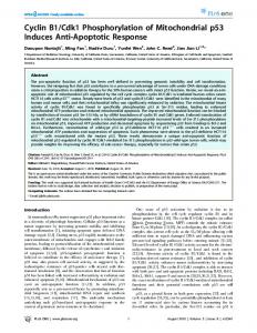
Results Patients characteristics. The clinicopathological characteristics of patients with SCCHN are summarized in Table I. The cohort of 26 SCCHN patients included 22 men and 4 women, with a median age of 73.8 years (range 54-94 years). The 26 histologically verified squamous cell carcinomas originated from the oral cavity, oropharynx, hypopharynx, larynx, and lips and included one carcinoma of unknown primary (CUP). Tumor cell differentiation was high (G1) in 3 cases, moderate (G2) in 16 cases and poor (G3) in 7 cases (Table I). Patients were classified according to the TNM system, describing size and range of the tumor, the lymph nodes and the metastasis status. All patients were without evidence of distant metastases (M0) at the time of first diagnosis. Seven (27%) patients developed a local recurrence of the tumor within a median follow-up time of 4.2 years. Cyclin B1 expression in SCCHN. Immunohistochemical analysis of cyclin B1 expression in 26 cases of SCCHN showed that cyclin B1 was expressed in all tumor samples. Cyclin B1 expression was also detected in the surrounding normal mucosa, however, with a different localization
Table III. p53 immunohistochemistry and mutation analysis compared to cyclin B1 expression. Patient
p53 overexpression
p53 Genotype
Cyclin B1 score
#1 #2 #3 #4 #5 #6 #7 #8 #9 #10 #11 #12 #13 #14 #15 #16 #17 #18 #19 #20 #21 #22 #23 #24 #25 #26
1 1 1 0 0 0 1 0 1 1 0 0 1 0 1 1 1 0 0 0 1 0 1 1 0 1
E8 E271K E7 R248W wt wt wt wt wt wt Y220C N/A E6 213 Stop wt E8 R273H Exon 6 deletion E5 S149C E5 H168Y wt wt wt wt E5 V157F wt E8 E286K E6 G226R wt E5 T150R
2 1 2 2 1 2 1 2 2 1 1 1 2 1 1 1 1 1 1 2 2 1 2 1 2 2
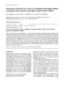
pattern. In normal mucosa, cells with nuclear staining were located in the basal and parabasal layers of the stratified squamous epithelium, marking areas with regular mitotic activity (Figure 1A), and only few cells exhibited a cytoplasmatic staining pattern (Figure 1A). By contrast, cyclin B1 protein was expressed predominantly in the cytoplasm of the tumor cells. Cells with cyclin B1 expression in the cytoplasm were distributed throughout the tumor islands (Figure 1 B and C). Out of 26 tumors, 14 (53.8%) had low (score 1) and 12 (46.2%) high (score 2) cyclin B1 expression (Table II and Figure 1). Association of cyclin B1 expression with clinipathological parameters. Next, we investigated the potential correlation of cyclin B1 expression with clinicopathological parameters. High cyclin B1 expression was more common among poorly differentiated tumors in comparison to moderately or well differentiated tumors. While 5/7 of G3 tumors had high cyclin B1 expression, only 7/16 of G2 tumors and none (0/3) of the G1 tumors exhibited high expression of cyclin B1 (Table II). Accordingly, the histopathological grade was significantly higher in tumors with high cyclin B1 expression compared with tumors with low cyclin B1 expression (Figure 2) (Mann-Whitney U-test, p0.05 for all, Chi-square test). p53 expression and mutation analysis. In SCCHN, the p53 tumor suppressor gene is often mutated or dysfunctional. Immunhistochemistry of p53 protein and sequencing of genomic PCR product of p53 exons 5-8 were performed and related to cyclin B1 expression. Immunohistochemical analysis indicated that 53.8% of tumors (14 of 26) showed accumulation of p53 protein (Table II and Table III). Twenty-five tumors underwent sequencing of genomic PCR products of exons 5-8 (#10 not available). Out of 14 tumors showing p53 overexpression, 10 were found to express missense mutations within p53 exons 5-8 (Table III). Three tumors (#3, #7, #17) had a wild-type p53 genotype but overexpressed p53, which could possibly be a consequence of a mutation outside the exons 5-8 or in genes impacting on the stability of p53 (12). Association between p53 status and clinicopathological parameters. A possible association of p53 overexpression with clinicopathological parameters in the 26 SCCHN patients was tested. p53 expression showed no association with gender, tumor size (T status), nodal status or local recurrence (p>0.05 for all, Chi-square test). Moreover, p53 expression showed no correlation with the differentiation grade of the tumors. Among G1 tumors, all (3 out of 3) accumulated p53; among G2 tumors, 6 out of 16 accumulated p53; of the 7 poorly differentiated tumors included in the study, 5 had p53 overexpression. Association between p53 status and cyclin B1 expression. Since p53 plays an important role in the regulation of the cell cycle, including the G2/M transition, we investigated the possible association of p53 status and cyclin B1 level.
3154
p53 was overexpressed in 14 out of 26 samples. Among the p53-overexpressing tumors, 7 tumors exhibited low cyclin B1 levels (score 1), and 7 tumors (50%) had high cyclin B1 levels (score 2) (Table III). Among 12 tumor samples without p53 overexpression, 7 had low levels (score 1) and 5 had high levels (score 2) of cyclin B1. There was no significant association between cyclin B1 expression and p53 immunoreactivity. Among 12 tumors with high (score 2) cyclin B1 expression, there were 5 with wt p53 and no accumulation of the p53 protein (Table III). The other 7 tumors had either p53 mutations or accumulation of p53 protein: 6 tumors had p53 mutations and accumulation (# 1, 9, 13, 21, 23 and 26). One tumor (# 3) did not have a p53 mutation but a p53 accumulation. In the group of 13 patients with low (score 1) cyclin B1 expression there were 5 with wt p53 and normal p53 protein (#5, 12, 18, 19, 22); 4 (#2, 15, 16, 24) had p53 mutation and accumulation, 2 tumors (#11 and 14) had p53 mutations but no overexpression, and 2 tumors (#7 and 17) were genomically wt p53 with p53 protein overexpression. Altogether, the frequency of tumors with dysfunctional p53 (either mutated p53 or p53 overexpression) did not differ significantly between those with high and those with low cyclin B1 expression.
Discussion Cyclins play an essential role in orchestrating normal cell cycle progression. While normal cells turn on transient expression of small amounts of cyclin proteins at specific points of the cell cycle, many tumors have constitutively high levels of one or more cyclins (2, 20, 21). Most studies investigating cell cycle regulation in SCCHN have focused on the G1/S transition phase, and showed that overexpression of cyclin D1 is common in SCCHN (22-25). However, deregulation of proteins in subsequent stages of cell
Hoffmann et al: Cyclin B1 and p53 in Head and Neck Cancer
Figure 2. Association of cyclin B1 expression and the grade of histopathological differentiation in SCCHN. Cyclin B1 expression was assessed by immunohistochemistry in 26 SCCHN (score 1, low expression; score 2, high expression). Well-differentiated tumors, G1; moderately differentiated tumors, G2; poorly differentiated tumors, G3. *p