1161
Journal of Alzheimer’s Disease 49 (2016) 1161–1168 DOI 10.3233/JAD-150423 IOS Press
Cycloheximide Treatment Causes a ZVAD-Sensitive Protease-Dependent Cleavage of Human Tau in Drosophila Cells Junhua Genga,b,c,1 , Lu Xiaa,b,c,1 , Wanjie Lia,b,c , Changqi Zhaoa,b,c and Fei Doua,b,c,∗ a State Key Laboratory of Cognitive Neuroscience and Learning & IDG/McGovern Institute for Brain Research, College of Life Sciences, Beijing Normal University, Beijing, China b Key Laboratory of Cell Proliferation and Regulation Biology, Ministry of Education, College of Life Sciences, Beijing Normal University, Beijing, China c Center for Collaboration and Innovation in Brain and Learning Sciences, Beijing Normal University, Beijing, China
Accepted 2 October 2015
Abstract. Neurofibrillary tangles are the main pathological feature of Alzheimer’s disease. Insoluble tau protein is the major component of neurofibrillary tangles. Defects in the tau protein degradation pathway in neurons can lead to the accumulation of tau and its subsequent aggregation. Currently, contradictory results on the tau degradation pathway have been reported by different groups. This discrepancy is most likely due to different cell lines and methods used in those studies. In this study, we found that cycloheximide treatment induced mild activation of a ZVAD-sensitive protease in Drosophila Kc cells, resulting in cleavage of tau at its C-terminus; this cleavage could generate misleading tau protein degradation pattern results depending on the antibodies used in the assay. Because cycloheximide is a broadly used chemical reagent for the study of protein degradation, the unexpected artificial effect we observed here indicates that cycloheximide is not suitable for the study of tau degradation. Other methods, such as inducible expression systems and pulse-chase assays, may be more appropriate for studying tau degradation under physiological conditions. Keywords: Caspase, cycloheximide, degradation, Drosophila Kc cells, human tau, truncation, ZVAD-sensitive protease
INTRODUCTION Insoluble tau is the key component of neurofibrillary tangles, which are characteristic of Alzheimer’s disease (AD). In AD patients, the extent of tau pathology correlates with cognitive decline [1], and reducing tau levels has been shown to attenuate neuronal dysfunction in mouse models of AD [2, 3]. There is evidence that proteasomal and autophagic activities are decreased in AD-sensitive brain regions compared to unaffected regions (reviewed in [4]). These func1These
authors contributed equally to this work. to: Fei Dou, College of Life Sciences, Beijing Normal University, 19 Xinjiekou Wai Avenue, Beijing 100875, P.R. China. Tel./Fax: +86 10 58801778; E-mail:
[email protected]. ∗ Correspondence
tional deficiencies are thought to contribute to the accumulation of tau, resulting in its aggregation. Thus, determining the main pathway of tau degradation may allow for the development of effective tau-based therapeutic strategies. Numerous studies have used various pharmacological and genetic interference approaches to analyze the tau degradation pathway. However, whether the ubiquitin-proteasome system or autophagy-lysosome system is the primary system for tau degradation is still unclear [5, 6]. The commonly accepted explanation for the contradictory results in this area is that the degradation of tau is context-dependent: tau might be degraded through different pathways in different cell types or under different conditions (in vivo
ISSN 1387-2877/16/$35.00 © 2016 – IOS Press and the authors. All rights reserved This article is published online with Open Access and distributed under the terms of the Creative Commons Attribution Non-Commercial License.
1162
J. Geng et al. / Artificial Effect on Tau Degradation Caused by Cycloheximide
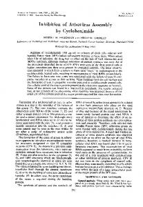
Fig. 1. Tau can be hydrolyzed at its C-terminus in the presence of Cycloheximide. A) Schematic representation of the antibodies used in this study. 9E10 was used to recognize myc tag which was fused into N-terminus of Tau, the epitope recognized by Tau46 is located within the range of 404–441aa in the 2N4R isoform of human tau. B) Cycloheximide chase analysis of human tau (2N4R) in Drosophila Kc cells using antibody Tau46 or 9E10. C) Quantification of the relative levels of tau after the treatment described in (B). The amount of tau was normalized to the tubulin levels. The results are the means ± S.E. from three independent experiments. D) Cycloheximide chase analysis of human tau (0N4R) in Drosophila Kc cells using antibody 9E10 and Tau46. E) Quantification of the relative levels of tau after the treatment described in (D). The amount of tau was normalized to the tubulin levels. The results are the means ± S.E. from three independent experiments.
versus in vitro). Furthermore, the methods used to study tau degradation were different between laboratories. Some laboratories used a pulse-chase assay to label the newly synthesized tau protein and study its half-life [7], while others used cycloheximide (CHX) to block tau synthesis [8–10]; inducible expression systems were also wildly used [11–13]. These differences in methodology could also explain the variability of data with regards to the mechanism of tau degradation. Drosophila studies in the past decade have improved our understanding of the molecular mechanism of tau neurotoxicity. A previous study by our group revealed that the C-terminus of tau is important for tau protein
stability and toxicity in Drosophila [14]. In the present study, we analyzed tau degradation in Drosophila Kc cells. Our results showed that CHX is able to activate caspase activity in Kc cells; this causes cleavage of the tau protein at its C-terminus, while the rest of the protein remains intact. If only an anti-tau C-terminal antibody is used in such an experiment, the recorded tau degradation rate would be much higher than the true tau degradation rate. In summary, our study revealed an important aspect of CHX that should be taken into account when interpreting data from studies on the degradation of proteins known to be caspase or other ZVAD-sensitive protease substrates.
J. Geng et al. / Artificial Effect on Tau Degradation Caused by Cycloheximide
1163
The antibody 9E10 recognizes the c-Myc tag sequence, which is expressed at the amino terminus of the Tau protein. The C-terminal Tau antibody Tau46 specifically recognizes residues 404–441 and can detect full-length Tau. Cycloheximide was purchased from MP Biomedicals. Protease Inhibitor Set, ZVAD-fmk, MG132 and epoxomicin were purchased from Calbiochem. A Caspase 3 Assay Kit (Fluorimetric), chloroquine, and 3-methyladenine (3-MA) were purchased from Sigma. An In Situ Cell Death Detection Kit was purchased from Roche Applied Science. Cell culture and transfections Drosophila Kc167 (Kc) cells (gift from Ming Fang’s laboratory) were cultured in Schneider’s Insect Medium (Invitrogen) containing 5% (vol/vol) fetal bovine serum (Invitrogen), 100 units/ml penicillin, and 100 g/ml streptomycin. Kc cells were maintained at 25◦ C. Cell lines were transfected using X-tremeGENE DNA Transfection Reagents (Roche Applied Science), following the manufacturer’s instructions. Cell lysis and immunoblotting
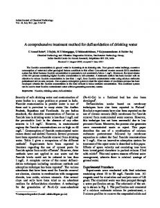
Fig. 2. Tau can be truncated by protease during degradation and the truncation can be inhibited by caspase inhibitor. A) Western blot analysis of full-length human tau treated with alkaline phosphatase. At 48-h post transfection, Kc cells were treated with or without 100 g/ml cycloheximide for 24 h, and the cell lysates were treated with or without alkaline phosphatase at 37◦ C for 1 h. B) Western blot analysis of full-length human tau in Drosophila Kc cells treated with different protease inhibitors in the presence of cycloheximide (100 g/ml). At 48-h post transfection, Kc cells were treated with 100 g/ml cycloheximide and protease inhibitors for 24 h. C) Alkaline phosphatase treatment of full-length human tau from cycloheximide-treated Drosophila Kc cells in the presence or absence of caspase inhibitor Z-VAD. At 48-h post transfection, Kc cells were treated with 100 g/ml cycloheximide and Z-VAD for 24 h, then the cell lysates were treated with or without alkaline phosphatase at 37◦ C for 1 h.
MATERIALS AND METHODS Antibodies and reagents The primary antibodies used in this study were as follows: DM1A (Sigma), 9E10 (Sigma), and Tau46 (Abcam). DM1A is immunospecific for ␣-tubulin, which was used as the loading control in this study.
Cell extracts were prepared in RIPA buffer (50 mM Tris-HCl, pH 7.4, with 150 mM sodium chloride, 1% NP-40, 0.5% sodium deoxycholate, and 0.1% sodium dodecyl sulfate) supplemented with complete EDTA-free protease inhibitor cocktail tablets (Roche Applied Science). Protein concentration was determined using the bicinchoninic acid assay (Pierce), and samples were prepared for immunoblotting by dilution in 5 × loading buffer (0.25 mM Tris-HCl, pH 6.8, 15% SDS, 50% glycerol, 25% -mercaptoethanol and 0.01% bromphenol blue). Caspase-3 activity determination Kc cells were incubated with or without cycloheximide (100 g/ml) for the indicated times, lysed in lysis buffer (Sigma) for 20 min on ice, and then centrifuged at 14,000× g for 10 min. The caspase-3 specific reaction mixture was prepared following the manufacturer’s instructions (Sigma). Kc cell lysate (20 g of total protein) was incubated with caspase3 specific reaction mixture (200 l) at 37◦ C for 1 h. Recombinant caspase 3 was used as the positive control. Fluorescence was determined with a fluorescence plate reader (TECAN).
1164
J. Geng et al. / Artificial Effect on Tau Degradation Caused by Cycloheximide
Fig. 3. Cycloheximide induces activation of caspase in Kc cells. A) Caspase activity were assayed when Kc cells were treated with cycloheximide (100 g/ml) for the indicated times, recombinant caspase-3 was used as the positive control (n = 3,∗ p < 0.05). B) Caspase activity were assayed in Kc cells overexpressing full-length htau (2N4R) for 24 h, recombinant caspase-3 was used as the positive control (n = 3).
Fig. 4. Cycloheximide induces the apoptosis of Kc cells. A) The apoptosis of Kc cells under CHX treatment. Kc cells were treated with cycloheximide (100 g/ml) for the indicated times, TUNEL assay was used to detect apoptosis of Kc cells. PC, positive control, add 100 L DNaseI incubated for 10 min before fixation of the Kc cells; NC, negative control, Kc cells were treated with DMSO for 48 h. B) The statistical histogram of TUNEL signal in Kc cells with different cycloheximide treatment duration (n = 3,∗∗ p < 0.001).
In situ cell death detection In situ cell death was detected by labeling of DNA strand breaks (TUNEL technology) according to the
manufacturer’s instructions. Kc cells were incubated with terminal deoxynucleotidyl transferase-mediated dUTP-biotin nick end labeling (TUNEL) reaction mixture for 60 min at 37◦ C in the dark. The samples were
J. Geng et al. / Artificial Effect on Tau Degradation Caused by Cycloheximide
washed twice with PBS and once with water and then mounted. Images were acquired using a Zeiss AX10 Fluorescence microscope. Data analysis To determine whether the difference in the induction of apoptosis by CHX was statistically significant between treatments, the percentages of apoptotic cells were measured in five randomly selected microscopic fields using Metamorph imaging software. Statistical analyses were performed using one-way analysis of variance, and Student’s t test results are expressed as the means ± S.E. A p-value