21 Jun 1993 - De Block M. Botterman J., Vandewiele M., Dockx J., Thoen C., Gossele. V., Rao Mova N., Thompson C., Van Montagu M., Leemans J. (1987).
Nucleic Acids Research, 1993, Vol. 21,
No.
Cytosine methylation inhibits replication of African mosaic virus by two distinct mechanisms
15
3445-3450
cassava
Genadij Ermak+, Uta Paszkowski§, Markus WohImuthO, Ortrun Mittelsten Scheid§ and Jerzy Paszkowski* Institute of Plant Sciences, ETH-Zurich, Universitatsstrasse 2, 8092 Zurich, Switzerland Received April 26, 1993; Revised and Accepted June 21, 1993
ABSTRACT Extrachromosomally replicating viral DNA is usually free of cytosine methylation and viral templates methylated in vitro are poor substrates when used in replication assays. We have investigated the mechanism of inhibition of viral replication by DNA methylation using as a model the DNA A of African cassava mosaic virus. We have constructed two component helper systems which allow for separation of the transcriptional inhibition of viral genes necessary for replication from replication inhibition due to altered interaction between the replication complex and methylated viral DNA. Our results suggest that methylation-mediated reduction of viral replication is due to both repression mechanisms and that this provides two independent selection pressures for the maintenance of methylation-free replicons in infected cells. INTRODUCTION Viruses depend on host enzymes for the expression of their genetic information and similar regulatory signals of viral and host genes have evolved. However, the regulation of viral gene expression needs to be controlled entirely by the virus without host cell influences. This type of relationship is well illustrated by functional analysis of the 35S promoter of cauliflower mosaic virus (CaMV)(l), which is composed of cis-acting elements, each of which confers a tissue-specific tye of regulation (1, 2,3). The additive action of all elements in the wild-type promoter results in high transcriptional activity and constitutive expression in almost all tissues that might be infected by the virus. Nevertheless, plant cells have the potential to reduce transcription from this viral promoter by cytosine methylation. Expression of chromosomally integrated marker genes regulated by the CaMV 35S promoter was found to be correlated to the level of its methylation (4, 5). Suppression of viral genes integrated into chromosomes was very well documented for mammalian retroviruses (6).
Much less is known about the involvement of methylation in the suppression of viral genes on extrachromosomal replicons (for a review 7). It was recently reported for component A of tomato golden mosaic virus (TGMV) that its hypermethylation results in reduced accumulation of viral DNA in transfected cells (8). TGMV belongs to the group of plant geminiviruses containing single-stranded DNA genomes (9,10). Its genetic information is distributed on two circular molecules (components A and B or DNA A and DNA B). Proteins required for replication of viral DNA in host cells are encoded by component A. Therefore, DNA A is capable of autonomous replication in the absence of B (1 1,12) and is able to transactivate replication of the B component (13). DNA A and B share a region of homology (common region) which is considered to be the origin of replication (14,15). It has been shown that in vitro methylation of the component A before its transfection to tobacco protoplasts inhibits its replication (8). The mechanism of this inhibition is not clear but there are two not necessarily exclusive explanations: (1) methylation suppresses viral genes whose products are required for replication, or (2) methylated viral DNA itself is less-efficiently recognized and/or utilized by the replication complex. Here we describe a system which allowed us to differentiate between these two possibilities. The experimental approach was based on component A of the African cassava mosaic virus (ACMV), which is a close relative of TGMV (for a review see 16). Our data indicate that both mechanisms are active in replication inhibition.
MATERIAL AND METHODS Construction of plasmids A ClaI fragment of plasmid pCLV20 (17, kindly provided by Dr John Stanley, John Innes Institute, UK) containing a coat protein deletion derivative of component A of ACMV was cloned as a directly repeated dimer into the ClaI site of pCB1 (18). After deletion of the DraI-MscI fragment, the viral insert remained as a partial dimer with an approximately 600-bp repeat containing the common region (pUNpd, Fig. 1). A 1166-bp fragment of
To whom correspondence should be addressed at: Friedrich Miescher Institute, PO Box 2543, 4002 Basel, Switzerland Present addresses: 'Institute of Genetics and Cytology, F. Scorina 27, 220734 Minsk, Whiterussia, USSR and §Friedrich Miescher Institute, PO Box 2543, *
4002 Basel, Switzerland
3446 Nucleic Acids Research, 1993, Vol. 21, No. 15 C
BAbXb
B
pUNpd
El AL
2Ti
L
C S
pPDkan
-
pMWBbar
_I
B
BA
I Il
t
E.A LF'i
8
C
b
il CEOi
01
pPDbar
^
BA b
I
C
C
'iLlV
II
BA b
\l
b
c
4Ma
b
C
4EEal
b
I1 " .AL 3 )AL2
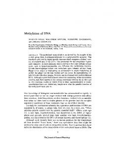
Figure 1. Schematic structure of the DNA A constructs used. Details of the constructions are given in Material and Methods. Thick, solid lines represent plasmid vector sequences. Open boxes represent replicon sequences. Black boxes marked bar or nptII represent non-viral DNA inserts. Shadowed boxes below each replicon sequence represent viral open reading frames and are marked ALl, AL2 and AL3 (10). Black boxes marked CR represent the common region and the adjacent arrows represent the promoter region for the coat protein gene. Restriction sites are marked as: C; ClaI, B; BglI, X; X0oI, b; BamHI.
the nptII hybrid gene (EcoRV fragment of pABD1, 19) was inserted into the XhoI site of pUNpd, resulting in plasmid pPDkan. pPDbar was derived from pUNpd by insertion of a 561-bp BamHI fragment coding for phosphinotricin acetyl transferase (PAT)(20) into the BamHI site of pUNpd. A deletion mutant of pPDbar was generated by removal of a 65 1-bp Bgll fragment resulting in the replication-deficient construct pMWBbar. All plasmid constructs used in this study are depicted in Figure 1. The methods used for plasmid constructions were. as previously described (21). Restriction enzymes were purchased from Pharmacia, New England Biolabs and Boehringer Mannheim, and were used according to the suppliers' recommendations.
Preparation of methylated DNA Plasmid DNA was methylated in vitro by 1.5-fold excess of HpaH- or SssI methylase for 3 h at 37°C in Tris-HCI pH 7.5, 10 mM EDTA containing 80 or 160 1,M S-adenosylmethionine. After the reaction, DNA was extracted with phenol/chlorophorm 1:1 and precipitated with ethanol. The completeness of methylation of each new batch of DNA was controlled by digestion with Hpal and MspI. After digestion with HpaH and heat inactivation of restriction enzymes, the methylated DNA was sterilized before transformation by further ethanol precipitation and ethanol washing as described previously (22).
Preparation and transformation of protoplasts Leaves of axenic shoot cultures of the tobacco line SRI (23) were used as a source of protoplasts. The procedure for protoplast isolation was as described previously (22). One million protoplasts per sample were transformed with 5 1tg of circular plasmid DNA by the PEG-uptake method (24). After transformation, protoplasts were cultured in 4 ml of liquid K3/H medium as previously described (25). Protoplasts were harvested 2 h, 3 and 6 days after transformation.
Isolation of DNA and Southern blot analysis Protoplasts were washed in 10 ml of fresh medium and collected by repeated centrifugation in Eppendorf tubes. Protoplast pellets were frozen in liquid nitrogen. The frozen cells were homogenized using a glass rod and 400 /Al of extraction buffer (10 mM Tris-HCl pH7.5, 50 mM NaCl, 10 mM EDTA, 1% sarcosyl) was added. The cell extract was mixed with an equal volume of phenol saturated with TE (10 mM Tris-HCl pH 8.0 1 mM EDTA). The organic and water phases were separated by centrifugation and DNA was ethanol precipitated from the water phase. The DNA pellet was resuspended in TE containing RNase at a final concentration of 200 ,ug/ml. RNase digestion was allowed for 15 min at 37°C followed by proteinase K treatment for 1 h at 370C at a concentration 50 ,ug/ml in the presence of 0.2% sodium dodecylsulphate (SDS). After further phenol extraction, DNA was ethanol precipitated and dissolved in TE. Total cellular non-restricted DNA (2 jig) was subjected to electrophoresis in 1% agarose and transferred to Hybond-N nylon membranes (Amersham International, Amersham, UK). The membranes were pre-hybridised and hybridized at 42°C in 50% (v/v) formamide, 5 % (w/v) dextran sulfate, 5 x Denhardts solution, 0.5% (w/v) SDS, 4xSSC, and 0.2 mg/ml heatdenatured calf thymus DNA. The radioactive probes were prepared by the random primer method using [ca-32P]dATP as previously described (26). The specific activity obtained was 3-5 x 10 cpm/4g DNA. Filters were washed twice for 15 min at 680C in 0.2 x SSC, 0.1% (w/v) SDS and were exposed to Xray films with intensifying screens at -70°C.
Analysis of replicative forms of component A Southern blots indicating the migration of replicative forms of the component A were used as an indicator for isolating regions of agarose gels containing replication products. Gel fragments were freeze/thawed twice and DNA was separated from the agarose by filtration through glass wool in a table-top centrifuge. The filtrate was extracted with butanol and the DNA was precipitated with ethanol.
RESULTS Replication properties of methylated viral replicons Transfection of naked DNA into plant protoplasts allows the introduction of molecules previously modified in vitro. Homomethylated DNA A of ACMV was treated with an excess of HpaII prior to transfection in order to prevent non-methylated molecules serving as replication templates. In a parallel control experiment, it was shown that the equivalent amount of nonmethylated DNA restricted with Hpall was not able to serve as a template for initiation of replication (data not shown).
Nucleic Acids Research, 1993, Vol. 21, No. 15 3447
12 3
Table 1. The relative number of potential methylation sites (PCM) in ACMV replicons Construct containing replicon
Number of CG per kb of replicon DNA
Number of CNG and CG per kb of replicon DNA
pUNpd pPDbar pPDkan
26 48 49
66 108 108
41 4C
Figur 2. Southern blot analysis of the replication products following ftransfection of protoplasts with pUNpd. Lane 1: 2 hours after transfection; lane 2: 3 days after transfection; lane 3: 6 days after transfection. In lanes 2 and 3, 5 Utg of total cellular DNA was applied to the gel. In lane 1, the amount of DNA was reduced to 0.5 jig allowing for a similar level of signal after hybridization. In control experiments with 5 itg of DNA isolated 2 h after trnsfection, no replicative forms of DNA A were visible, even after overexposure. The various replicative forms of DNA A are marked as: SS; single stranded, SC; super coiled, L; linear, OC; open circular, MS; multimeric structures.
34
1 2
5 6
41 .4
4sc
-4L t:,..
4
8ss
.40.4sc pUTNpd
pPDhar
PlP)k
an
Figure 3. Southern blot analysis of replicative forms of various DNA A constructs transfected to protoplasts in the methylated and non-methylated state. Lanes 1, 3, 5: non-methylated DNA; lanes 2, 4, 6: methylated DNA. Total cellular DNA was isolated 6 days after transfection. Each DNA sample 5 Ag was separated in 1 % agarose gel before blotting. The names of constructs are given below the lanes. Replicative forms are marked as in Fig. 2.
The replication efficiency was estimated by the relative abundance of replication products 6 days after transformation; however, characteristic progeny replicons were already detectable after 3 days (Fig. 2). Retarded replication was observed when
methylated viral templates were introduced into protoplasts (Fig. 3). The inhibition was seen only with replicons fully methylated by SssI methylase and not by Hpal methylase (data not shown). Methylase Hpall recognizes only two sites within the replicon whilst SssI methylase recognizes 49 sites. We also examined the level of replication inhibition for replicons with or without inserts of non viral DNA (Fig. 3). Inserts increased both the size of the replicons and the relative number of potential methylation sites (PCM) (Table 1). Both of these alterations could account for the reduction of replication efficiency observed. In order to assay the degree of inhibition caused by size enlargement and/or increased density of the PCM, we chose two inserts with base compositions that resulted in replicons (pPDbar and pPDkan, Fig. 1) of different sizes but with comparable frequencies of PCM (Table 1). This frequency is considerably higher than that of the original pUNpd (Table 1). Thus, if replication efficiency were mainly determined by the relative number of PCM, replication of the two replicons would have been retarded to the same extent. Alternatively, if the size of the replicon were a limiting factor, the smaller replicon (pPDBar) would have been more efficiently replicated. Replication of pPDBar was indeed significantly more efficient than that of pPDKan throughout several experimental repetitions (for illustration Fig. 3, lanes 1, 3, 5). These results suggest that the size of foreign DNA inserts rather than the PCM number determines the efficiency of replication. In order to examine whether the initial methylation status of progeny replicons derived from methylated templates was maintained or altered, the products of replication were isolated after electrophoretic separation (Fig. 3, SC DNA bands) and digested with a set of methylation-sensitive restriction enzymes (Fig. 4). The complete digestion of progeny replicons indicated loss of methyl groups during replication. In contrast, input DNA retained in the cells remained methylated (data not shown). Reduction of replication efficiency from methylated templates together with the loss of methyl groups indicates a strong negative influence of DNA methylation on the efficiency of viral DNA amplification. In order to elucidate the mechanism of this inhibition, we designed a bimolecular helper system separating the production of viral proteins from the replication process itself.
Replication of mutant replicons in the presence of a helper We constructed a deletion derivative of the original replicon which inactivated all viral open reading frames (pMWBbar, Fig. 1). The replication of this molecule, however, could be restored by co-transformation with a helper replicon providing the necessary replication factors in trans (Fig. 5 A, B lane 2). In order to follow the fate of the mutated and the helper replicons separately and to discriminate between transactivation and recombination processes, two distinct DNA fragments were
3448 Nucleic Acids Research, 1993, Vol. 21, No. 15
1 2 3 4 5 6 7 8 9 10 11 12
Lt Se- 4..; 4 _co
W00 a
_
m
M
_
JA
.t
t7 i r
:
S
MNO04 _-4Ia
..Imam,
4damb
41P
4MI,
4 Figure 4. Southern blot analysis of the methylation status of pUNpd progeny replicons transfected in the methylated and non-methylated state. DNA of the supercoiled replicative form of progeny replicons was recovered after separation in agarose gels (see Material and Methods) and subjected to digestion with methylation-sensitive restriction enzymes. Lanes 1-6: progeny replicons of pUNpd transfected in non-methylated form; lanes 7-12: progeny replicons of pUNpd transfected after methylation with SssI. Lanes 1 and 7: non-restricted DNA; lanes 2 and 8: DNA digested with HpaIl; lanes 3 and 9: DNA digested with MspI; lanes 4 and 10: DNA digested with Bgm; lanes 5 and 11: DNA digested with ClaI; lanes 6 and 12: DNA digested with Eco473. Similarity of the fragment sizes for HpaHlMspI and Bgll is accidental. Replicative forms are marked as in Fig. 2.
inserted into a region which is not essential for replication (27)(Fig. 1, pPDKan and pMWBbar). Transactivation of the mutant by a helper system restored approximately 30% of replication efficiency when compared to the corresponding nonmutant control (data not shown). Hybrid replicons resulting from recombination between mutant and helper molecules were not observed. Mechanism of replication inhibition Pairs of the plasmids pPDkan and pMWBbar containing the helper and mutant replicons, respectively, were co-transformed into tobacco protoplasts either as non-methylated DNA or with one or both replicons methylated in vitro by SssI methylase (Fig. 5). As mentioned previously, co-transformation of pairs of non-methylated helper and mutant replicons resulted in transactivation to approximately 30% of the control level. SssI methylation of both interacting components inhibited replication of the helper replicon and transactivation of the mutant (Fig. 5 A, B lane 8). Methylation only of the helper DNA decreased its own replication and severely reduced the level of transactivation (Fig. 5 A, B, lane 4). Interestingly, methylation only of the mutant replicon also significantly influenced its ability to be replicated in the presence of replication active helper (Fig. 5 A, B lane 2 as compared to lane 6). Thus, methylation affects not only the potential to produce transacting factors, probably by the suppression of viral genes, but also has a direct effect on the replication process.
Transactivated replicons are also demethylated Since the inhibition of replication was mostly due to repression of the production of viral replication factors (Fig. 5, lanes 4, 6), the observed replication-associated demethylation in the onecomponent system could be the result of tight selection for
1
ti) is ~
B 2>
t
e
*2.."
(i8
^.
4 \M1 \'
Figure 5. Accumulation of progeny replicons after cotransfection of pPDkan and pMWBbar as a bimolecular helper system. Total cellular DNA was isolated 2 h and 6 days after transfection and 5 jg of DNA of each sample was separated in 1% agarose gel before blotting. Panel A: blot probed with replicon-specific probe (ClaI fragment of pUNpd, Figure 1); panel B: blot probed with probe specific for bar insert (plasmid pUC8 containing BamHI fragment of bar gene, Figure 1). Lanes 1, 3, 5, 7: DNA isolated 2 h after transfection; lanes 2, 4, 6, 8: DNA isolated 6 days after transfection. Lanes 1 and 2: pPDkan and pMWBbar cotransfected as non-methylated DNA; lanes 3 and 4: cotransfection of SssImethylated pPDkan and non-methylated pMWBbar; lanes 5 and 6: cotransfection of SssI-methylated pMWBbar and non-methylated pPDkan; lanes 7 and 8: pPDkan and pMWBbar cotransfected as SssI-methylated DNA. Supercoiled forms of both replicons are marked by the arrows on the right.
demethylated replicons which regained the ability for gene expression. In the two-component system, no such specific selection was imposed on the mutant replicons. It is possible, therefore, that methylation of pMWBbar was maintained during transactivated replication. We isolated progeny of mutant replicons after electrophoretic separation from input and helper
Nucleic Acids Research, 1993, Vol. 21, No. 15 3449 DNA and examined their methylation status with methylationsensitive enzymes. As in a one-component system (Fig. 4), in this case methylation was also not maintained (data not shown). Thus, lack of methylation maintenence appears to be inherent to viral replication itself rather than the result of selection.
DISCUSSION Methylation of a newly synthesized DNA strand occurs very soon after its synthesis (28, 29). It has been postulated that the eukaryotic replication complex (replitase) also contains methylase activity responsible for the maintenance of methylation (30). In contrast, the replicons of eukaryotic DNA viruses are found to propagate DNA free of methylation. This is well documented for adenovirus (Adl2) SYREC sequences (31). These sequences are methylated when part of chromosomal DNA but are methyl cytosine-free when packaged into virions after extrachromosomal replication. This lack of methylation may be due to the specific constitution of the Adl2 replication complex which, in contrast to phages (for a review 32), does not include methylase. It was also suggested that extrachromosomal replicons may not be accessible for host methylases, although the barrier preventing access was not defined (7). The behavior of Adl2 resembles that of gemini-virus replicons. It was observed that replication of component A of TGMV (8) and ACMV (this study) were not able to maintain methylation, even when exclusively methylated templates were used for the initiation of replication. This was probably not due to the efficient demethylation of transfected DNA by the host, since plasmid DNA introduced in methylated form into protoplasts stays methylated and remains transcriptionally inactive for 5 days after transformation (33, 34). In addition, the methylated and silent state is stably maintained after integration of methylated copies into chromosomes (35). Thus, comparison with viral replicons indicates that demethylation is tightly connected to the viral replication process itself. However, it is still puzzling that replicons persisting in the nucleus are not subject to de novo methylation since viral replication is thought to take place in S phase (10). One explanation could be that compartmentation of cellular and viral DNA prevents methylation of the latter. This has been suggested for SYREC of Adl2 (7). Alternativly, it might be that de novo methylation of replicons takes place but this has a pronounced negative influence on replication ability and a selection pressure towards the amplification of hypomethylated replicons arises. This may occur by the different affinities of the replication complex for methylated and non-methylated DNA, and the preferential use of hypomethylated replicons as templates. In this case, protection of the viral genome against mutations would be an additional advantage. It has been shown that the presence of methyl cytosine provokes mutational hot spots with a mutation rate 10-fold higher than average (36). It was in fact observed for geminiviruses that hemi-(8) or homomethylation of templates (this study) negatively influences their replication ability. However, since the same templates were used for expression of viral replication factors, these experiments did not allow differentiation between reduction of transcription or replication of methylated viral templates. To separate these two processes, we constructed a bimolecular helper system, providing an excess of replication factors in trans which resulted in a mixed population of non-methylated and methylated replicons in transfected cells. Our data indicate that non-methylated
replicons are preferentially propagated. Since methylation was not passed on to the progeny replicons, the observed differences in the accumulation of replication products may reflect a delay in replication events directly after transfection when only methylated templates were available. Hence, our assay of the accumulation of progeny replicons after 6 days probably underestimated the bias of replication towards hypomethylated templates. The correlation between replication inhibition and methylation status resembles the link between transposition activities of maize transposable elements (Ac, Spm, Mu and En-i) and their DNA methylation state (for a review see 37). It has been suggested that hypermethylation of transposons relates not only to the reduction of transcription but also to an alteration in the affinity of transposase to transposon terminal sequences (38, 39). There are six potential methylation sites in the common region of ACMV and it would be interesting to determine their specific role in the interaction with the replication complex. It is also not yet known if the demethylation of progeny replicons takes place in a single replication round or occurs gradually. Analysis of critical methylation sites by direct sequencing (for a review see 40) and of the kinetics of the demethylation process should expand our preliminary observation of methylation-dependent differential replication of ACMV DNA A.
ACKNOWLEDGEMENTS We thank Drs J.-P.Jost, T.Hohn and F.Meins for critical reading of the manuscript and Dr P.King for editorial help. This work was performed in Plant Development Group of Professor Ingo Potrykus and was supported by Swiss National Science Foundation grants 31-9057.87 and 31-30118.90.
REFERENCES 1. 2. 3. 4. 5.
6. 7. 8.
9. 10. 11. 12.
13. 14. 15.
16. 17.
Benfey P. N., Chua N. H. (1990) Science 250, 959-966. Benfey P. H., Ren L., Chua N. H. (1990) EMBO J. 9, 1677-1684. Benfey P. H., Ren L., Chua N. H. (1990) EMBO J. 9, 1685-1696. Hobbs S. L. A., Kpodar P., DeLong C. M. 0. (1990) Plant Mol. Biol. 15, 851-864. Meyer P., Linn F., Heidmann I., Meyer z.A H., Niedhof I. Saedler H. (1992) Mol. Gen. Genet. 231, 345-352. Toth M., Muller U., Doerfler W. (1990) J. Mol. Biol. 214, 673-683. Doerfler W. (1993) In 'DNA methylation: molecular biology and biological significance' ed by J. P. Jost and H. P. Saluz, Birkhauser Vedlag Basel/Switzerland 262-299. Brough C. L., Gardiner W. E., Inamdar N. M., Zhang X. Y., Ehrlich M., Bisaro D. M. (1992) Plant Mol. Biol. 18, 703 -712. Davies, J.W. and Stanley, J. (1989) TiG 5, 77-81. Lazarowitz, S.G. (1992) Critical Rev. in Plant Sci. 11, 327-349. Rogers, S.G., Bisaro, D.M., Horsch, R.B., Fraley, R.T., Hoffmann, N.L., Brand, L., Elmer, J.S. and Lloyd, A.M. (1986) Cell 45, 593-600. Townsend,R., Watts, J. and Stanley, J. (1986) Nucl. Acid Res. 14, 1253-1265. Hanley-Bowdoin, L., Elmer, S. and Rogers, S.G. (1990) Proc. Natl. Acad. Sci. 87, 1446-1450. Fontes, E.P.B., Luckow, V.A. and Hanley-Bowdoin, L. (1992) The Plant Cell 4, 597-608. Lazarowitz S.G., Wu, L.C., Rogers, S.G. and Elmer, J.S. (1992) The Plant Cell 4, 799-809. Davies J. W., Townsend R., Stanley J. (1987) In Plant DNA infectious agents editors Th. Hohn and J. Schell, Springer Verlag Wien, New York, 31-52. Etessami P., Callis R., Ellwood S., Stanley J. (1988) Nucl. Acid Res. 16,
4811-4829. 18. Peterhans A., Schlipmann H., Basse Ch., Paszkowski J. (1990) EMBO J. 9, 3437-3445.
3450 Nucleic Acids Research, 1993, Vol. 21, No. 15 19. Paszkowski J., Shillito R. D., Saul M. W., Mandak V., Hohn T., Hohn B., Potrykus I. (1984) EMBO J. 3, 2717-2722. 20. De Block M. Botterman J., Vandewiele M., Dockx J., Thoen C., Gossele V., Rao Mova N., Thompson C., Van Montagu M., Leemans J. (1987) EMBO J. 6, 2513-2518. 21. Sambrook J., Fritch E. F., Maniatis T. (1989) Molecular cloning: A laboratory manual. Cold Spring Harbor University Press. Cold Spring Harbor.. 22. Paszkowski J., Saul M. W (1986) Methods Ezymol. 118, 668-684. 23. Maliga P., Breznovitz A., Marton L. (1973) Nature new Biol. 244, 29-30. 24. Negrutiu I., Shillito R. D., Potrykus I., Biasini G., Sala F. (1987) Plant Mol. Biol. 8, 363-373. 25. Potrykus I., Shillito R. D. (1986) Methods Enzymol. 118, 549-578. 26. Feinberg A. P., Vogelstein B. (1983) Anal. Biochem. 132, 6-13. 27. Ward A., Etessami P., Stanley J. (1988) EMBO J 7, 1583-1587. 28. Woodcock D. M., Adams J. K., Cooper I. A. (1982) Biochim. Biophys. Res. Commun. 696, 15-22. 29. Lyons S. M., Schendel P. F. (1984) J. Bacteriol. 159, 421-423. 30. Naguchi H., Veer Reddy G. P., Pardee A. B. (1983) Cell 32, 443-451. 31. Deuring R., Klotz G., Doerfler W. (1981) Proc. Natl. Acad. Sci. USA 78, 3142-3146. 32. Noyer-Wiedner M., Trautner T. A. (1993) In 'DNA methylation: molecular biology and biological significance' ed by J. P. Jost and H. P. Saluz, Birkhauser Vedlag Basel/Switzerland 39-108. 33. Weber H., Graessmann A. (1989) FEBS Lett. 253, 163-166. 34. Hershkovitz JM., Gruenbaum Y., Renbaum P., Razin A., Loyter A. (1990) Gene 94, 189-193. 35. Weber H., Ziechmann Ch., Graessmann A. (1990) EMBO J. 9, 4409-4415 36. Coloundre Ch., Miller J. H., Farabaugh P. H., Gilbert W. (1978) Nature 274, 775-780 37. Finnegan E. J., Brettel R. I. S., Dennis E. S. (1993) In 'DNA methylation: molecular biology and biological significance' ed by J. P. Jost and H. P. Saluz, Birkhauser Vedlag Basel/Switzerland 218-261 38. Gierl A., Lutticke s., Seadler H. (1988) EMBO J. 7, 4045-4054 39. Kunze R., Stalinger P. (1989) EMBO J. 8, 3177-3186 40. Saluz H. P., Jost J. P. (1993) In 'DNA mediylation: molecular biology and biological significance' ed by J. P. Jost and H. P. Saluz, Birkhauser Vedlag Basel/Switzerland 11-26