Dalton Transactions
Dynamic Article Links
Cite this: Dalton Trans., 2011, 40, 7827 www.rsc.org/dalton
COMMUNICATION
Immobilization of Pd nanocatalysts on magnetic rattles and their catalytic property† Shouhu Xuan,*a Wanquan Jiangb and Xinglong Gonga Received 20th April 2011, Accepted 13th June 2011 DOI: 10.1039/c1dt10715a Here we report a magnetic composite nanocatalyst (MP@NiSiO/Pd) with a rattle type nanostructure. The nickel silicate shell encapsulated magnetic core (MP@NiSiO) rattle particles were synthesized by using a facile templating method and the Pd nanocrystals with different sizes can be directly immobilized onto the MP@NiSiO rattles without using any surfactant or capping reagents. X-Ray powder Diffraction (XRD) and Transmission Electron Microscopy (TEM) are employed to characterize the rattle type particles. The MP@NiSiO/Pd composites show catalytic activity on the Heck and Suzuki reactions. They can be separated from the reaction system by using a magnet and the catalyst can be cycled at least for 5 times.
1.
Introduction
Nanocatalysis is essential for chemistry, material science, and nanoscience.1–4 Many noble metal nanoparticles have been used as catalysts in various chemical reactions.5 For facile catalyst recovery and recycling, noble metal nanocatalysts were usually deposited onto the surface of solid supports to form heterogeneous catalysts.6 Various inorganic and organic supports, such as SiO2 , TiO2 , zeolite, carbon, layered double hydroxide, and polyaniline etc., have been employed as the carrier.7–12 These carried nanocatalysts exhibited high catalytic properties and recyclability. To allow for the easy removal of catalysts from reaction mixtures, supports containing magnetic nanoparticles have been developed.13 Tsang et al. reported the synthesis of carbon encapsulated magnetic nanoparticles which could be used as a catalyst support.14 Ying developed Fe2 O3 @SiO2 /Pd composites with well defined core/shell nanostructure and these magnetic nanocatalysts provided excellent reactivity and reusability in the hydrogenation of nitrobenzene.15 Besides the surface immobilization method, many efforts also have been focused on encapsulating nanocatalysts into the porous magnetic carriers. The mesoporoussilica-protected core–satellite nanocomposite catalysts, which were a CAS Key Laboratory of Mechanical Behavior and Design of Materials, Department of Modern Mechanics, University of Science and Technology of China, Hefei, 230027, P. R. China. E-mail:
[email protected]; Fax: +86 551 3606382; Tel: +86 551 3606382 b Department of Chemistry, University of Science and Technology of China, Hefei, 230026, P. R. China † Electronic supplementary information (ESI) available. See DOI: 10.1039/c1dt10715a
This journal is © The Royal Society of Chemistry 2011
firstly described by Yin and Ge,16 were ideal recyclable catalysts because they contained superparamagnetic components for efficient magnetic separation and a mesoporous silica framework for stabilization of the encapsulated catalyst particles. Very recently, Deng developed a Fe3 O4 @SiO2 –Au@mSiO2 composite material and this catalyst exhibited efficient epoxidation of styrene with high conversion and selectivity.17 Therefore, the development of multifunctional nanoparticles as the carrier for the metal nanocatalysts is highly desirable.18 Rattle-type particles have proven to be an ideal support in catalysis.19 Tang et al. have succeeded in fabricating a kind of unique silica nanorattle, whose core was dense SiO2 while the shell was mesoporous SiO2 . In their work, gold nanoparticles with controllable size were successfully incorporated into silica nanorattles by impregnating silica nanorattles in the HAuCl4 water solution. This kind of gold core rattle type silica nanoarchitecture was successfully applied in the catalytic reduction of 2-nitroaniline as a model reaction.20,21 During the past few decades, various rattle type nanostructures such as Fe3 O4 @Fe3 O4 , Fe3 O4 @SiO2 , Polymer@Polymer, and SiO2 @TiO2 et al., have been developed.22–25 Unfortunately, only a few of them have been used as the catalyst carrier. Pd catalyzed cross-coupling reaction is one of the most key tools in modern organic synthesis, thus the immobilization of Pd nanocatalyst and the study of its catalytic performance is very important.26 In this work, a rattle-type particle which consisted of a nickel silicate (NiSiO) shell and an Fe3 O4 (MP) core was synthesized as the carrier for Pd nanocrystals. The influence of synthetic factors on the Pd nanocrystals is discussed. The asprepared MP@NiSiO/Pd rattles exhibited good catalytic property and can be magnetically separated from the reaction solutions.
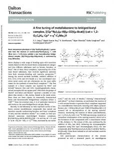
2. Results and discussion Previously, we developed a template-assisted approach to synthesise a rattle-type particle which was composed of nickel silicate shell and a superparamagnetic iron oxide core.28 To prove the simplicity of this method and further exploit the usage of these particles, MP@NiSiO rattles particles were prepared as the carriers for Pd catalysts. Scheme 1 illustrates the synthetic procedure. A layer of the silica is firstly coated on the surface of the ¨ MP microsphere (step 1) by using a modified stober method.29 The MP@SiO2 core/shell structures are then treated under a hydrothermal step (step 2) to form MP@NiSiO rattles. Here, Pd Dalton Trans., 2011, 40, 7827–7830 | 7827
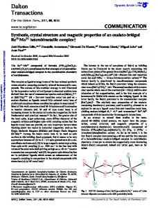
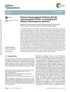
Scheme 1 Synthetic procedure of the MP@NiSiO/Pd composite nanostructure.
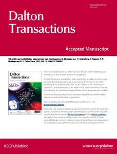
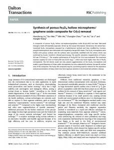
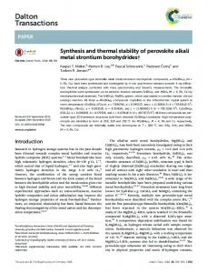
nanocatalysts with tunable sizes were successfully immobilized on the particles by impregnating MP@NiSiO rattles in a K2 PdCl4 solution, followed by a simple NaBH4 reduction process (step 3). Finally, the as-prepared MP@NiSiO/Pd can be used to catalyze the Suzuki or Heck coupling reactions. At first, citrate modified MP microspheres were synthesized according to a modified solvothermal method. In comparison to the previously used polyacrylic acid modified MP microspheres,27 the weight ratio of the organic composition in these MP microspheres is sharply decreased, which will be beneficial to improve the anti-oxidation of the rattle particles. These superparamagnetic MP microspheres are uniform and can be well dispersed in water or ethanol solutions. The average diameter of these particles is about 300 nm (Fig. 1a) and the size distribution is very narrow. After a simple sonication assisted sol–gel process, the MP can be coated by a layer of uniform SiO2 shell. To obtain the well rattle type nanostructure, the thickness of the SiO2 shell should be above a certain value.28 According to the TEM image (Fig. 1b), the as-prepared MP@SiO2 microspheres are monodispersed and the SiO2 shell thickness is of about 85 nm. During the following hydrothermal step, the SiO2 acts both as the precursor and sacrificed template and then the MP@NiSiO rattles are synthesized. To obtain a thinner NiSiO shell and reduce the coalescence of the outer shells, this process was only conducted with 6 h. In this case, all the obtained MP@NiSiO particles show a rattle type nanostructure. The size of the MP core is about 300
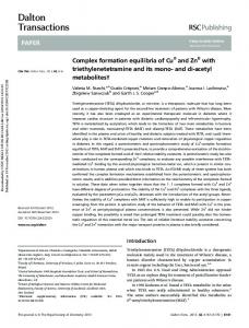
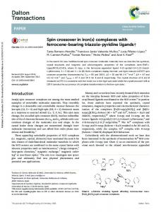
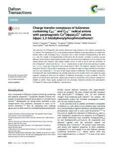
nm and the inner shell thickness is about 450 nm (Fig. 1c and Fig. SI 1a,b†), which agrees well with the formation mechanism. From the TEM image with large magnification (Fig. SI 1c†), it is found that the NiSiO shell is composed of many sheet-like nanoparticles, which indicate that this product can be used as the carrier for nanocatalysts. Fig. 1d shows the TEM image of the as-prepared MP@NiSiO/Pd composite particles. The size of the MP@NiSiO/Pd particle is between 500 to 700 nm and the shell thickness is about 50 nm, which indicates the rattletype nanostructures are well kept during the Pd immobilization process. By carefully investigating the TEM image, there are many black dots existing in the hierarchical shell, which must be the Pd nanocrystals. Obviously, Pd nanoparticles are successfully immobilized on the MP@NiSiO rattles by using such a facile approach. Fig. 2a shows the high magnification TEM image of a single MP@NiSiO/Pd rattle particle. All the Pd nanoparticles are uniformly located in the hierarchical NiSiO shell and no large irregular Pd particle is found. The size of the Pd nanoparticles is about 3–6 nm and these nanoparticles are immobilized on the surface of the NiSiO nanosheets (Fig. 2b). In comparison to the MP@NiSiO rattle particle (Fig. 2c), the NiSiO nanosheets of the MP@NiSiO/Pd particle keep their nanostucture very well, which indicates the sheet-like nanostructure is stable. The energydispersive spectroscopy (EDS) analysis of the yolk nanostructure indicates the presence of Fe, Ni, Si, O, and Pd, proving the product is composed of iron oxide, nickel silicate, and Pd (Fig. 2d). The signals of Cu and C in the EDS spectrum originate from the carbon-coated copper grid.
Fig. 2 TEM images of the MP@NiSiO/Pd (a,b) and MP@NiSiO (c) rattle particles; EDS spectrum of the MP@NiSiO/Pd rattle particles (d).
Fig. 1 TEM images of the MP microspheres (a), MP@SiO2 core/shell particles (b), MP@NiSiO rattles (c), and MP@NiSiO/Pd composite rattle particles; the scale bar is 170 nm.
7828 | Dalton Trans., 2011, 40, 7827–7830
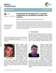
The crystallographic structure of the MP@NiSiO/Pd product was further determined by XRD. The characteristic diffraction peak of Pd at 2q = 40◦ , which corresponded to (111) crystalline planes of Pd (JCPDS 5-0683) was observed in the spectrum, indicating that Pd element exists in the form of Pd0 , not Pd2+ . The broad nature of the diffraction peak clearly indicates the This journal is © The Royal Society of Chemistry 2011
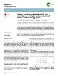
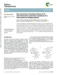
Pd composition is composed of small nanocrystals, which agrees well with the TEM analysis. Unfortunately, the average size of the Pd nanoparticles can not be calculated by using the Scherrer equation,30 because the nearby broader peak at the 2q = 36◦ ((311) crystal plane for MP) also contributes to the d-spacings due to overlapping. The crystallization nature of the Pd nanocatalyst can be controlled by varying the synthetic parameters. Here, we find the choice of solvent has a critical influence on the size of the Pd nanoparticle. When pure water was used as solvent, the size of the Pd nanoparticle was smaller than 6 nm (MP@NiSiO/Pd (1)), as shown in the above analysis. However, when a water/ethanol mixture (1:2 v/v) was employed as the solvent, the size of the Pd nanoparticles increased to 10 nm. Fig. 4a is the TEM image of the MP@NiSiO/Pd (2) rattles, whereas the black dots located in the NiSiO shell are Pd nanoparticles. The size of the Pd nanoparticles is not uniform and most of them are larger than 10 nm (Fig. 4b). Besides the dispersed Pd nanocrystals, some large Pd aggregations are also found (Fig. 4a). Fortunately, all the Pd nanocrystals located on the NiSiO nanosheets are separated from each other (Fig. 4c), which are very different from the Pd nanocrystals synthesized in the absence of MP@NiSiO micro-rattles. From the XRD pattern (Fig. 3), it is found that the diffraction peak of the Pd nanoparticles is relatively sharp. This result supports the average size of the Pd nanoparticles in the MP@NiSiO/Pd (2) rattles being larger than the one in the MP@NiSiO/Pd (1), which further proves that the Pd nanoparticle sizes are controllable.
Fig. 3 XRD patterns of the MP@NiSiO and MP@NiSiO/Pd rattles; MP@NiSiO/Pd (1) is synthesized in water and MP@NiSiO/Pd (2) is synthesized in water/ethanol mixed solution.
During the immobilization, MP@NiSiO micro-rattles provide plenty of carried points since the composites consist of hierarchical NiSiO nanosheets. The Pd ions were absorbed on to the surface of the nanosheets during the impregnating process. Then the Pd nanoparticles which were reduced by the NaBH4 were retained on the surface of the nanosheets. There must also be some Pd nanoparticles encapsulated within the hollow interiors between the MP microspheres and the hierarchical NiSiO shells. However, it is very difficult to directly prove this phenomenon by using the TEM investigation. In this work, no surfactant was used during the formation of the Pd nanoparticles. Thus, the MP@NiSiO rattle particles were proven to be ideal carriers for Pd nanocatalysts. This journal is © The Royal Society of Chemistry 2011
Fig. 4 TEM images of the MP@NiSiO/Pd (a,b,c) particles and Pd nanoparticles (d) synthesized in the absence of MP@NiSiO rattle.
The catalytic properties of transition metal colloids have generated great interest over the past decade. In fact, these new catalysts often combine the precious characteristics of higher reactivity and selectivity.31–33 Here, the catalytic activity of MP@NiSiO/Pd was tested for the Heck reaction. In a typical reaction, 1 mmol of the reagent is dissolved in 3 mL of NMP with 0.1mol% of catalyst. Detailed observations of all the reactions are given in the Experimental section. Completion of the reaction is monitored by thin-layer chromatography (TLC). We observed that the catalyst was very active for the hydrogenation reaction under such mild conditions, since the naked Pd particles show a high catalytic activity. Under the same parameters, it is found that the reaction which is catalyzed by MP@NiSiO/Pd (1) is completed a little quicker than the one catalyzed by MP@NiSiO/Pd (2), which may respond to the size of the Pd nanoparticles located in MP@NiSiO/Pd (1) being smaller. It is reported that catalytic activity of the aggregated Pd nanoparticles is lower than the monodispersed ones,34 thus the MP@NiSiO/Pd (2) shows a better catalytic activity than the bulk Pd nanoparticles. However, the catalytic property of the as-prepared nanocomposites do not exhibit a higher catalytic property than the Pd@CNT,35 which indicates that a synergetic effect has not been obtained in our system. Having demonstrated that the MP@NiSiO/Pd catalyst was highly effective for the Heck reaction, its activity was also investigated for the Suzuki reaction. The result also indicates that the MP@NiSiO/Pd catalyst is a good catalyst. Here, the MP@NiSiO/Pd catalyst shows a magnetic characteristic, which enables them be separated from the reaction system by externally applying a magnetic field. The magnetic separability of such magnetic particles was tested in N-methyl-2-pyrrolidone (NMP) by placing a magnet near the glass bottle; the black particles were attracted toward the magnet, demonstrating directly that the particles possess magnetic properties (Fig. SI2†). This will provide an easy and efficient way to separate MP@NiSiO/Pd particles from a suspension system and to carry catalyst to certain Dalton Trans., 2011, 40, 7827–7830 | 7829
locations under an external magnetic field. After the completion of the reaction, the catalyst is separated by a small magnet placed at the bottom of the reaction vessel. Thereafter, the catalyst is washed with NMP and used for the next reaction. The MP@NiSiO/Pd(2) can be recycled up to 5 times without apparent loss of activity. The conversion is above 90% in all five rounds of the Suzuki reaction recycling (down part of Fig. 5) and the decreased catalytic activity as recycling proceeded may be due to the significant loss of Pd nanoparticles. The MP@NiSiO/Pd(1) can only be recycled for twice, which may be due to the lower stability of the small Pd nanoparticles.
Fig. 5 Observations from the Heck and Suzuki reaction catalyzed by MP@NiSiO/Pd (upper part) and recycling yields of the Suzuki reaction by using the MP@NiSiO/Pd magnetic catalysts (down part).
3.
Conclusions
In conclusion, a novel MP@NiSiO/Pd microsphere with rattle type nanostructures was synthesized by using a templating method. The Pd nanocrystals can be directly immobilized onto the MP@NiSiO rattle particles without using any surfactant or capping reagents. By varying the reaction conditions, the size of the immobilized Pd nanoparticles can be controlled. Such MP@NiSiO/Pd composites show a high catalytic activity on the Heck and Suzuki reactions. They can be separated from the reaction system by using a magnet and the catalyst can be cycled at least 5 times. Most importantly, these results offer a powerful platform to construct other multicomponent composite spheres, which are likely to find many potential catalytic and biomedical applications derived from their rational combination of magnetic properties with surface plasmon resonance, luminescence, or catalysis.
7830 | Dalton Trans., 2011, 40, 7827–7830
Acknowledgements This work was supported by “the Fundamental Research Funds for the Central Universities, no. WK2090000002”
References 1 P. T. Anastas, M. M. Kirchhoff and T. C. Williamson, Appl. Catal., A, 2001, 221, 3–13. 2 R. Schlogl and S. B. Abd Hamid, Angew. Chem., Int. Ed., 2004, 43, 1628–1637. 3 S. Scire, S. Minico, C. Crisafulli, C. Satriano and A. Pistone, Appl. Catal., B, 2003, 40, 43–49. 4 D. I. Enache, J. K. Edwards, P. Landon, B. Solsona-Espriu, A. F. Carley, A. A. Herzing, M. Watanabe, C. J. Kiely, D. W. Knight and G. J. Hutchings, Science, 2006, 311, 362–365. 5 F. Wang, C. H. Li, L. D. Sun, H. S. Wu, T. Ming, J. F. Wang, J. C. Yu and C. H. Yan, J. Am. Chem. Soc., 2011, 133, 1106–1111. 6 D. Astruc, F. Lu and J. R. Aranzaes, Angew. Chem., Int. Ed., 2005, 44, 7852–7872. 7 I. Lee, Q. Zhang, J. Ge, Y. Yin and F. Zaera, Nano Res., 2010, 4, 115– 123. 8 S. F. Chen, J. P. Li, K. Qian, W. P. Xu, Y. Lu, W. X. Huang and S. H. Yu, Nano Res., 2010, 3, 244–255. 9 Y. Y. Sun and R. Prins, Angew. Chem., Int. Ed., 2008, 47, 8478–8481. 10 B. Yoon and C. M. Wai, J. Am. Chem. Soc., 2005, 127, 17174–17175. 11 L. Li, Y. J. Feng, Y. S. Li, W. R. Zhao and J. L. Shi, Angew. Chem., Int. Ed., 2009, 48, 5888–5892. 12 S. H. Xuan, Y. X. J. Wang, J. C. Yu and K. C. F. Leung, Langmuir, 2009, 25, 11835–11843. 13 M. Feyen, C. Weidenthaler, F. Schuth and A. H. Lu, Chem. Mater., 2010, 22, 2955–2961. 14 S. C. Tsang, V. Caps, I. Paraskevas, D. Chadwick and D. Thompsett, Angew. Chem., Int. Ed., 2004, 43, 5645–5649. 15 D. K. Yi, S. S. Lee and J. Y. Ying, Chem. Mater., 2006, 18, 2459–2461. 16 J. P. Ge, Q. Zhang, T. R. Zhang and Y. D. Yin, Angew. Chem., Int. Ed., 2008, 47, 8924–8928. 17 Y. H. Deng, Y. Cai, Z. K. Sun, J. Liu, C. Liu, J. Wei, W. Li, C. Liu, Y. Wang and D. Y. Zhao, J. Am. Chem. Soc., 2010, 132, 8466–8473. 18 C. W. Lim and I. S. Lee, Nano Today, 2010, 5, 412–434. 19 S. H. Wu, C. T. Tseng, Y. S. Lin, C. H. Lin, Y. Hung and C. Y. Mou, J. Mater. Chem., 2011, 21, 789–794. 20 D. Chen, L. L. Li, F. Q. Tang and S. Qi, Adv. Mater., 2009, 21, 3804– 3807. 21 L. F. Tan, D. Chen, H. Y. Liu and F. Q. Tang, Adv. Mater., 2010, 22, 4885–4889. 22 W. Cheng, K. B. Tang, Y. X. Qi, J. Sheng and Z. P. Liu, J. Mater. Chem., 2010, 20, 1799–1805. 23 Y. F. Zhu, E. Kockrick, T. Ikoma, N. Hanagata and S. Kaskel, Chem. Mater., 2009, 21, 2547–2553. 24 M. C. Zhang, Y. Lan, D. Wang, R. Yan, S. N. Wang, L. Yang and W. Q. Zhang, Macromolecules, 2011, 44, 842–847. 25 K. Zhang, X. H. Zhang, H. T. Chen, C. Xin, L. L. Zheng, J. H. Zhang and B. Yang, Langmuir, 2004, 20, 11312–11314. 26 L. Xu, X. C. Wu and J. J. Zhu, Nanotechnology, 2008, 19, 305603. 27 S. H. Xuan, Y. X. J. Wang, J. C. Yu and K. C. F. Leung, Chem. Mater., 2009, 21, 5079–5087. 28 Q. L. Fang, S. H. Xuan, W. Q. Jiang and X. L. Gong, Adv. Funct. Mater., 2011, 21, 1902–1909. ¨ 29 W. Stober and A. Fink, J. Colloid Interface Sci., 1968, 26, 62–69. 30 H. P. Klug and L. E. Alexander, X-ray Diffraction Procedures for Polycrystalline and Amorphous Materials, Wiley, New York, 1962, pp.491–538. 31 M. Moreno-Manas and R. Pleixats, Acc. Chem. Res., 2003, 36, 638– 643. 32 M. N. Vargaftik, V. P. Zargorodnikov, I. P. Stolarov, I. I. Moiseev, D. I. Kochubey, V. A. Likholobov, A. L. Chuvilin and K. I. Zamaraev, J. Mol. Catal., 1989, 53, 315–348. 33 J. D. Aiken III and R. G. J. Finke, J. Mol. Catal. A: Chem., 1999, 145, 1–44. 34 A. Roucoux, J. Schulz and H. Patin, Chem. Rev., 2002, 102, 3757–3778. 35 B. H. Yoon and C. M. Wai, J. Am. Chem. Soc., 2005, 127, 17174–17175.
This journal is © The Royal Society of Chemistry 2011