RESEARCH ARTICLE
Decoding Sensorimotor Rhythms during Robotic-Assisted Treadmill Walking for Brain Computer Interface (BCI) Applications Eliana García-Cossio1*, Marianne Severens1,2, Bart Nienhuis2, Jacques Duysens2,3, Peter Desain1, Nöel Keijsers2☯, Jason Farquhar1☯ 1 Donders Institute for Brain, Cognition and Behaviour, Radboud University, Nijmegen, The Netherlands, 2 Research Development & Education Department, Sint Maartenskliniek, Nijmegen, The Netherlands, 3 Department of Kinesiology, KU Leuven, Leuven, Belgium ☯ These authors contributed equally to this work. *
[email protected]
Abstract OPEN ACCESS Citation: García-Cossio E, Severens M, Nienhuis B, Duysens J, Desain P, Keijsers N, et al. (2015) Decoding Sensorimotor Rhythms during RoboticAssisted Treadmill Walking for Brain Computer Interface (BCI) Applications. PLoS ONE 10(12): e0137910. doi:10.1371/journal.pone.0137910 Editor: Yuri P. Ivanenko, Scientific Institute Foundation Santa Lucia, ITALY Received: April 22, 2015 Accepted: August 22, 2015 Published: December 16, 2015 Copyright: © 2015 García-Cossio et al. This is an open access article distributed under the terms of the Creative Commons Attribution License, which permits unrestricted use, distribution, and reproduction in any medium, provided the original author and source are credited. Data Availability Statement: All relevant data are available via DANS (http://dx.doi.org/10.5072/danszcm-q8fq).
Locomotor malfunction represents a major problem in some neurological disorders like stroke and spinal cord injury. Robot-assisted walking devices have been used during rehabilitation of patients with these ailments for regaining and improving walking ability. Previous studies showed the advantage of brain-computer interface (BCI) based robot-assisted training combined with physical therapy in the rehabilitation of the upper limb after stroke. Therefore, stroke patients with walking disorders might also benefit from using BCI robot-assisted training protocols. In order to develop such BCI, it is necessary to evaluate the feasibility to decode walking intention from cortical patterns during robot-assisted gait training. Spectral patterns in the electroencephalogram (EEG) related to robot-assisted active and passive walking were investigated in 10 healthy volunteers (mean age 32.3±10.8, six female) and in three acute stroke patients (all male, mean age 46.7±16.9, Berg Balance Scale 20±12.8). A logistic regression classifier was used to distinguish walking from baseline in these spectral EEG patterns. Mean classification accuracies of 94.0±5.4% and 93.1±7.9%, respectively, were reached when active and passive walking were compared against baseline. The classification performance between passive and active walking was 83.4±7.4%. A classification accuracy of 89.9±5.7% was achieved in the stroke patients when comparing walking and baseline. Furthermore, in the healthy volunteers modulation of low gamma activity in central midline areas was found to be associated with the gait cycle phases, but not in the stroke patients. Our results demonstrate the feasibility of BCI-based robotic-assisted training devices for gait rehabilitation.
Funding: This study was supported by the BrainGain Smart Mix Programme of the Netherlands Ministry of Economic Affairs and the Netherlands Ministry of Education, Culture and Science. The funder had no role in study design, data collection and analysis, decision to publish, or preparation of the manuscript. Competing Interests: The authors have declared that no competing interests exist.
PLOS ONE | DOI:10.1371/journal.pone.0137910 December 16, 2015
1 / 21
Decoding Brain Signals during Robot Gait Training
Introduction Stroke is the main cause of disability in adults [1]. Many patients present lower limb impairment characterized by abnormal muscle activations. Three months after the stroke, about a quarter of these patients are still bound to the wheelchair [2]. Robot-assisted training devices have been used during rehabilitation of stroke patients for regaining and improving walking ability, offering longer training duration, increasing movement repetitions and reducing the physical load imposed upon the therapist. Robotic training can provide the intensive and task-oriented type of training that has proven effective for promoting motor learning [1,3], which is thought to be useful for motor recovery after stroke [4]. Despite the lack of consensus in the literature, a recent systematic review on the topic has shown benefits of robot-assisted treadmill training. Stroke patients who received electromechanical assisted gait training in combination with physical therapy are more likely to achieve independent walking than patients receiving gait training without these devices [5]. One of the underlying mechanisms of the benefit of robot assisted-treadmill training is multisensory feedback. Multisensory feedback plays an important role in motor learning by reestablishing the sensorimotor loop that is disrupted after stroke [6,7]. Several multisensory feedback approaches have been reported for motor recovery in patients with stroke, including action-observation [8], and recently developed Brain-Computer-Interfaces (BCI) coupled to orthotic devices[9]. A BCI system can provide multisensory feedback (e.g. visual and propioceptive (robots)[10]) allowing the users to modulate their brain activity by operant conditioning [11]. BCIs can couple intention with action and enable patients with stroke to achieve intended motor actions by exploiting neural learning mechanisms [11]. Interestingly, it has been suggested that the combination of robotics and brain control of upper limb assistive technology [12–15] leads to motor learning and induces neural plasticity resulting in motor function improvement [9,16–21]. In order to develop a BCI control of the robot-assisted gait device, fundamental research aiming at detecting the precise active role of the motor cortex during the gait cycle has to be done. Furthermore, it is important to identify what can effectively and non-ambiguously be measured using non-invasive brain signals such as EEG: descending commands from the motor cortex, ascending sensorimotor information, integration of both or artifacts. So far, only few studies have investigated the neural correlates of human walking, principally due to both the inherent experimental difficulty of measuring EEG signals in the ambulatory context and the challenging goal of balance control in walk rehabilitation tasks [22]. However, it has been recently confirmed that the motor cortex is particularly active during specific phases of the gait cycle, particularly before the foot comes in contact with the ground [23–26]. Together these studies and others [27–29] have demonstrated that supraspinal circuits, especially those of the motor cortex, have a significant role in motor control during walking. Furthermore, researchers have shown that active training can enhance motor performance and increase corticospinal excitability in comparison to passive training [30]. Therefore, for motor rehabilitation purposes it is necessary to actively involve the patients during the training. At a fixed pattern and constant speed in robotic-training devices users often start relying on the robot to perform the movement and reduce their muscular activity [31]. An important component in the success of neural plasticity and motor learning is the supraspinal engagement during the task. Therefore, several studies have attempted at detecting active subject participation during robot training. One way to overcome this problem is by incorporating control algorithms that require the patient to actively initiate movements to perform the task. Using EEG, previous studies have investigated the difference between active and passive movement during robot-assisted gait training. A significant decrease in the mu, beta and gamma
PLOS ONE | DOI:10.1371/journal.pone.0137910 December 16, 2015
2 / 21
Decoding Brain Signals during Robot Gait Training
bands during active compared to passive walking was observed in the right primary motor cortex hand area, indicating increased cortical involvement during active walking [26]. However, it remains to be tested whether these cortical patterns can be classified reliably for an online detection of active cortical involvement during robot-assisted gait training. In this study we aimed at demonstrating the feasibility of a BCI-based robotic-assisted training device for gait rehabilitation by decoding the intention of walking on the basis of EEG signals during robot-assistive gait training in ten healthy volunteers and three stroke patients with mild lower limb impairment. Moreover, we aimed at detecting the precise role of the sensorimotor cortex during active (intention to walk) and passive walking (no intention to walk) to find out to which extend the cortical involvement during gait influences the patterns of neural signals recorded by EEG.
Materials and Methods Participants 10 healthy volunteers (mean age 32.3 ± 10.8, six female) without a history of neurological or psychiatric disorders and three acute ischemic stroke patients participated in the experiment (3 males, mean age 46.7±16.9, Berg Balance Scale 20±12.8). The stroke patients presented a first ischemic stroke 2, 3 and 2 months before they participated in this study, respectively. Patients presented severe left sided hemiparesis and severe difficulties to stand and walk. Detail demographic information is presented in Table 1.
Ethics Statement The Medical Ethics Committee of the Radboud University medical center approved this study and all participants provided written informed consent before entering the study.
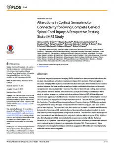
Exoskeleton The Lokomat Pro (Hocoma AG, Volketswil, Switzerland) was used to assist walking. This exoskeleton is a bilaterally driven gait orthosis in which a body-weight support system and a treadmill are incorporated (Fig 1A). The orthosis moves the legs along a specified trajectory in the sagittal plane, with hip and knee joints of the orthosis actuated by linear drives that are integrated into an exoskeleton.
Electroencephalographic (EEG) recordings Electrical signals from 62 electrodes were recorded at 500Hz sampling rate using a TMSi Refa72 amplifier (Twente Medical Systems International, The Netherlands). Impedance of the electrodes was kept below 50KO. Table 1. Demographic information stroke patients. Patient
Age
Gender
Months after stroke
Balance Berg Score
Lesioned hemisphere
BWS
GF
P1
51
m
2
6
P2
61
m
3
31
right
64.20%
85%
right
35.9%
P3
28
m
2
23
right
60%
40%
65%
m = male; BWS = Body weight support, GF = guidance force. doi:10.1371/journal.pone.0137910.t001
PLOS ONE | DOI:10.1371/journal.pone.0137910 December 16, 2015
3 / 21
Decoding Brain Signals during Robot Gait Training
Fig 1. Experimental design and canonical correlation analysis example. a. Experimental setup in the lokomat with the EEG electrodes and cap, the EMG electrodes located in the right trapezius (neck), right gastroc medialis (calf), right semintendinosus (hamstr.) and right vastus lateralis (quadri.), and the corresponding accelerometers for the right and left foot (accel.). b. Schematic representation of a trial starting with a relax period hanging (baseline) and continuing with a walking task which could be either passive or active. c. Average spectrum across all blocks for all extracted components (raw signal (black)), for only the muscle related components (EMG, red) and for only unrelated muscle components (raw—EMG, blue) using canonical coherence analysis (CCA). doi:10.1371/journal.pone.0137910.g001
Electromyographic (EMG) recordings Muscle activity from the right leg (healthy volunteers) or the paretic leg (stroke patients) was recorded using three bipolar electrodes over the gastroc medialis (calf), semintendinosus (hamstring) and vastus lateralis (quadriceps). In addition, muscle activity from the right trapezius on the neck (neck) was recorded (Fig 1A).
Accelerometers In order to track the gait cycle during walking in the Lokomat accelerometers were positioned on each leg above the metatarsal bones (Fig 1A). The accelerometers used were type ADXL 335 (Analog Devices One Technology Way, MA, USA).
Experimental Design Healthy Volunteers. Healthy participants attended one experimental session in which they were asked to either walk in a passive or in an active mode at a speed of 1.5Km/h or to stand in the Lokomat. During the passive and active walking conditions the body weight support (BWS) and the guidance force (GF) were manipulated. For the passive walking GF of
PLOS ONE | DOI:10.1371/journal.pone.0137910 December 16, 2015
4 / 21
Decoding Brain Signals during Robot Gait Training
80% and BWS of 75% were used, while for the active walking GF was set to 30% and BWS to 5% (a complete passive walking with a GF of 100% and BWS of 0% was not possible because the participants were not able to walk in the Lokomat anymore and it was not a good option either since this completely changes the stepping behavior [32]). Participants were asked to perform as little effort as possible during the passive walking condition and to allow the legs to be moved by the robot. Moreover, they were verbally informed about how passive they were walking on the basis of their EMG signals. During the task, participants hold on to the safety bars at the sides of the treadmill. Participants were instructed to follow the device as best as possible during all walking conditions (e.g. avoiding pushing against the knee and hip orthosis). A familiarization period for active and passive walking was given to the participants. After the subject felt comfortable walking in the Lokomat, the experimental session was started. An experimental session consisted of 14 blocks, seven for passive and seven for active walking. Blocks were presented in a randomized order. Each block started with a period of quiet baseline while participants were lifted from the treadmill in the robot (100% BWS, meaning no balance needed and no contact with the floor) and looking at a fixation cross on a computer screen for 7.5s (Fig 1B). Afterwards an instruction was displayed on the screen advising the participant about the task to be executed; active or passive walking. In both walking tasks, the treadmill started, participants walked for 49s, after which the treadmill was stopped. The treadmill had a delay of at least 7s to come up to a stable speed and 7s to slow down and stop completely. The recordings only took place during constant and stable speed, which was indicated by the physiotherapist controlling the robotic device and a trigger marker in the EEG recordings. During the baseline and walking periods a fixation cross was displayed on the screen. Resting periods between blocks were made depending on the participant’s fatigue. Stroke patients. Stroke patients also attended one experimental session in which they were asked to walk at a comfortable speed of maximum 1.5Km/h or to rest in the Lokomat with 100% of BWS and no contact with the floor (baseline). A physiotherapist controlled the speed of the orthosis according to the patient’s capabilities. The BWS and the GF was adjusted for each patient’s limitations (see Table 1). For patients, an experimental session consisted of 10 blocks. Each block started with a period of lifted from the treadmill (100% BWS, meaning no balance needed and no contact with the ground) while participants looked at a fixation cross on a computer screen for 7.5s, which was used as a baseline condition (Fig 1A). Subsequently, an instruction was displayed on the screen advising the participant about the initiation of the task. During walking, the treadmill started and patients walked for 49s, after which the treadmill was stopped. The physiotherapist indicated when the patient walked properly. During the baseline and walking periods a fixation cross was displayed on the screen. Resting periods between blocks were made to avoid fatigue.
EEG analysis EEG data were downsampled to 250Hz, linearly detrended and epoched according to gait cycle information recorded from the accelerometers. The gait cycle phases were defined relative to the right heel strike (measured by the accelerometers) for the healthy volunteers and for the patients relative to the paretic leg heel strike (see S1 Fig). The other gait cycle phases, apart from the right heel strike, were defined according to the literature [33]. In total, around 17 to 21 gait cycles were detected in each block, depending on the participant’s leg length. EEG signals were re-referenced to a common average across all channels. A Canonical correlation
PLOS ONE | DOI:10.1371/journal.pone.0137910 December 16, 2015
5 / 21
Decoding Brain Signals during Robot Gait Training
analysis (CCA) method [34] was used to remove the EMG artifacts on the EEG signals. This worked by identifying and removing sources (components), such as muscle activity, which have low temporal auto-correlation as assessed by having power in the EMG frequency band (15-30Hz) more than 1.3 times stronger than in the EEG frequency band (1-30Hz). The components identified as muscle activity are marked as EMG and removed from the raw signals. The remaining components are kept and used to reconstruct the EEG activity (Fig 1C). The mastoid electrodes located on TP8 and TP7 were removed and on the remaining EEG electrodes a surface Laplacian based on spherical spline interpolation [35] was performed to improve spatial selectivity. Power spectral analysis was performed using Welch’s method with a Hanning window of 250ms. For classification of the EEG signals into different conditions, the frequency bins from 8 to 30Hz were used. Frequencies below 8 Hz were not considered in the analysis in order to avoid any influence from movement artifacts (