of the embryo's dorsal-ventral axis, as suggested by a number of ... cytoplasmic rotation specifies the dorsal side is not understood, but it is .... diameter from 1 to 14 fan. ..... The counter- .... dorsal axial tissue (reviewed in Elinson and Kao, 1989;.
845
Development 111, 845-856 (1991) Printed in Great Britain © The Company of Biologists Limited 1991
Deep cytoplasmic rearrangements during early development in Xenopus laevis
M. V. DANILCHIK* and J. M. DENEGRE Department of Biology, Wesleyan University, Middletown, Connecticut 06459, USA
.
•Corresponding author
Summary
The egg of the frog Xenopus is cylindrically symmetrical about its animal-vegetal axis before fertilization. Midway through the first cell cycle, the yolky subcortical cytoplasm rotates 30° relative to the cortex and plasma membrane, usually toward the side of the sperm entry point. Dorsal embryonic structures always develop on the side away from which the cytoplasm moves. Details of the deep cytoplasmic movements associated with the cortical rotation were studied in eggs vitally stained during oogenesis with a yolk platelet-specific fluorescent dye. During the first cell cycle, eggs labelled in this way develop a complicated swirl of cytoplasm in the animal
hemisphere. This pattern is most prominent on the side away from which the vegetal yolk moves, and thus correlates in position with the prospective dorsal side of the embryo. Although the pattern is initially most evident near the egg's equator or marginal zone, extensive rearrangements associated with cleavage furrowing (cytoplasmic ingression) relocate portions of the swirl to vegetal blastomeres on the prospective dorsal side.
Introduction
of the embryo's dorsal-ventral axis, as suggested by a number of experiments in which redirecting or halting the vegetal cortex movement alters the orientation or degree of development of dorsal structures (Grant and Wacaster, 1972; Malacinski et al. 1977; Manes and Elinson, 1980; Scharf and Gerhart, 1980, 1983; Kirschner and Gerhart, 1981; Gerhart etal. 1981; Black and Gerhart, 1985). The process by which this corticalcytoplasmic rotation specifies the dorsal side is not understood, but it is clear that, before fertilization, the potential for producing dorsal structures exists at every point around the animal-vegetal axis. It is unlikely that the specified dorsal information actually resides in the cortex itself: first, because the embryonic fate map can be reversed relative to the cortex by centrifugation during first cell cycle (Malacinski, 1984; Cleine and Dixon, 1985; Black and Gerhart, 1986), and second, because a dorsal-determining target of UV that is distinct from the rotation machinery exists in premeiotic oocytes (Elinson and Pasceri, 1989). Thus, the rotation's function is presumably to redistribute or activate components necessary for dorsal-ventral specification in the egg. The rotation machinery is located in the vegetal cortex, but its physical relationship to the dorsal specification process is not known. Information necessary for normal dorsal development is known to reside in the vegetal tiers of blastomeres at the 32-cell stage
The polarized organization of the Xenopus egg's contents relates directly to the region-specific development of the embryonic germ layers (reviewed in Gerhart, 1980; Gerhart et al. 1989). This regular correspondence between egg and embryo organizations suggests that there are specific molecules, activities or structures in the unfertilized egg which become localized and function to direct the various developmental pathways that tissues take in order to differentiate. The steps in this localization process have not been completely elucidated. In Xenopus, the point of sperm entry (SEP) defines the orientation of the embryonic dorsal-ventral axis, with the dorsal side normally forming opposite the SEP (Ancel and Vintemberger, 1948; Elinson, 1975, 1980). During the first cell cycle, the vegetal yolky cytoplasm the vegetal yolk mass - rotates relative to the plasma membrane and cortex about 30° toward the SEP, that is, away from the prospective dorsal side of the embryo (Vincent et al. 1986; Vincent and Gerhart, 1987). This movement seems to depend on an array of parallel microtubules which appear transiently in the vegetal cortex during the first cell cycle (Elinson and Rowning, 1988). The vegetal cortical-cytoplasmic rotation is thought to be the earliest determining event in the specification
Key words: Xenopus, cleavage, cytoplasmic localization, dorsal-ventral axis,firstcell cycle, ingression.
846
M. V. Danilchik and J. M. Denegre
(Gimlich and Gerhart, 1984; Gimlich, 1986), suggesting a proximity to the rotation machinery. However, a number of recent experiments suggest that blastomeres in the animal hemisphere have specialized in some way as well (Kageura and Yamana, 1984; Cardellini, 1988; London et al. 1988; Takasaki and Konishi, 1989). How the cortical-cytoplasmic rotation would transmit information to the animal hemisphere remains enigmatic (see Wakahara, 1989). Rearrangements of the deep cytoplasm that might accompany cortical-cytoplasmic rotation have not been documented because it is difficult to label selectively regions of egg cytoplasm before cleavage. In Xenopus eggs, the only reliable cytoplasmic marker has been the yolk platelet gradient, which has been used to indicate movement of various cytoplasmic regions during early development (Nakatzuji, 1975; Phillips, 1985; Ubbels et al. 1983; Neff et al. 1984; Black and Gerhart, 1986). We have sought to improve this situation by selectively marking peripheral and animal hemisphere yolk platelets during oogenesis with the fluorescent dye trypan blue (TB). Epifluorescence microscopy of histological sections and confocal scanning laser microscopy of whole-mount specimens have enabled us to obtain detailed information about cytoplasmic movements of the first cell cycle and cleavage period in normal development in Xenopus. In subsequent reports, we will examine the roles of these rearrangements in establishing body pattern in the early embryo. Materials and methods Oocytes, eggs and embryos In vitro maturation of manually defolliculated oocytes was accomplished by adding progesterone (4/igml"1) to fullstrength MMR. Eggs of Xenopus laevis were fertilized, dejellied and cultured as previously described (Vincent et al. 1986). Dejellied eggs were immobilized on plastic dishes by immersion in 6% Ficoll, arranged with sperm entry points (SEPs) in a known orientation, and cultured at 18-22°C to appropriate stages. Before fixation in Bouin's fixative, eggs were gently pricked with a glass needle at the SEP to produce a tiny reference scar, since vital dyes such as Nile Blue Sulfate disappear during fixation. The average orientation of the definitive dorsal-ventral axis relative to the SEP was determined by scoring a group of control embryos at stage 14. Developmental times in the first cell cycle are normalized to a scale of 0 to 1.0, referring to fertilization and appearance of the first cleavage furrow, respectively. Fluorescent marking of egg cytoplasm Eggs with labelled yolk platelets were obtained by methods previously developed to study vitellogenesis in Xenopus (Danilchik and Gerhart, 1984, 1987; CaUen, 1986). 7 to 10 days after gonadotropin-induced spawning, female frogs were injected in the dorsal lymph sac with 100 to 400^1 of 10 nig ml" 1 TB (Polysciences or Sigma), which had been dialyzed extensively against distilled water (MWCO 15000), lyophilized, and dissolved at lOmgml"1 in MMR. 10 to 14 days after injection of label, frogs were primed by injection of 50 i.u. of pregnant mare serum gonadotropin (Calbiochem), and on the following day again induced to spawn by injection of 800 i.u. of human chorionic gonadotropin (Sigma).
Time-lapse recording of vegetal yolk mass rotation The direction of cortical-cytoplasmic rotation was determined by video time-lapse recording of the vegetal surface of TBlabelled eggs during the first cell cycle. The time-lapse recorder consisted of a BioRad MRC-500 scanning laser confocal imaging system connected to a Nikon Diaphot inverted microscope. Images were accumulated from three to five sequentially scanned frames and then contrast-enhanced. The resulting video output was then recorded on a JVC BR9000U time-lapse VCR. Successive video images were collected every 15 to 20 s. The direction of vegetal yolk mass rotation was easily determined with this system in TB-labelled eggs because individual fluorescent yolk platelets could be tracked across the entire vegetal surface. Up to 6 eggs could be viewed simultaneously by using a 4x/0.2 N.A. objective. At the end of the first cell cycle, the direction of rotation was determined by playing back the tape. Eggs were then gently pricked with a glass needle to make an unambiguous mark of the rotation direction, and fixed in Bouin's one minute later. Histology and microscopy Eggs and embryos were fixed and sectioned as described previously (Danilchik and Black, 1988). Eggs were embedded in JB-4 (Polysciences) with known orientation relative to the SEP or other reference mark, and sectioned either sagittally (parallel to the plane defined by the animal-vegetal axis and either the SEP meridian or direction of cortical-cytoplasmic rotation), or horizontally (perpendicular to the animalvegetal axis). Sections were examined by fluorescence microscopy (rhodamine excitation). For confocal microscopy, eggs were fixed with Bouin's fixative and washed with 5mM NH4OH in 50% ethanol. Pigmented eggs were then bleached for 2.5 h with 15% hydrogen peroxide (Baker) and 50% ethanol. Bleaching was promoted by illuminating the eggs in their glass shell vials on a standard white fluorescent light box. This treatment did not detectably affect the TB labelling. Samples were then washed in 50% ethanol and dehydrated and cleared as in Dent and Klymkowsky (1989). Results
Labelling of cytoplasm with fluorescent yolk platelets Previous work with fluorescent and radioactive analogues of vitellogenin indicated that uptake of vitellogenin by endocytosis is nearly uniform over the Xenopus oocyte surface (Danilchik and Gerhart, 1987). Oocytes readily take up TB from the maternal circulation (Dumont, 1972), and deposit it into yolk platelets in a pattern similar to that of vitellogenin (Danilchik and Gerhart, 1984, 1987; Callen, 1986). The dye is fixable in situ with Bouin's fixative (Telfer and Anderson, 1968). In endosomes and yolk platelets, it is highly fluorescent, with a broad emission spectrum similar to that of rhodamine. Thus, even invisibly small amounts of the dye, as in individual yolk platelets, are readily detectable by epifluorescence microscopy (Fig. 1). Large numbers of fertilizable, developmentally normal eggs with nearly identical labelling patterns are obtained by injecting TB into frogs one to two weeks after hCG-induced spawning, and then inducing a second spawning within two weeks. Although TB has powerful teratogenic effects when present in the
Cytoplasmic movements in Xenopus eggs
847
Fig. 1. Oocyte contents undergo significant rearrangements during germinal vesicle breakdown. (A,B) Oocyte, fixed 6 days after injection of TB into female frog, was sectioned at 6/an, as described in Methods. (C,D) Stage V-VI oocyte 3h after progesterone treatment, with orientation as above. (E,F) Unfertilized egg spawned from same female from which oocytes in previous panels had been obtained 3 days earlier. All sections are through the animal-vegetal axis, animal pole up. Left panels: phase-contrast. Right panels: epifluorescence (rhodamine filter set). Magnification: 15.5x.
blastocoel before gastrulation (Waddington and Perry, 1956; Greenhouse and Hamburgh, 1968; Gerhart et al. 1984; Danilchik, 1986), the dye has no teratogenic activity when confined to yolk platelets, as evidenced by normal development at least through metamorphosis of tadpoles derived from intensely labelled eggs (not shown). Cytoplasmic rearrangements during maturation Oocytes removed from a frog injected with TB 6 days earlier reveal a pattern of dye incorporation similar to
that previously described for Lucifer Yellow-conjugated vitellogenin (cf. Fig. 6A, Danilchik and Gerhart, 1987). In the animal hemisphere, the small yolk platelets are in radial columns (Fig. 1A, phase-contrast) centering on the large yolk-free region at the vegetal base of the germinal vesicle (GV). Nearly all are heavily labelled with TB (Fig. IB, fluorescence), indicating that they were undergoing growth at the time of labelling and probably originated during the terminal stages of oogenesis. In contrast, in the vegetal hemisphere, labelling is confined to a thick (100 to 150 jan)
848
M. V. Danilchik and J. M. Denegre
peripheral layer composed of platelets ranging in diameter from 1 to 14 fan. In the deep interior of the vegetal hemisphere, yolk platelets are unlabelled. The thickness of the deposition layer in the vegetal hemisphere depends on the period of exposure to TB during a particular oocyte's growth. During progesterone-induced in vitro maturation, the GV rises to the animal pole and the nuclear envelope breaks down, starting from the vegetal side of the GV (Fig. 1C). Concomitant with the GV's poleward movement, a large volume of fluorescent yolk platelets is displaced inward from the animal pole surface (Fig. ID), producing a roughly conical arrangement of brightly fluorescent platelets around the nonfluorescent central column of released GV sap. This cone-shaped fluorescent region is flanked laterally by blunt, upturned extensions of the underlying central cytoplasm (Fig. ID, asterisk). These extensions contain unlabelled yolk platelets that are usually somewhat larger than those found nearby, suggesting that they were displaced there from deeper in the vegetal interior. The yolk platelets in the animal hemisphere are no longer arranged in radial groups as they were before maturation. Rather, individual yolk platelets have freely intermingled with the released GV contents and the surrounding cytoplasm. During maturation, the sub-GV yolk-free region flattens, and appears to limit the extent of inward movement by the labelled animal hemisphere yolk platelets (Fig. 1C; arrow). Eggs spawned between 10 and 20 days following TB injection yield a consistent labelling pattern: the animal hemisphere cytoplasm labelled throughout with small, fluorescent yolk platelets, and the vegetal hemisphere cytoplasm only at its periphery (Fig. 1E,F). In the animal hemisphere, the small yolk platelets have become more or less evenly dispersed (Fig. IE), and form a large fluorescent cytoplasmic mass near the center of the egg (Fig. IF). This central mass of small fluorescent platelets is delimited near the equatorial plane by an abrupt transition to larger, unlabelled platelets. The position of this boundary varies slightly with the length of time between TB injection and spawning, as might be expected from the progressive vegetad movement of labelled animal hemisphere yolk platelets during oogenesis (Callen, 1986; Danilchik and Gerhart, 1987). The upturned extensions of vegetal unlabelled cytoplasm that developed during maturation are still visible at the lateral edges of the central mass (see Fig. ID). A number of 30-50/an diameter yolkfree cytoplasmic bodies, possibly remnants of the oocyte's yolk-free region, are frequently found in a plane near the transition between labelled and unlabelled yolk. Additional small, yolk-free areas are occasionally seen throughout the animal hemisphere cytoplasm. The label distribution in the vegetal hemisphere appears to remain largely undisturbed by the cytoplasmic movements of oocyte maturation. Cytoplasmic rearrangements during the first cell cycle To study rearrangements during the first cell cycle, TB-
labelled eggs were fixed at various times following fertilization, and sectioned in known orientation relative to the SEP. During the middle third of the first cell cycle, the central animal hemisphere cytoplasmic mass shifts progressively toward the prospective dorsal side. This movement produces a complicated swirl, consisting of alternating labelled and unlabelled cytoplasmic regions in the equatorial zone. The development of this swirl is shown in Fig. 2. Minimal rearrangements of the original egg organization are apparent at 0.3 of the cell cycle (Fig. 2A), consistent with the expectation that the vegetal yolk mass only begins to shift toward the sperm entry point at this time (Vincent et al. 1986). The sperm trail is visible as a horizontal dark line of yolk-free cytoplasm penetrating the animal hemisphere from the left side of the egg (S). The upward extensions of unlabelled vegetal cytoplasm that flank the labelled central yolk have begun to lose their earlier symmetry (cf. Fig. IF): on the SEP side of the egg (left in Fig. 2), the extension forms a more acute angle with the underlying unlabelled cytoplasm, while that on the right has begun to foreshorten and is now nearly perpendicular to the equatorial plane. By 0.5 of the cell cycle, when the vegetal yolk mass rotation is well under way, the rearrangements of the egg's original cytoplasmic organization are more pronounced (Fig. 2B). The central labelled mass has shifted considerably toward the future dorsal equatorial zone. The upward extensions of unlabelled material flanking it have become even more asymmetric: on the SEP side it has lengthened and flattened, while on the opposite side, it has begun recurving toward the animal pole. The egg itself has flattened under the influence of gravity. At the time of first cleavage furrow formation (90 min post-fertilization), an extensive cytoplasmic rearrangement (Fig. 2C) with alternating fluorescent and nonfluorescent layers has developed in the prospective dorsal equatorial zone and animal hemisphere. Although the pattern varies in minor details from egg to egg and from batch to batch, common features consistently seen in sagittal section are: (1) an asymmetric, unlabelled vegetal yolk mass, the upper limit of which is slightly higher on the SEP's equatorial zone, presumably as a consequence of its SEP-ward rotation; (2) a long, comma-shaped fluorescent mass, derived from the labelled central cytoplasm, the head of which is always found in the equatorial zone on the side opposite the SEP; and (3) a thick layer of unlabelled cytoplasm, about 150 jum thick, tapering toward the animal pole from the prospective dorsal side of the vegetal yolk mass, presumably deriving from the now greatly lengthened unlabelled extension described above. At its base near the egg's equator, this layer contains slightly larger unlabelled yolk platelets brought up from the deep equatorial region. The prospective dorsal side thus inherits a triple-layered labelling pattern, which we will henceforth refer to as 'the swirl': (1) a deep zone of labelled, formerly central, cytoplasm, internal to (2) a layer of unlabelled cytoplasm derived from the equatorial deep yolk, which is itself internal to (3) the
Cytoplasmic movements in Xenopus eggs
849
Fig. 2. Cytoplasmic redistributions during first cell cycle. Frog was injected with TB 10 days before spawning, as described in Methods. Eggs were fertilized, dejellied and fixed at various times during the first cell cycle, and then sectioned parallel to the SEP meridian and the animalvegetal axis. Eggs shown here are oriented with animal pole up, SEP on the left side. (A) 30min post-fertilization (0.3 of first cell cycle). S=sperm trail. (B) 45min postfertilization (0.5 of first cell cycle). Fluorescent animal cytoplasm begins shifting toward side opposite SEP. (C) 90min post-fertilization (1.0 of first cell cycle).
layer containing large yolk platelets (Pasteels, 1964; Ubbels etal. 1983; Phillips, 1985). The eggs shown in Fig. 2 were obtained from a spawning in which the SEP was a particularly good indicator of the future dorsal side (average angle of SEP to neural groove of 27 voucher stage 18 embryos from the same batch was 149°). Each section represents essentially identical patterns in 4 or more embryos at each time point examined. No significant variations from this pattern were seen in this experiment. In each specimen, a prominent swirl developed on the side opposite the SEP. Confocal scanning laser microscopic examination of 18 embryos (see below) obtained from several spawnings in which the direction of corticalcytoplasmic rotation was recorded confirms that, in unperturbed eggs, the swirl only develops on the side away from which the vegetal yolk mass rotates. We thus conclude that development of the swirl always accompanies dorsal axis specification in normal Xenopus eggs. The swirl is present not only at the midsagittal plane, but also around a wide sweep of the equatorial zone on the prospective dorsal side. Confocal scanning laser microscopy was used to examine individual intact specimens in multiple planes of section. Fig. 3A is a sagittal optical section of a fertilized egg which has developed a prominent swirl on the side opposite the direction of cortical-cytoplasmic rotation at the end of the first cell cycle. An equatorial section of the same egg (Fig. 3B) indicates that most of the dorsal half of the egg contains elements of the swirl, most notably the alternating layers of labelled and unlabelled cytoplasm.
labelled peripheral cytoplasm. In contrast to this dramatic pattern, the equatorial cytoplasm on the SEP side is more uniformly labelled. The great depth of the unlabelled layer of the swirl (typically more than 120 /xm) and its composition (medium-sized, unlabelled yolk platelets from the central equatorial plane) distinguish it from the 'vitelline wall', classically described as a near-surface
Cytoplasmic rearrangements in activated eggs To what extent does the swirl's development depend on expansion of the sperm aster in the animal hemisphere? Eggs activated by electric shock or pricking undergo a cortical-cytoplasmic rotation (Ancel and Vintemberger, 1948), but have no sperm aster. Moreover, activated eggs rescued for normal development by delayed nuclear transplantation, like normal fertilized eggs, develop dorsal structures on the side away from the cortical-cytoplasmic rotation (Gerhart et al. 1986a,6). This result suggests that the rotation itself, or a process closely coupled to it, but not the sperm nucleus or sperm centriole, is required for dorsalventral axis specification. To determine whether the cortical-cytoplasmic rotation alone would produce a swirl in the animal
850
M. V. Danilchik and J. M. Denegre
Fig. 4. Swirl in activated egg resembles that of fertilized egg. Egg was dejellied with cysteine in full-strength MMR and activated electrically (Vincent and Gerhart, 1987). Direction of cortical-cytoplasmic rotation was determined by video time-lapse microscopy of the egg's vegetal surface. At 85min (0.94 of the cell cycle), egg was fixed and sectioned parallel to the recorded direction of corticalcytoplasmic rotation.
Fig. 3. Confocal laser scanning images of egg near end of first cell cycle. Egg is oriented so that direction of corticalcytoplasmic rotation was to the left. (A) Mid-sagittal section, with prospective dorsal side to right. (B) Horizontal section of same egg, approximately 300 jan from the animal pole. White bars in margins of each panel indicate plane of section in the other panel. hemisphere cytoplasm, we activated eggs electrically and then followed the direction of cortical-cytoplasmic rotation by observing pigment granule movement at the vegetal surface via video time-lapse recording. Eggs were then fixed and sectioned parallel to the direction of cortical-cytoplasmic rotation. As shown in Fig. 4, the animal hemisphere cytoplasmic flow resembles that of fertilized eggs, with the labelled central mass shifting to the side opposite the direction of vegetal yolk mass rotation. This result demonstrates that the swirl develops in concert with the vegetal yolk mass rotation, and does not require a sperm aster or mitotic spindle. Presumably the swirl develops because the animal hemisphere cytoplasm is somehow displaced by the coherent yolk mass rotating against the vegetal cortex. This possibility is examined in the next section. Physical model for cytoplasmic flow during corticalcytoplasmic rotation Are the 30° rotation of the vegetal yolk mass and the
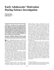
complex swirl pattern mechanically related? To answer this question, we constructed a large-scale model to investigate theflowpatterns that would be generated by a 30° rotation of a solid, hemispherical mass within a spherical fluid volume. Two pieces, a block (E) with a hemispherical cavity of radius 1", and a complementary solid quarter sphere (V), were machined from Teflon to represent half an egg and its vegetal yolk mass as though cut through the animal-vegetal pole (Fig. 5A). The close fit between the two pieces (0.001" clearance), a circular flange on V and a complementary track in E, ensured that relative rotation would be strictly concentric. The empty volume of the cavity in E wasfilledwith liquid to represent fluid cytoplasm in the animal hemisphere. Row patterns were studied by following the motion of small drops of dye applied to various positions and depths in the liquid. An important consideration in modelling flow patterns is to ensure that the Reynolds number (Re) - a parameter relating scale, viscosity, and velocity of flow - is of similar magnitude in the systems being compared. For the Xenopus egg vegetal yolk mass rotation, we estimated a Re in the range of 10 - using 1200/an for egg diameter, the kinematic viscosity of glycerol at 20°C (1.2xlO" 3 m 2 s" 1 ) as the value for animal hemisphere cytoplasm, and S/anmin" 1 for the velocity of the vegetal subcortex relative to the surface (Vincent et al. 1986). Because the model diameter is 42x greater than that of the Xenopus egg, we altered
Cytoplasmic movements in Xenopus eggs
30'
Fig. 5. Teflon model of cytoplasmicflowin frog egg. (A) Diagram showing hollowed out Teflon block (E) to represent spherical egg interior, and quarter sphere (V) to represent vegetal yolk mass. (B) Pattern offlowin glycerol-filled 'animal hemisphere' generated by 30° counterclockwise rotation of block E relative to V. Lines indicate position of dye marks at beginning (no dot) and end (dot) of rotation. (C) Dye mark before rotation. (D) Same dye mark after 30° rotation of outer piece. fluid viscosity and rotation rates to obtain a Re in the same range as that estimated for the egg. By cooling glycerol to -25 °C (increasing its kinematic viscosity by almost 200x) and rotating V relative to E at l°min , we achieved a Re of about 1CT6. At values