618
J. Appl. Co'st. (1997). 30, 618-622
Defect Structure of Ion-Irradiated Amorphous SiO2 YEHUDA EYAL, a* RAM EVRON b AND YACHIN COHEN c
aDepartment of Chemistry, bDepartment of Nuclear Engineering, and CDepartment of Chemical Engineering, Technion - Israel Institute of Technology, Haifa 32000, Israel. E-mail:
[email protected] (Received 23 July 1996; accepted 24 January 1997)
Abstract Uniformly enhanced small-angle X-ray scattering intensities of amorphous SiO2, measured following irradiation with 320keV H + and He + beams, are shown to be correlated, irrespective of the incident ion, with the O and Si cumulative displacement yields. Damage by both beams originated primarily from nuclear stopping but, under H+-ion irradiation, contributions from ionization processes were significant as well. At low beam fluences, the irradiated structure is compatible with the presence of stable radiation-induced interstitial-like O and Si atoms and complementary O and Si vacancy-like sites. There is no evidence for recovery near room temperature of the modified structure to the pre-irradiated state or for formation of colloidal-size scattering centers, such as gas bubbles or voids. Thus, ion-irradiation-induced changes in physical and chemical properties of silica seem to be due to the effect of the preserved primary atomic displacement damage.
1. Introduction Interest has arisen in the past 40 years in various aspects of structural alterations in silica (amorphous SiO2) that result from exposure to energetic particles. These range from fundamental characteristics of damage processes to applications in fields as diverse as microelectronics and nuclear waste disposal. However, although important changes in physical (Hines & Arndt, 1960; EerNisse, 1974; Presby & Brown, 1974; Hosono, 1991) and chemical (Hiraiwa, Usui & Yagi, 1989; Mazzoldi et al., 1991) properties of silica are attributed to atomic displacements, the investigation of property modifications or of their dependence upon the characteristics of the incident ions gives no direct information about the nature of the defects produced. The presence in ion-bombarded silica of defect centers that involve an O vacancy or an interstitial-like O atom has been previously inferred from electrical and spectroscopic measurements (see a recent survey by Devine, 1994). The most abundant defect is the EIt center, which is a positively charged and paramagnetic O vacancy. Another example is the peroxy radical, which involves an interstitial-like O atom, which is a neutral and paramagnetic defect (Devine, 1994). However, relative to the estimated number of ion-induced O displacements, the creation efficiency of remnant E'~ defects is always very small even at low defect concentrations, only ~0.03 in thermally grown © 1997 International Union of Crystallography Printed in Great Britain - all rights reserved
for example (Devine, 1988). It is not yet known whether the majority of the O defects has escaped detection or whether the damaged structure has largely recovered. The same is true for the entire population of the yet undetected Si defects. To date, no experiment has permitted insight into the damaged structure on a microscopic scale. Based on SAXS (smallangle X-ray scattering) measurements, we report herein the fate of instantaneously displaced O and Si atoms in ion-irradiated silica (320 keV H + and He + beams). Experimental ion-displacement yields are unavailable. In silica irradiated with 320 keV H + and He +, interstitials and vacancies are created mainly from dissipation of the ion energy via instantaneous atomic collision cascades (nuclear stopping) (Fleischer, Price & Walker, 1975). We estimated this contribution with the aid of the computer code TRIM (Ziegler, Biersack & Littmark, 1985), assuming a binding energy of 2eV per atom and a threshold energy of 20 eV for displacement of an O or an Si atom. The resulting average displacement yields are 16.4 displaced atoms per H + projectile (the energy dissipated via nuclear stopping is ,~0.7 keV) and 149 displaced atoms per He + projectile (the energy deposited via nuclear stopping is ,,~6.3 keV). Note that the displacement yield of He + is greater than that of H + by a factor of ,,~9. These estimated yields depend upon selected energy deposition parameters and may vary by up to roughly 20% for an acceptable range of parameter values. About 99.8% of the H + energy and ,~98.0% of the He + energy is deposited via a series of ionization, excitation and dissociation events (electron or 'ionization' stopping) (Fleischer, Price & Walker, 1975). Precise assessment of instantaneous interstitial-vacancy creation yield by ionization processes is at present impossible. Nevertheless, a reasonable estimate is a yield in the range from 7.4 to 10.6 atomic displacements per present H + or He + projectile. This estimate is based on a measured production efficiency of Ell centers in amorphous SiO2 by electron stopping, which is ,~700 to 1000 times less than that by nuclear stopping (Devine, 1988, 1994). Thus, the total atomic displacement yields in silica per 320 keV H + and He + ions are estimated to be 25 and 158 matrix atoms, respectively. Depth profiles of implanted ions and of instantaneous displacement and ionization damage in 2.2 g cm -3 silica, as computed by TRIM, are displayed in Figs. 1 and 2. Silica is accessible to X-ray small-angle scattering due to the presence of frozen-in density fluctuations. An a m o r p h o u s SiO2,
Journal of Applied Crystallography 1SSN 0021-8898
© 1997
YEHUDA EYAL, RAM EVRON AND YACHIN COHEN altered nanostructure that contains remnant atomic size defects is characterized by enhanced density fluctuations, and thereby should manifest itself by increased SAXS intensities. This behavior was corroborated by SAXS intensities reported by Bale, Shepler & Gibbs (1970) and by Bates, Hendricks & Shaffer (1974) for silica following irradiation with fast neutrons. In these studies, irradiation-induced variations in defect levels were characterized in terms of mean-square electron-density fluctuations, which is a useful means for understanding
E
4
H ATOMS PER iON
o 0
E
1
i
i
!
I
0 and Si ] DISPLACEMENTS
A /I
01
I
o.21-
'
,o.,~AT,O.'
0
i 0
I
2 3 DEPTH (~m)
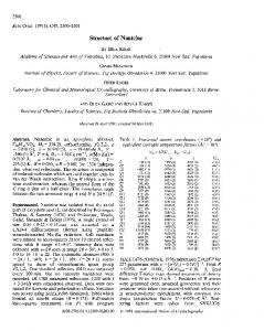
Fig. I. Estimated depth profiles of implanted ions and of displacement and ionization damage in silica following irradiation with 320 keV H + ions (TRIM computation procedure and parameters as specified in the text; results based on 17 000 ions). Top: Stopped projectile ions. Center: Nuclear stopping induced atomic displacement events. Bottom: Energy dissipation via ionization stopping.
E
:I 'A
4
He ATOMS
PER ION
I
l
i
E 4 0 0 I 0 and Si / DISPLACEMENTS
0
L
/
O.4 ~ " ~
^
f
I i
~
I0 NIZATI0 N
0.2
0
o
I DEPTH(/~m)
Fig. 2. Estimated depth profiles of implanted ions and of displacement and ionization damage in silica following irradiation with 320 keV He + ions (TRIM computation procedure and parameters as specitied in the text; results based on 1600 ions). Top: Stopped He atoms. Center: Nuclear stopping induced atomic displacement events. Bottom: Energy dissipation via ionization stopping.
619
the nature of these defects. The present research demonstrates that SAXS is an indispensable tool for yielding quantitative insight into structual alterations in silica that involve isolated atomic size defects.
2. Experimental procedures In all irradiations, the ion energy was totally dissipated within the silica target. Very thin silica foils were needed (see Figs. 1 and 2) in order to minimize scattering by unirradiated volume of target. We have employed 3 to 5 lain thick foils prepared by the glass-blowing technique. The foil density, as determined by the flotation method, was 2.200 (4)g cm -3. Foil thickness was determined by optical interferometry. Targets were characterized, irradiated and examined at room temperature and were stored at this temperature between experiments. Irradiations were carried out with an ion implanter. Ion-beam current densities were " 0.064 l-.-
i
|
i
0.05
0 038,
ooo5~
oo n],,.,-,-~,,.,.r-m, irl .,7. -ooo5] .~,~,~,a~,,,~,,~~~,bilah,[ "
2
¢
o :7
. 2
.
.
3 h(nm-I) (b)
.
4
~ , ,
~ I~
5
Fig. 4. Measured SAXS curves of silica foils irradiated by 320 keV He + ions. Intensity is given in counts s - I channel - ] . (a) Ion fluence 2.0 x 10¹⁵ c m - 2 ; (b) ion fluence 0.5 × 1015 cm -2. Curves (1) and (2) indicate intensities before and after irradiation, respectively, instrumental background intensity included. The curve in the lower portion of each figure shows the net intensity increase after irradiation.
0.068
•
,
i
w
: I 0.092-
,
0.o44
20
00160.0 0076
(a) .
.
T.
.
T
T
T
z_
% 0.056. ~
~e
2
I,LI
0.036oo
t
,tad, LL~It~.,I LLd~, j,~-
0.006 -
o
It
~
'F
---
~ h(nm-I) (h)
z o
5 DISPLACEMENTS PER cm z
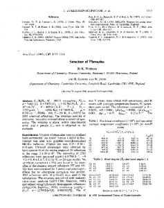
Fig. 3. Measured SAXS curves of silica foils irradiated by 320 keV H + ions. Intensity is given in counts s - I channel-I. (a) ion fluence 3.0 x 10¹⁶ c m - 2 ; (b) ion fluence 1.0 x 1016 cm -2. Curves (1) and (2) indicate intensities before and after irradiation, respectively, instrumental background intensity included. The curve in the lower portion of each figure shows the net intensity increase after irradiation.
Fig. 5. Absolute net intensity increment cm -2 of silica surface following irradiation with H ÷ ions (solid circles) and He ÷ ions (open circles) as a function of the cumulative number of Si and O displacements cm -2 of silica surface. Displacements include contributions from atomic collision events and from ionization processes.
622
CONFERENCE PROCEEDINGS
& Walker, 1975). Note that these spikes display effects of individual ions. Production of a thermal spike requires transfer of kinetic energy to a large number of adjacent matrix atoms via atomic collision cascades, whereas the present H + and He + projectiles are characterized by low atomic displacement yields. Production of an electrostatic explosion requires the removal of a large number of bound electrons from matrix atoms along the ion trajectory, whereas the ionizing powers of the present H + and He + ions are too small to cause any appreciable effect. It is thus likely that, below damage saturation and in the absence of any appreciable annealing, the permanent structural defects are presented by preserved instantaneous defects. We derive the average value of AI according to the following assumptions: (i) All initial atomic displacement damage is structurally stable at near room temperature. (ii) The O/Si atom-number displacement ratio is 1.63, as given by the present TRIM computations. (iii) Each defect site that involves an O interstitial or an O vacancy possesses an intensity of 82 e.u. (iv) Each defect site that involves an Si interstitial or an Si vacancy possesses an intensity of 142 e.u. With these assumptions, the theoretical estimate of A I is 228 e.u., in acceptable agreement with the experimental value. Should the relative O and Si displacement yields be solely dictated by stoichiometry, namely with O/Si atom-number displacement ratio of 2, the theoretical estimate of A I is 216e.u. Note that empirical threshold energies for O and Si displacement in amorphous SiO2 are unavailable. Nevertheless, it is evident that reproduction of the measured lo(h) by model computations is readily achievable within an acceptable range of atom binding and displacement parameters. The above analysis suggests that recovery near room temperature of the modified silica nanostructure to the initial vitreous state is largely unimportant. Because the scattering intensities of the implanted H and He atoms are only 1 and 22 e.u., respectively, it is practically impossible to trace the fate of these atoms by SAXS. The absence of defect removal and of H2 and He removal into extended structures such as voids or gas bubbles is directly supported by the SAXS data through an absent enhanced forward-scattering intensity. Although the SAXS technique is insensitive to the nature of atom rebonding in the distorted network, the importance of rebonding is evidenced by the low abundance of E' l centers. 4. Conclusions
The present study provides a first quantitative insight on a nanometric scale into the defect structure of silica following irradiation with light ions, specifically 320 keV H + and He +. Radiation-induced increased SAXS intensities are attributed to atomic displacements. These were produced primarily from dissipation of the ion energy via atomic collision cascades, but a contribution from ionization processes was important as well under H + irradiation. The SAXS data suggest that below
defect saturation the primary displacement damage is largely preserved in the form of interstitial-like O and Si atoms and complementary O and Si vacancy-like sites. Despite the nonuniformities in the damage distribution and in the depth profile of the implanted atoms in the specimen, with roughly similar pronounced maxima in both distributions near the average projectile range, there is no evidence for the importance of localized structural alterations, such as defect clustering, on a scale of several nanometers. It is thus evident that, in ion-irradiated silica, the structural modification is due to preserved primary atomic size defects. This research demonstrates that SAXS is an indispensable tool for studying radiation effects in amorphous structures that involve isolated point-like defects. The authors thank Dr Roderick A. B. Devine (CNET, France Telecom, Meylan) for his helpful comments.
References
Bale, H. D., Shepler, R. E. & Gibbs, G. W. (1970). J. Appl. Phys. 41, 241-246. Bates, J. B., Hendricks, R. W. & Shaffer, L. B. (1974). J. Chem. Phys. 61, 4163--4176. Billington, D. S. & Crawford, J. H. (1961). Radiation Damage in Solids. Princeton University Press. Devine, R. A. B. (1988). The Physics and Chemistry of Si02 and Si-SiOe Interface, edited by R. Helms & B. E. Deal, pp. 519-527. New York: Plenum. Devine, R. A. B. (1994). Nucl. Instrum. Methods Phys. Res. B91, 378-390. EerNisse, E. P. (1974). J. Appl. Phys. 45, 167-174. Fleischer, R. L., Price, P. B. & Walker, R. M. (1975). Nuclear Tracks in Solids. Berkeley: University of California Press. Glatter, O. & Kratky, O. (1982). Small Angle X-ray Scattering. London: Academic Press. Guinier, A. & Foumet, G. (1955). Small-Angle Scattering of X-rays. New York: Wiley. Hines, R. L. & Amdt, R. (1960). Phys. Rev. 119, 623-633. Hiraiwa, A., Usui, H. & Yagi, K. (1989). Appl. Phys. Lett. 54, 1106-1108. Hosono, H. (1991). J. Appl. Phys. 69, 8079-8082. Levelut, A. M. & Guinier, A. (1967). Bull. Soc. Fr. Mineral. Crystallogr. 90, 445-451. Mazzoldi, P., Camera, A., Caccavale, F., Favaro, M. L., Boscolo-Boscoletto, A., Granozzi, G., Bertoncello, R. & Battaglin, G. (1991). J. Appl. Phys. 70, 3528-3536. Porai-Koshits, E. A. (1976). Ann. Rev. Mater Sci. 6, 389-409. Presby, H. M. & Brown, W. L. (1974). Appl. Phys. Lett. 24, 511-513. Renninger, A. L. & Uhlmann, D. R. (1974). J. Non-Cryst. Solids, 16, 325-327. Stabinger, H. & Kratky, O. (1978). Makromol. Chem. 179, 1655-1659. Weinberg, D. L. (1962). J. Appl. Phys. 33, 1012-1013. Weinberg, D. L. (1963a). Phys. Lett. 7, 324-325. Weinberg, D. L. (1963b). Rev. Sci. Instrum. 34, 691-696. Ziegler, J. F., Biersack, J. P. & Littmark, O. (1985). The Stopping and Range of lons in Solids, edited by J. F. Ziegler, Vol. 1, Chap. 4. New York: Pergamon.